Download presentation
Presentation is loading. Please wait.

1
Radiographic Interpretation Review: Anatomic Landmarks, Caries, Bone loss & Dental Materials Also processing/operator errors

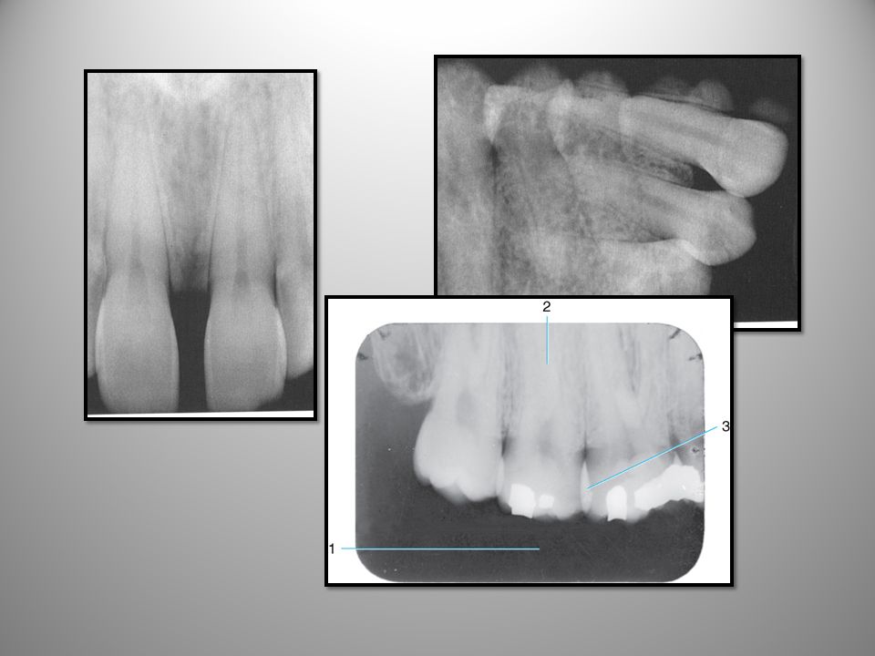
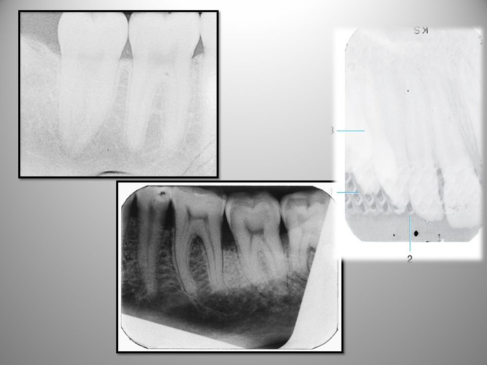
2
1-2 parts of the tooth again; 3/6 pulp; 4 PDL; 5 lamina dura; 7 cancellous bone (trabeculation)
What is #1- 7 pointing to?

4
Radiographic Anatomy Basics: Canine Area
3 4 2 1 Activity: What are the DM on tooth #8?

5
Radiographic Anatomy Basics: Anterior Region
2 3 Also can see the nasal concha; nasal septum clearly

6
Radiographic Anatomy Basics: Premolar Area
Activity: Can you identify 1-6 in this picture?

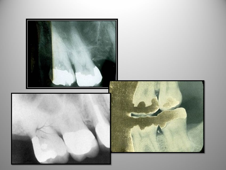
7
Radiographic Anatomy Basics: Molar Area
8

8
Radiographic Anatomy Basics: Molar Area
Activity: What is the thin radiopaque line here? 3 Soft tissue outline of maxillary tuberosity Look at typo– see that the beam will penetrate the anatomic structures and will show up on the film– coronoid process; zygomatic arch

9
Radiographic Anatomy Basics: Lower anterior

10
Radiographic Anatomy Basics: Lower Canine Area

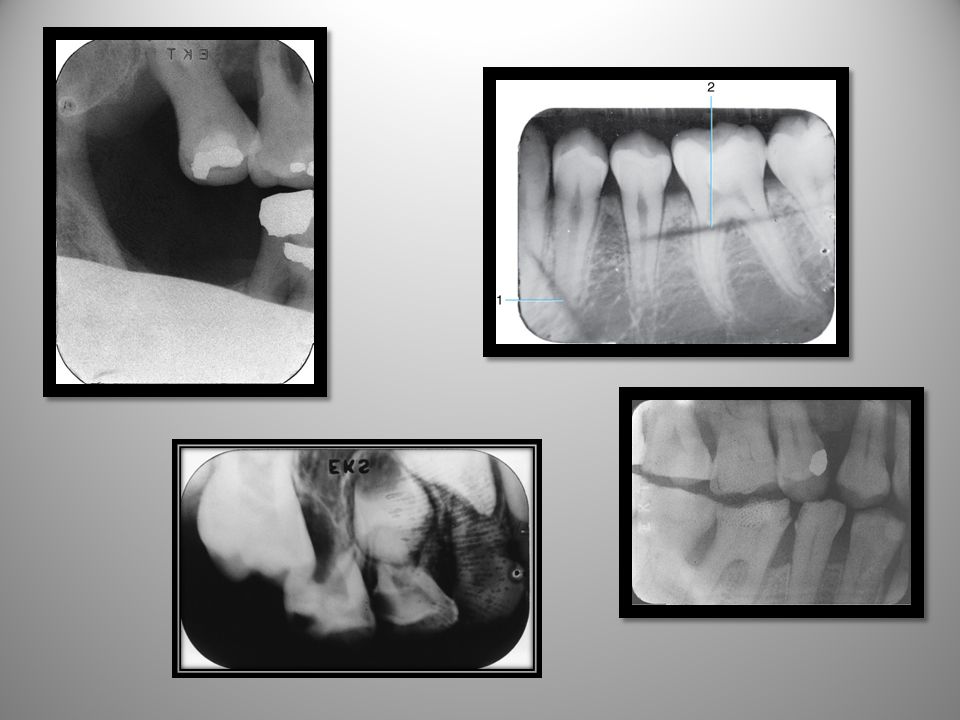
11
Radiographic Anatomy Basics: Lower premolar- molar area
Activity: Why do you see the cortical bone or inferior border of the mandible here?

12
Radiographic Anatomy Basics: Molar Area

13
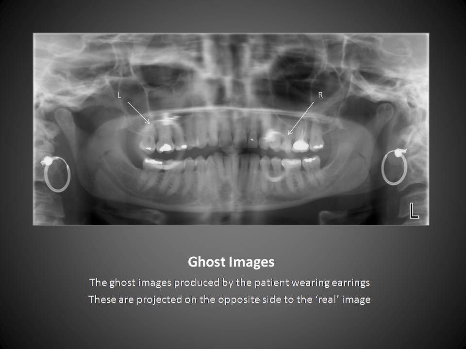
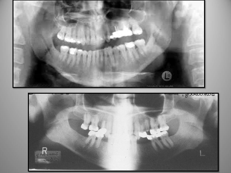
Anatomy: Panoral Radiograph 10

14
Activity: What was the operator error here (excessive smile)?
")
15
8 7 oblique ridge (ext) coronoid process
 8. coronoid process")
16
FIND: orbit, anterior nasal spine; nasal conchae; hard palate; maxillary sinus; floor of maxillary sinus; median palatine suture; submandibular fossa; bite block; air or open spaces; mandibular canal; impacted #17

18
Be sure to review chapter 27 & 29 in rad book for normal anatomy
Be sure to review chapter 27 & 29 in rad book for normal anatomy. Bones will be pictures similar to anatomy book and on PP. Identify: hard palate; mandibular notch; art eminence; lip line; mand canal; hyoid bone; nasal septum; condyle; zygomatic process; #14; mental foramen; ant nasal spine; ramus; glenoid fossa; ext auditory meatus

19
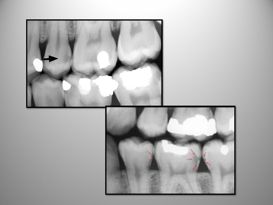
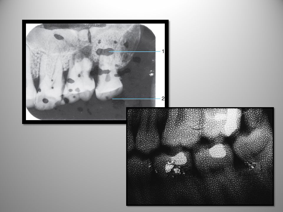
Dental Caries & Materials

20
Dental Caries & Materials

22
Dental Caries ROOT

25
Skim through chapter 35 in rad book on periapical lesions

26
Radiographic Appearance: Dental Restorative Material

27
Radiographic Appearance: Dental Restorative Material

28
Restorative Materials

29
Restorative Materials
Be sure to look in book on page shows implants, lingual bar, all porc crowns, ss crowns, etc….

31
Restorative Materials

32
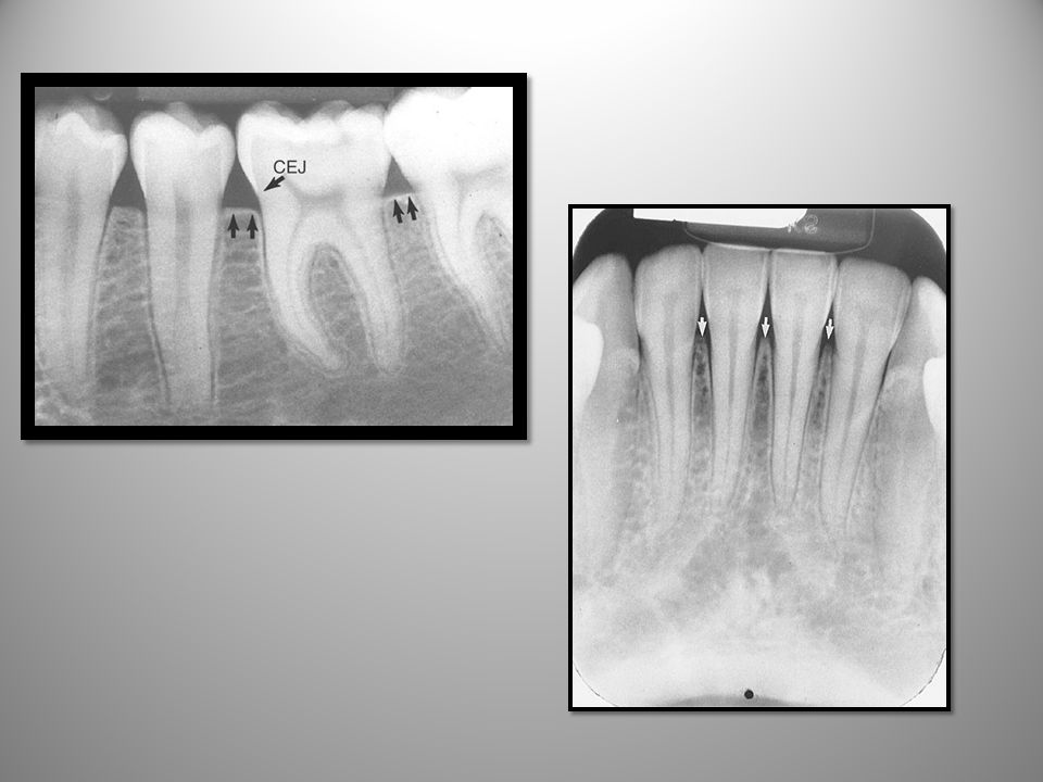
Bone Levels Normal bone where? Compared to CEJ? CEJ CEJ
In health– no radiolucency at crestal bone- LD can be flat in molar and pointed in anterior- but should be continuous; PDL should also be continuous

Similar presentations


 Process = Prominence or extension.>")


