Download presentation
Presentation is loading. Please wait.

1
Physiology of respiratory system. External breathing

2
General functions of respiratory system The respiratory system comprises of the nose, mouth, throat, larynx, trachea, bronchi and lungs. The function of the respiratory system is to facilitate gaseous exchange to take place in the lungs and tissue cells of the body. Oxygen is required by cells in the body to allow various metabolic reactions to take place and to produce energy and is therefore essential to life. The respiratory system may be defined as the organs and tissues through which air is passed into and out of the body to allow the necessary gaseous exchanges to take place.

3
Oral and nasal cavity

4
Functions of air conductive pathway

5
Lungs

7
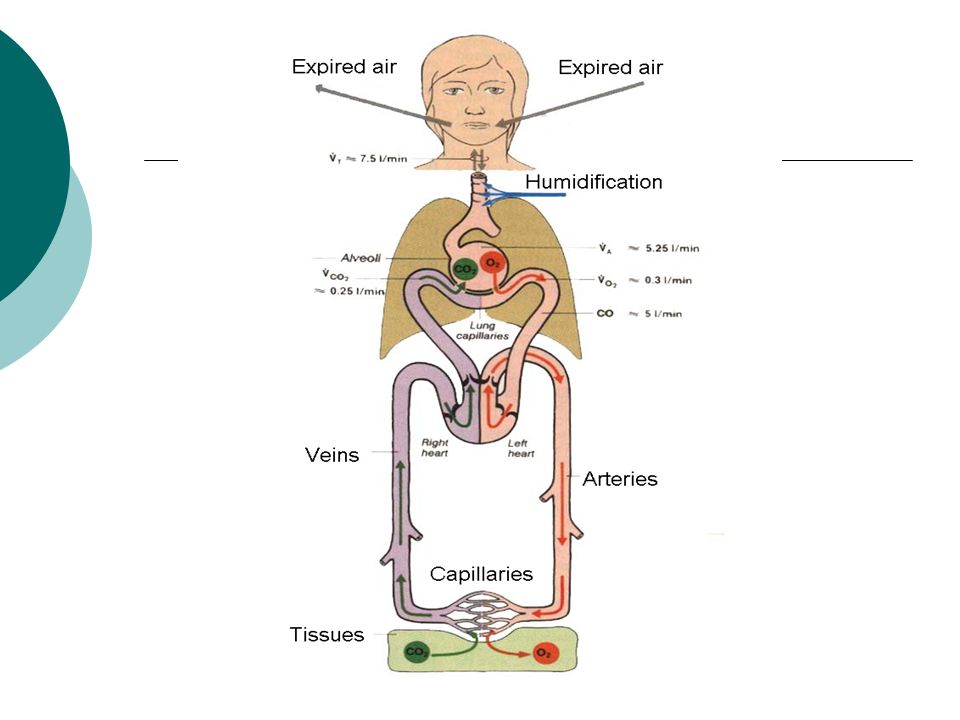
External and internal respiration External respiration is the means by which oxygen from the air passes into the blood stream for transportation to the tissue cells and carbon dioxide is collected and transferred back to the lungs and expelled from the body. Internal respiration involves the vital chemical activities which take place in every living cell requiring oxygen and glycogen to combine and release energy, water and carbon dioxide. The normal rate of inspiration and expiration, the respiration rate, is about 16 times a minute in an adult.

9
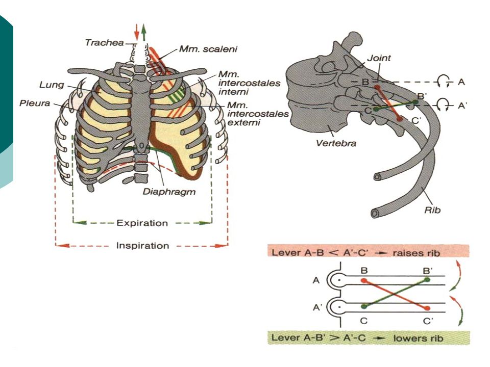
Biomechanism of breathing Breathing is an active process - requiring the contraction of skeletal muscles. The primary muscles of respiration include the external intercostal muscles (located between the ribs) and the diaphragm (a sheet of muscle located between the thoracic & abdominal cavities). The external intercostals plus the diaphragm contract to bring about inspiration: Contraction of external intercostal muscles > elevation of ribs & sternum > increased front- to-back dimension of thoracic cavity > lowers air pressure in lungs > air moves into lungs Contraction of diaphragm > diaphragm moves downward > increases vertical dimension of thoracic cavity > lowers air pressure in lungs > air moves into lungs: To exhale: relaxation of external intercostal muscles & diaphragm > return of diaphragm, ribs, & sternum to resting position > restores thoracic cavity to preinspiratory volume > increases pressure in lungs > air is exhaled
 and the diaphragm (a sheet of muscle located between the thoracic & abdominal cavities). The external intercostals plus the diaphragm contract to bring about inspiration: Contraction of external intercostal muscles > elevation of ribs & sternum > increased front- to-back dimension of thoracic cavity > lowers air pressure in lungs > air moves into lungs Contraction of diaphragm > diaphragm moves downward > increases vertical dimension of thoracic cavity > lowers air pressure in lungs > air moves into lungs: To exhale: relaxation of external intercostal muscles & diaphragm > return of diaphragm, ribs, & sternum to resting position > restores thoracic cavity to preinspiratory volume > increases pressure in lungs > air is exhaled.")
12
Effect of Rib and Sternum Movement on Thoracic Volume

13
EffectEffect of Rib and Diaphragm Movement on Thoracic Volume

14
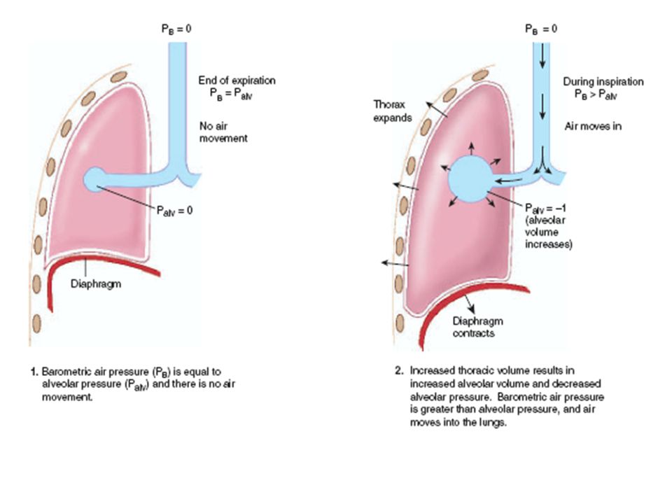
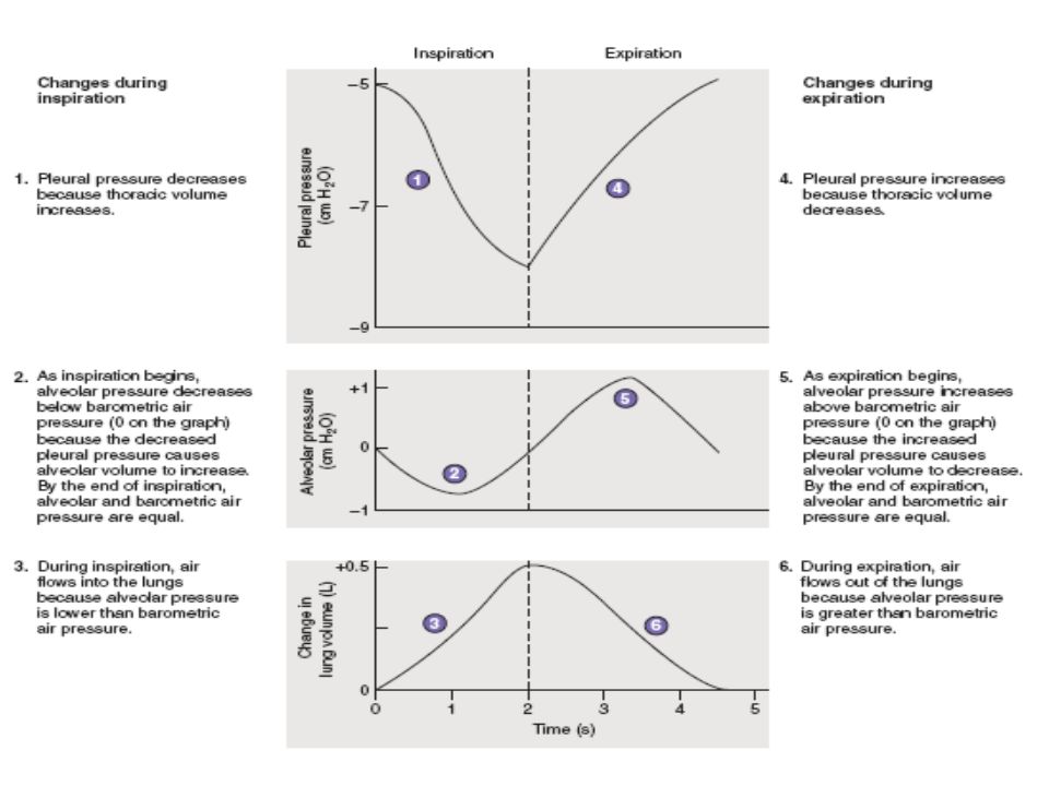
Pressure in lungs As the external intercostals & diaphragm contract, the lungs expand. The expansion of the lungs causes the pressure in the lungs (and alveoli) to become slightly negative relative to atmospheric pressure. As a result, air moves from an area of higher pressure (the air) to an area of lower pressure (our lungs & alveoli). During expiration, the respiration muscles relax & lung volume descreases. This causes pressure in the lungs (and alveoli) to become slight positive relative to atmospheric pressure. As a result, air leaves the lungs.
 to become slightly negative relative to atmospheric pressure. As a result, air moves from an area of higher pressure (the air) to an area of lower pressure (our lungs & alveoli). During expiration, the respiration muscles relax & lung volume descreases. This causes pressure in the lungs (and alveoli) to become slight positive relative to atmospheric pressure. As a result, air leaves the lungs..")
17
Surface tension in lungs The walls of alveoli are coated with a thin film of water & this creates a potential problem. Water molecules, including those on the alveolar walls, are more attracted to each other than to air, and this attraction creates a force called surface tension. This surface tension increases as water molecules come closer together, which is what happens when we exhale & our alveoli become smaller (like air leaving a balloon). Potentially, surface tension could cause alveoli to collapse and, in addition, would make it more difficult to 're-expand' the alveoli (when you inhaled). Both of these would represent serious problems: if alveoli collapsed they'd contain no air & no oxygen to diffuse into the blood &, if 're-expansion' was more difficult, inhalation would be very, very difficult if not impossible. Fortunately, our alveoli do not collapse & inhalation is relatively easy because the lungs produce a substance called surfactant that reduces surface tension.
. Potentially, surface tension could cause alveoli to collapse and, in addition, would make it more difficult to re-expand the alveoli (when you inhaled). Both of these would represent serious problems: if alveoli collapsed they d contain no air & no oxygen to diffuse into the blood &, if re-expansion was more difficult, inhalation would be very, very difficult if not impossible. Fortunately, our alveoli do not collapse & inhalation is relatively easy because the lungs produce a substance called surfactant that reduces surface tension..")
19
Spirometer, Lung Volumes, and Lung Capacities

20
Spirometry

21
CardiopulmonaryCardiopulmonary circulation

22
Mechanism of gas transport Primary function is to obtain oxygen for use by body's cells & eliminate carbon dioxide that cells produce. Includes respiratory airways leading into (& out of) lungs plus the lungs themselves Pathway of air: nasal cavities (or oral cavity) > pharynx > trachea > primary bronchi (right & left) > secondary bronchi > tertiary bronchi > bronchioles > alveoli (site of gas exchange) The exchange of gases (O2 & CO2) between the alveoli & the blood occurs by simple diffusion: O2 diffusing from the alveoli into the blood & CO2 from the blood into the alveoli. Diffusion requires a concentration gradient. So, the concentration (or pressure) of O2 in the alveoli must be kept at a higher level than in the blood & the concentration (or pressure) of CO2 in the alveoli must be kept at a lower lever than in the blood. We do this, of course, by breathing - continuously bringing fresh air (with lots of O2 & little CO2) into the lungs & the alveoli.
 lungs plus the lungs themselves Pathway of air: nasal cavities (or oral cavity) > pharynx > trachea > primary bronchi (right & left) > secondary bronchi > tertiary bronchi > bronchioles > alveoli (site of gas exchange) The exchange of gases (O2 & CO2) between the alveoli & the blood occurs by simple diffusion: O2 diffusing from the alveoli into the blood & CO2 from the blood into the alveoli. Diffusion requires a concentration gradient. So, the concentration (or pressure) of O2 in the alveoli must be kept at a higher level than in the blood & the concentration (or pressure) of CO2 in the alveoli must be kept at a lower lever than in the blood. We do this, of course, by breathing - continuously bringing fresh air (with lots of O2 & little CO2) into the lungs & the alveoli..")
24
Role of Pulmonary Surfactant Surfactant decreases surface tension which: 1)increases pulmonary compliance (reducing the effort needed to expand the lungs) 2)reduces tendency for alveoli to collapse
increases pulmonary compliance (reducing the effort needed to expand the lungs) 2)reduces tendency for alveoli to collapse")
25
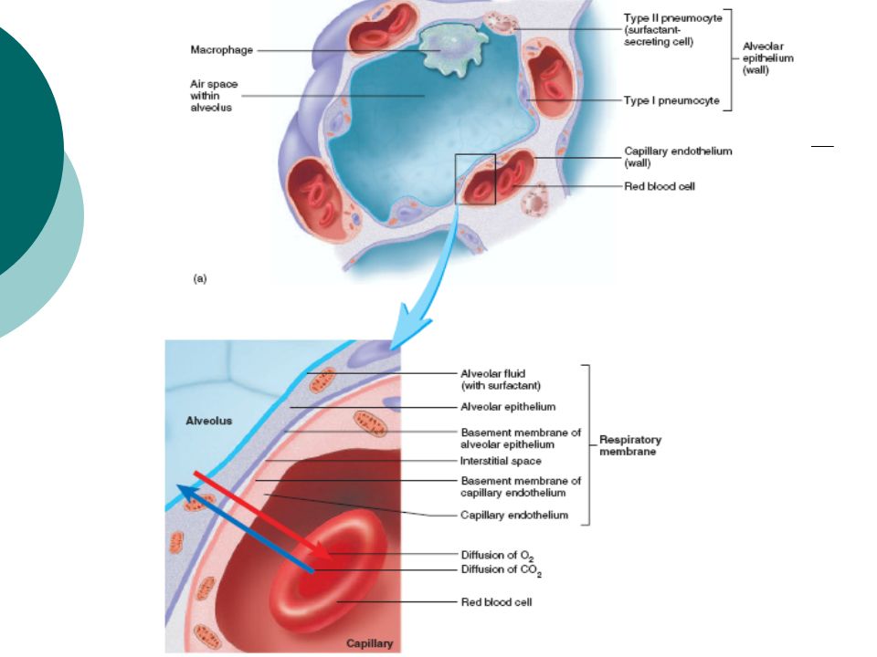
Exchange of gases Exchange of O2 & CO2 between external environment & the cells of the body efficient because alveoli and capillaries have very thin walls & are very abundant (your lungs have about 300 million alveoli with a total surface area of about 75 square meters) Internal respiration - intracellular use of O2 to make ATP and occurs by simple diffusion along partial pressure gradients
 Internal respiration - intracellular use of O2 to make ATP and occurs by simple diffusion along partial pressure gradients")
27
Role of the partial pressure of gases it's the individual pressure exerted independently by a particular gas within a mixture of gasses. The air we breath is a mixture of gasses: primarily nitrogen, oxygen, & carbon dioxide. So, the air you blow into a balloon creates pressure that causes the balloon to expand (& this pressure is generated as all the molecules of nitrogen, oxygen, & carbon dioxide move about & collide with the walls of the balloon). However, the total pressure generated by the air is due in part to nitrogen, in part to oxygen, & in part to carbon dioxide. That part of the total pressure generated by oxygen is the 'partial pressure' of oxygen, while that generated by carbon dioxide is the 'partial pressure' of carbon dioxide. A gas's partial pressure, therefore, is a measure of how much of that gas is present (e.g., in the blood or alveoli). The partial pressure exerted by each gas in a mixture equals the total pressure times the fractional composition of the gas in the mixture. So, given that total atmospheric pressure (at sea level) is about 760 mm Hg and, further, that air is about 21% oxygen, then the partial pressure of oxygen in the air is 0.21 times 760 mm Hg or 160 mm Hg.
. However, the total pressure generated by the air is due in part to nitrogen, in part to oxygen, & in part to carbon dioxide. That part of the total pressure generated by oxygen is the partial pressure of oxygen, while that generated by carbon dioxide is the partial pressure of carbon dioxide. A gas s partial pressure, therefore, is a measure of how much of that gas is present (e.g., in the blood or alveoli). The partial pressure exerted by each gas in a mixture equals the total pressure times the fractional composition of the gas in the mixture. So, given that total atmospheric pressure (at sea level) is about 760 mm Hg and, further, that air is about 21% oxygen, then the partial pressure of oxygen in the air is 0.21 times 760 mm Hg or 160 mm Hg..")
29
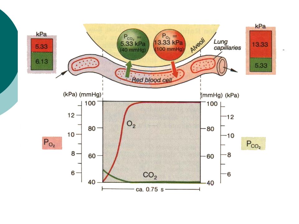
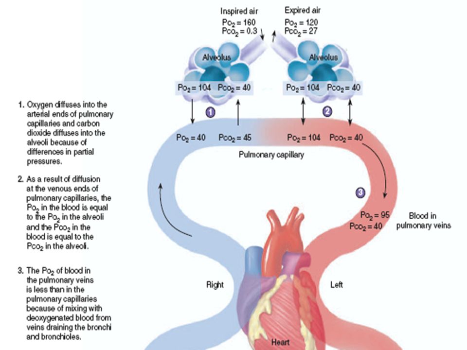
Level of the partial pressure of main gasesin the human body Partial Pressures of O2 and CO2 in the body (normal, resting conditions): Alveoli PO2 = 100 mm Hg PCO2 = 40 mm Hg Alveolar capillaries Entering the alveolar capillaries PO2 = 40 mm Hg (relatively low because this blood has just returned from the systemic circulation & has lost much of its oxygen) PCO2 = 45 mm Hg (relatively high because the blood returning from the systemic circulation has picked up carbon dioxide)
: Alveoli PO2 = 100 mm Hg PCO2 = 40 mm Hg Alveolar capillaries Entering the alveolar capillaries PO2 = 40 mm Hg (relatively low because this blood has just returned from the systemic circulation & has lost much of its oxygen) PCO2 = 45 mm Hg (relatively high because the blood returning from the systemic circulation has picked up carbon dioxide)")
31
While in the alveolar capillaries, the diffusion of gasses occurs: oxygen diffuses from the alveoli into the blood & carbon dioxide from the blood into the alveoli. Leaving the alveolar capillaries PO2 = 100 mm Hg PCO2 = 40 mm Hg Blood leaving the alveolar capillaries returns to the left atrium & is pumped by the left ventricle into the systemic circulation. This blood travels through arteries & arterioles and into the systemic, or body, capillaries. As blood travels through arteries & arterioles, no gas exchange occurs. Entering the systemic capillaries PO2 = 100 mm Hg PCO2 = 40 mm Hg Body cells (resting conditions) PO2 = 40 mm Hg PCO2 = 45 mm Hg
 PO2 = 40 mm Hg PCO2 = 45 mm Hg.")
32
Changes in the Partial Pressures of Oxygen and Carbon Dioxide

33
Because of the differences in partial pressures of oxygen & carbon dioxide in the systemic capillaries & the body cells, oxygen diffuses from the blood & into the cells, while carbon dioxide diffuses from the cells into the blood. Leaving the systemic capillaries PO2 = 40 mm Hg PCO2 = 45 mm Hg Blood leaving the systemic capillaries returns to the heart (right atrium) via venules & veins (and no gas exchange occurs while blood is in venules & veins). This blood is then pumped to the lungs (and the alveolar capillaries) by the right ventricle.
 via venules & veins (and no gas exchange occurs while blood is in venules & veins). This blood is then pumped to the lungs (and the alveolar capillaries) by the right ventricle..")
34
The oxygen-hemoglobin dissociation curve The oxygen-hemoglobin dissociation curve 'shifts' under certain conditions. These factors can cause such a shift: 1)lower pH 2)increased temperature 3)more 2,3-diphosphoglycerate 4)increased levels of CO2
lower pH 2)increased temperature 3)more 2,3-diphosphoglycerate 4)increased levels of CO2.")
35
Oxygen- Hemoglobin Dissociation Curve at Rest

36
These factors change when tissues become more active. For example, when a skeletal muscle starts contracting, the cells in that muscle use more oxygen, make more ATP, & produce more waste products (CO2). Making more ATP means releasing more heat; so the temperature in active tissues increases. More CO2 translates into a lower pH. That is so because this reaction occurs when CO2 is released: CO2 + H20 -----> H2CO3 -----> HCO3- + H+ & more hydrogen ions = a lower (more acidic) pH. So, in active tissues, there are higher levels of CO2, a lower pH, and higher temperatures.
. Making more ATP means releasing more heat; so the temperature in active tissues increases. More CO2 translates into a lower pH. That is so because this reaction occurs when CO2 is released: CO2 + H > H2CO > HCO3- + H+ & more hydrogen ions = a lower (more acidic) pH. So, in active tissues, there are higher levels of CO2, a lower pH, and higher temperatures..")
37
At lower PO2 levels, red blood cells increase production of a substance called 2,3-diphosphoglycerate. These changing conditions (more CO2, lower pH, higher temperature, & more 2,3-diphosphoglycerate) in active tissues cause an alteration in the structure of hemoglobin, which, in turn, causes hemoglobin to give up its oxygen. In other words, in active tissues, more hemoglobin molecules give up their oxygen.
 in active tissues cause an alteration in the structure of hemoglobin, which, in turn, causes hemoglobin to give up its oxygen. In other words, in active tissues, more hemoglobin molecules give up their oxygen..")
38
Carbon Dioxide Transport and Chloride Movement in Tissues

39
Carbon Dioxide Transport and Chloride Movement in Lungs

41
Role of Pulmonary Surfactant Surfactant decreases surface tension which: 1)increases pulmonary compliance (reducing the effort needed to expand the lungs) 2)reduces tendency for alveoli to collapse
increases pulmonary compliance (reducing the effort needed to expand the lungs) 2)reduces tendency for alveoli to collapse")
45
Changes in the Partial Pressures of Oxygen and Carbon Dioxide

46
Oxygen- Hemoglobin Dissociation Curve at Rest

47
Carbon Dioxide Transport and Chloride Movement in Tissues

48
Carbon Dioxide Transport and Chloride Movement in Lungs

49
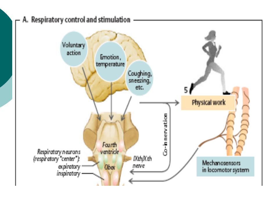
Respiratory Structures in the Brainstem VideoVideo

51
Location of the carotid and aortic bodies. Note that each carotid body is quite close to a carotid sinus, the major arterial baroreceptor. Both right and left common carotid bifurcations contain a carotid sinus and a carotid body.

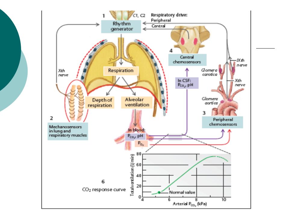
53
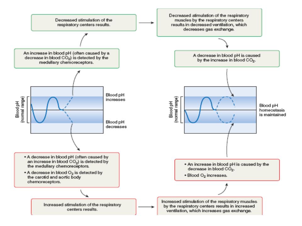
Pathways by which increased arterial PCO2 stimulates ventilation. Note that the peripheral chemoreceptors are stimulated by an increase in H concentration, whereas they are also stimulated by a decrease in PO2.

54
Summary of factors that stimulate ventilation during exercise

55
Ventilation changes during exercise. Note (1) the abrupt increase at the onset of exercise and (2) the equally abrupt but larger decrease at the end of exercise.
 the abrupt increase at the onset of exercise and (2) the equally abrupt but larger decrease at the end of exercise..")
Similar presentations


>")