Download presentation
Presentation is loading. Please wait.

1
Lower limb injuries Richard Hardern

2
Content Knee, ankle, foot Anatomy History and examination Treatment of limb threatening problems

3
Not a case for the Emergency Nurse Practitioner!

4
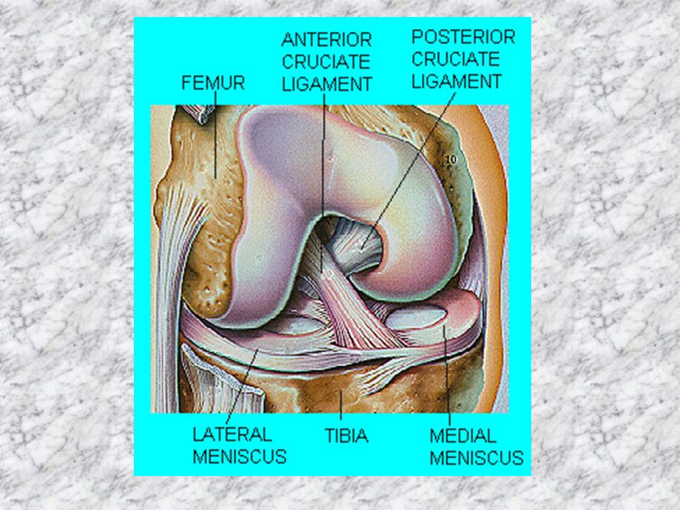
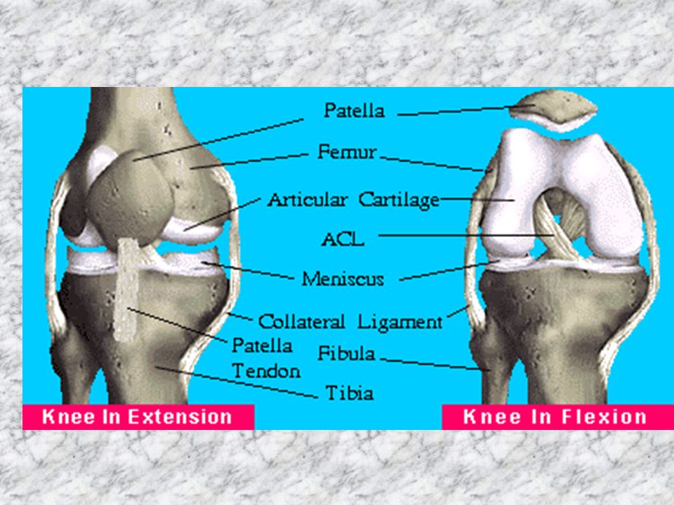
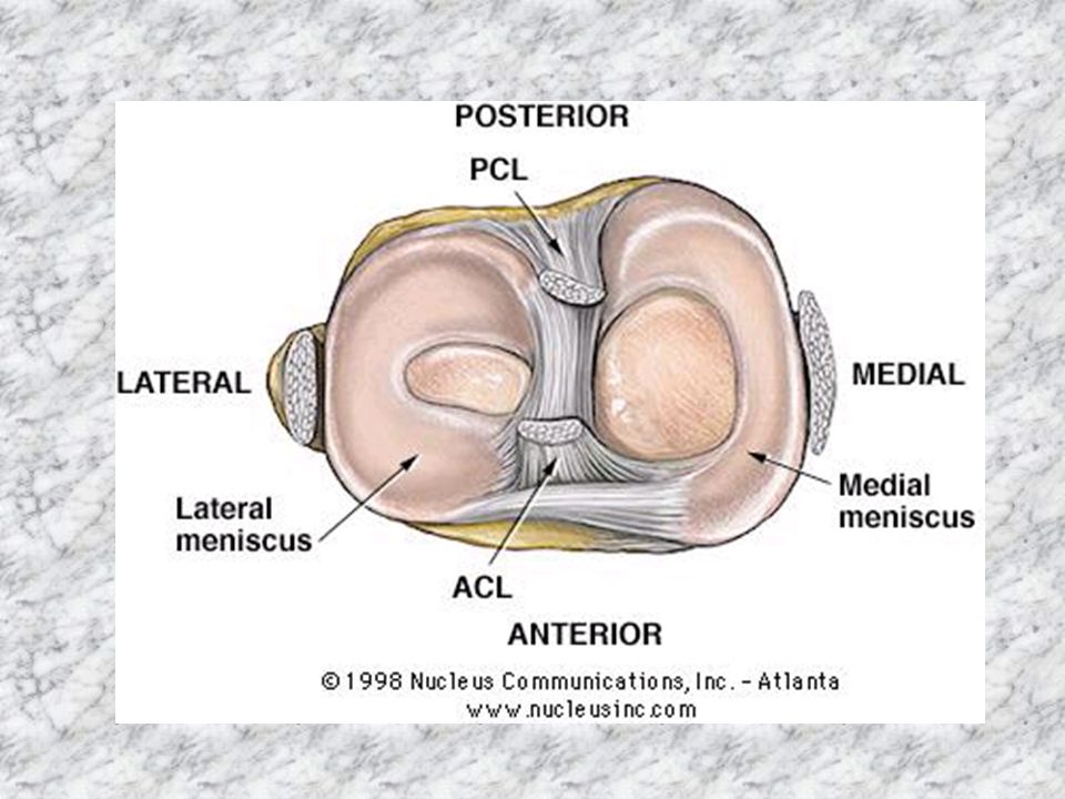
Knee anatomy Bones Ligaments: cruciate and collateral Menisci

9
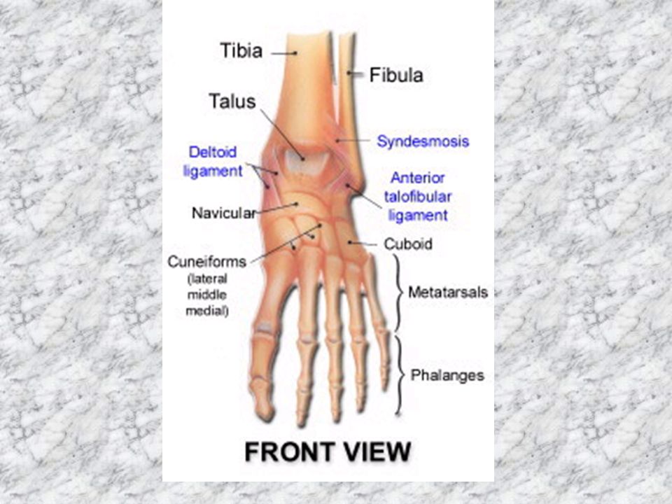
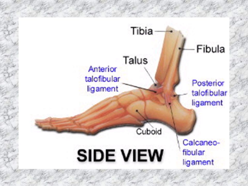
Ankle anatomy Bones Ligaments: medial & lateral Tendons

13
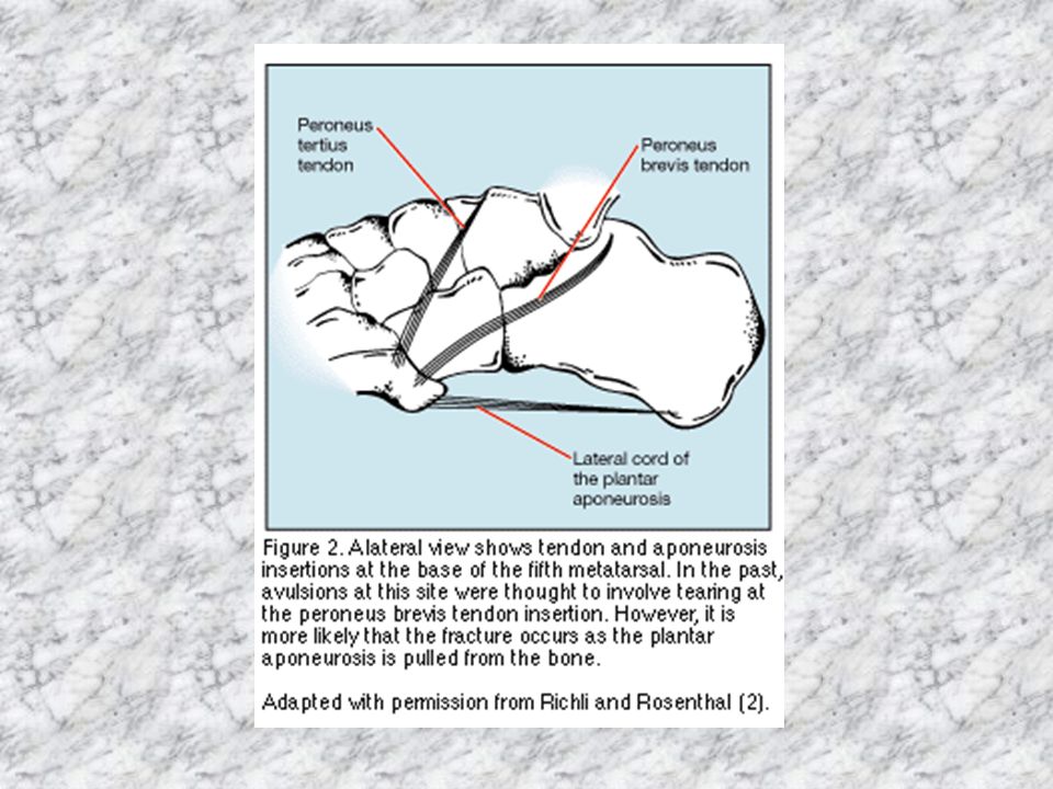
Peroneus brevis

14
Gastrocnemius

15
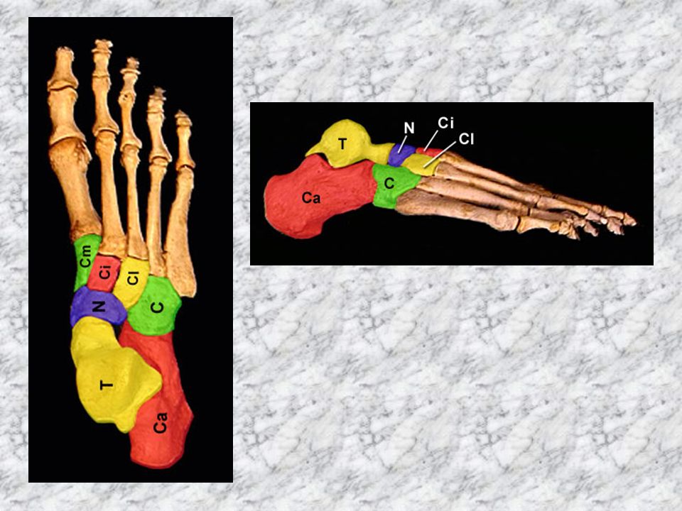
Foot anatomy Bones

17
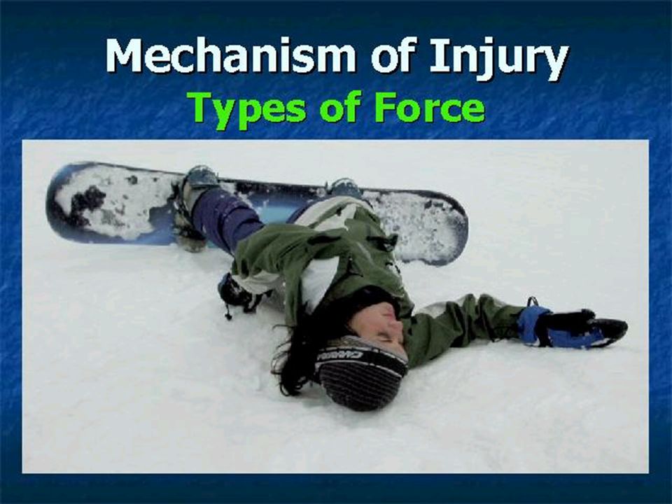
History & examination Mechanism of injury

19
General Considerations Always inquire about the mechanism of injury. Always inquire about the effect on function. A lways do the following in this order: Inspection Palpation Range of Motion (active before passive)
.")
20
Knee: look Skin- scars, redness Muscle- wasting of quads (compare diameter of thigh if quads wasted) Bone/joint- Effusion, Varus Valgus deformity( measure intermalleolar distance if valgus), Watch them walking too at some point (even if only from WR into examination cubicle)
 Bone/joint- Effusion, Varus Valgus deformity( measure intermalleolar distance if valgus), Watch them walking too at some point (even if only from WR into examination cubicle)")
21
Knee: feel Skin - Temperature, back of hand Muscle- Ask patient to contract quads Bone/joint- Effusion fluid displacement test, patellar tap test (may be negative if tense effusion) Joint line tenderness (with knee bent) Patellar tendon MCL,LCL Popliteal swellings
 Joint line tenderness (with knee bent) Patellar tendon MCL,LCL Popliteal swellings")
23
Knee: move Active then passive- Flexion (135 degrees normal) Extension (put hand behind knee) Feel for crepitus
 Extension (put hand behind knee) Feel for crepitus")
24
Knee: special tests - collaterals

25
Knee: special tests - cruciates ACL

26
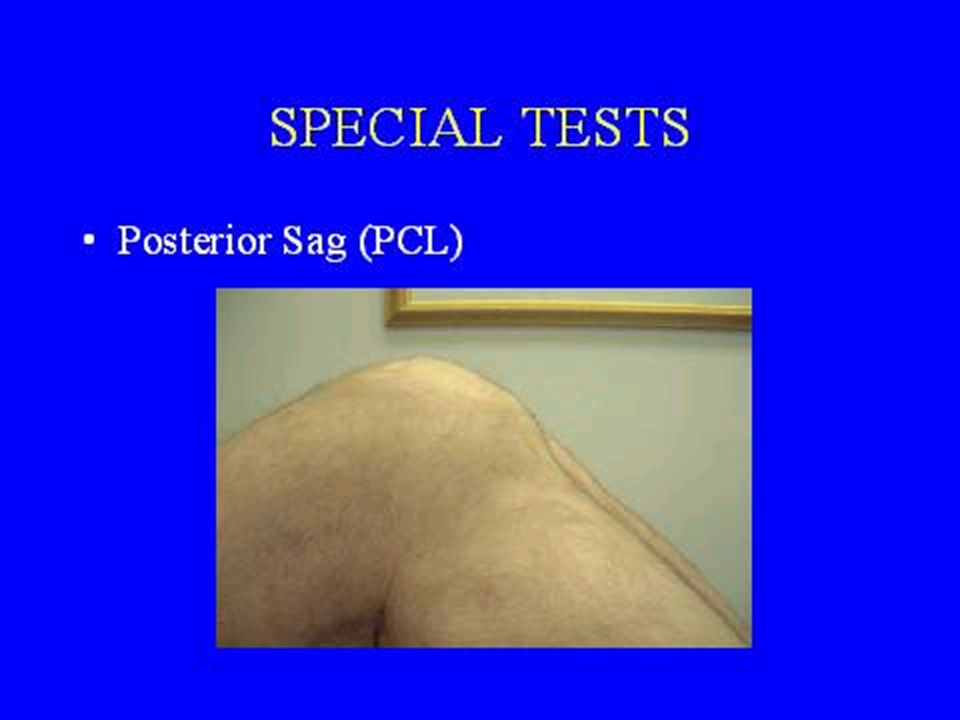
PCL

28
Knee: special tests - menisci

30
Knees: active resisted extension

31
Ankle/foot examination Look –Knee distally –Walking too (at some point)
")
32
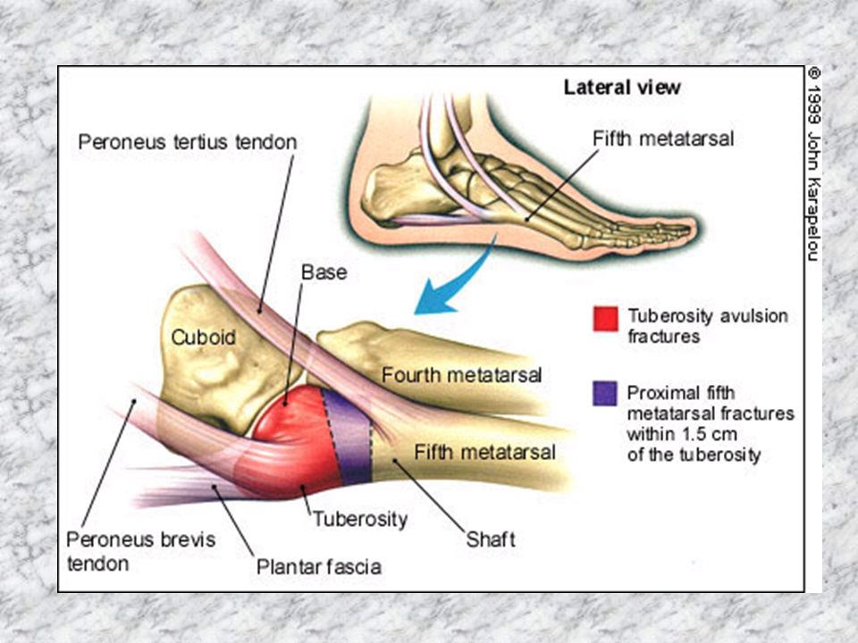
Ankle/foot examination Feel –Knee distally –Medial & lateral (include base 5 th MT) –Leave tender area until last
 –Leave tender area until last")
33
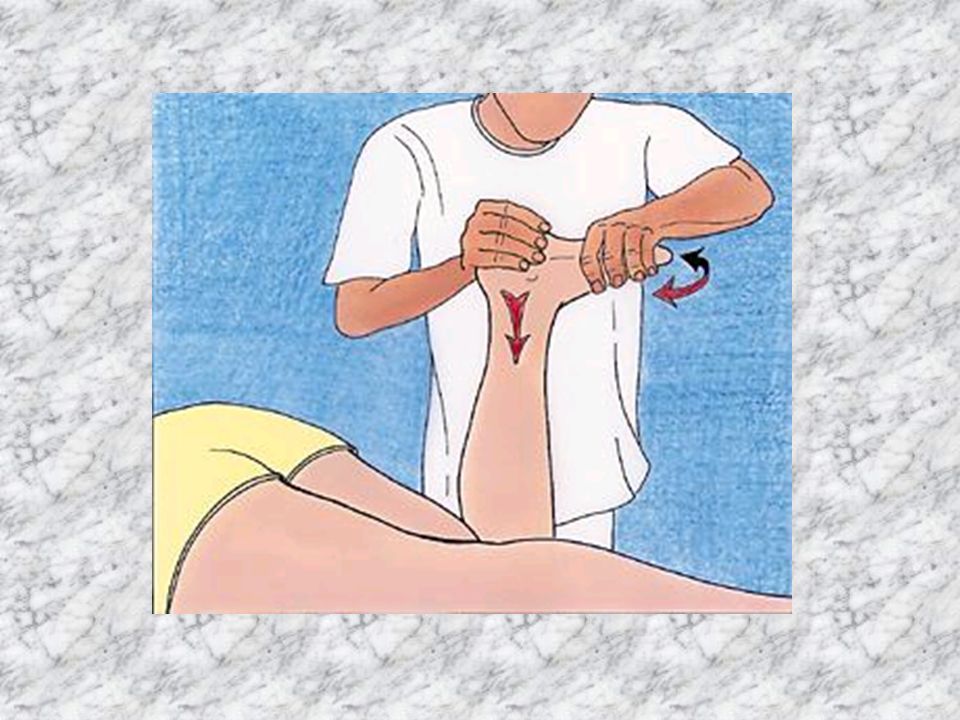
Ankle / foot examination Move –Ankle –Midtarsal –Stability test: anterior drawer

34
Anterior draw test

39
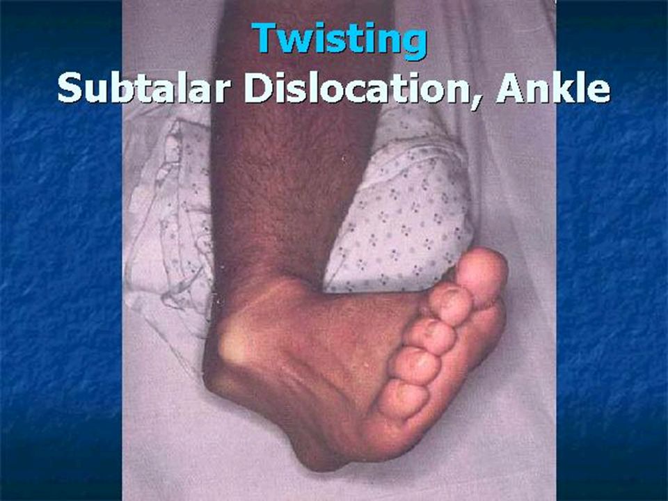
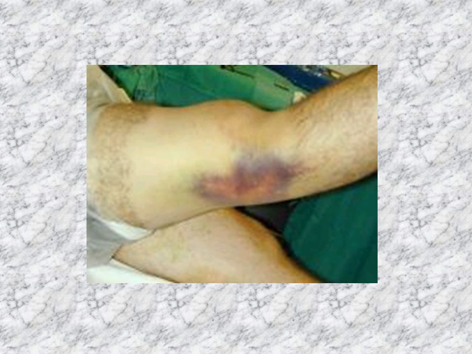
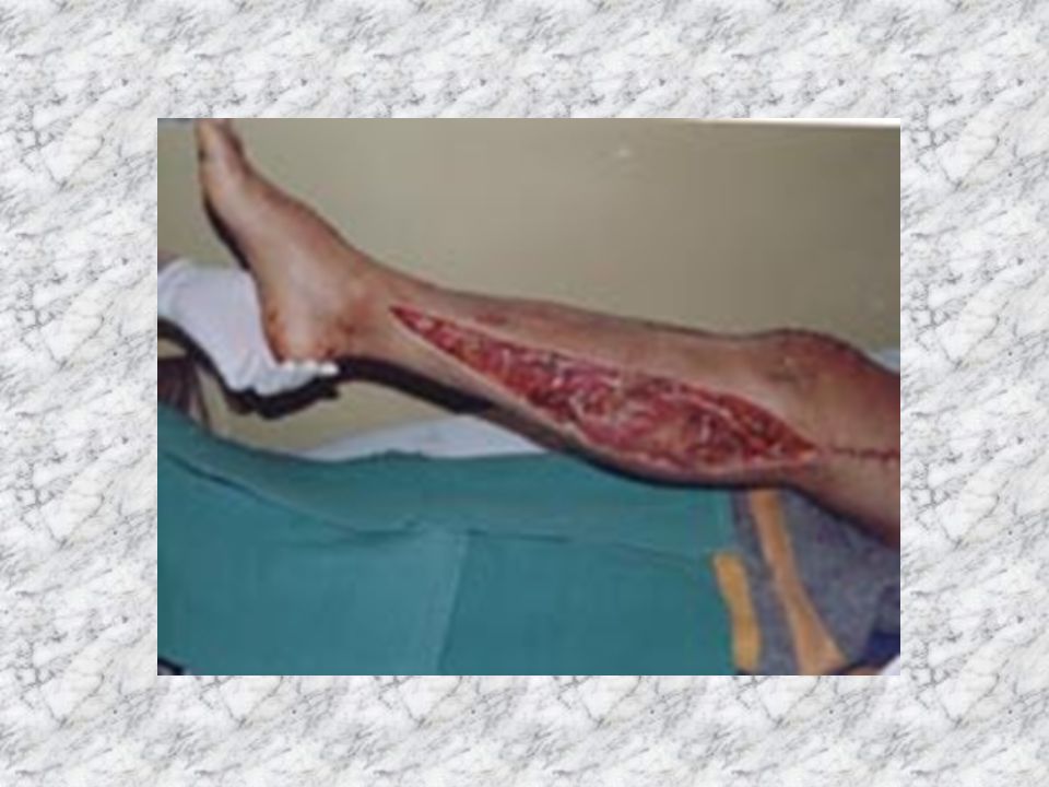
Emergency problems –Dislocation (not patellar) –Compartment syndrome
 –Compartment syndrome")
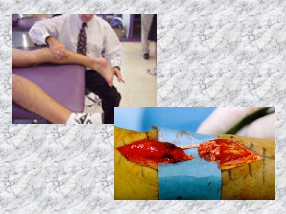
44
Skin medially is at risk. If skin becomes broken/necrotic, # becomes an open one. Risks of complications much greater (especially infection). Needs emergent reduction (with analgesia). Damage to popliteal artery if dislocated knee
. Needs emergent reduction (with analgesia). Damage to popliteal artery if dislocated knee.")
45
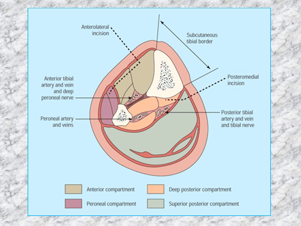
Compartment syndrome The pain may be intensely out of proportion to the injury, especially if no bone is broken. There may also be a tingling or burning sensation (paresthesias) in the muscle. The muscle may feel tight or full. If the area becomes numb or paralysis sets in, cell death has begun and efforts to lower the pressure in the compartment may not be successful in restoring function. Pain worse if affected muscle passively stretched. Pulses not lost (until very late).
 in the muscle. The muscle may feel tight or full. If the area becomes numb or paralysis sets in, cell death has begun and efforts to lower the pressure in the compartment may not be successful in restoring function. Pain worse if affected muscle passively stretched. Pulses not lost (until very late)..")
Similar presentations


>")


 Medial condyle (8 left) Intercondylar fossa (7 left)>")
