Download presentation
Presentation is loading. Please wait.

1
THE KNEE JOINT

2
BONES OF THE KNEE

3
FEMUR Lateral condyle (6 left) Medial condyle (8 left) Intercondylar fossa (7 left)
 Medial condyle (8 left) Intercondylar fossa (7 left)")
4
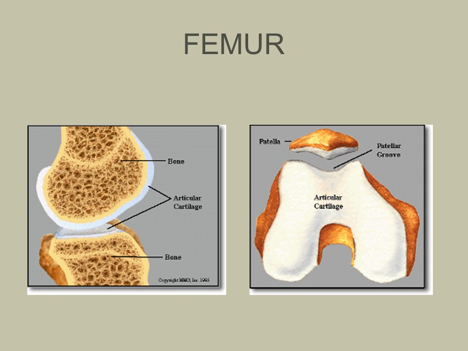
FEMUR

6
TIBIA Medial condyle Lateral condyle Tibial Tuberosicty Medial Malleolus

7
TIBIA

8
Gerdy’s tubercle IT band insertion Anterior medial surface Insertion for semitedonosis, semimembranosis, gracilis and sartorius

9
FIBULA No connection with the femur Head Lateral malleolus

10
PATELLA A ‘sesamoid’ (floating) bone Protection Mechanical advantage to quads. Without the patella, 30% more force would be required by the quads

11
PATELLA

12
THE KNEE 1. Femur - medial condyle 2. Femur - lateral condyle 3. Tibia - medial condyle 4. Tibia - lateral condyle 5. Anterior medial surface 6. Tibial tuberoscity 7. Gerdy’s tubercle (IT band insertion) 8. Head of fibula 9. Patella
 8. Head of fibula 9. Patella.")
13
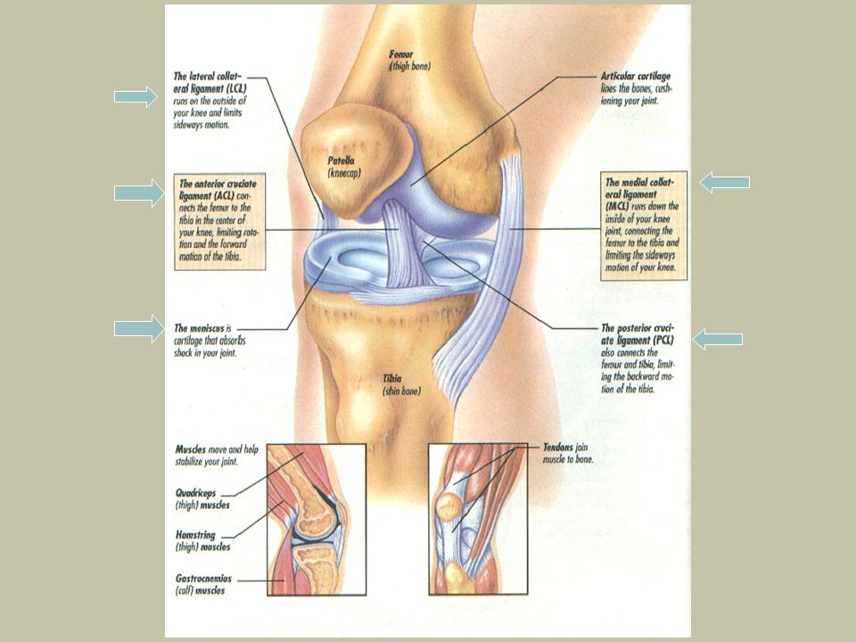
KNEE LIGAMENTS AND CARTILAGE

14
COLLATERAL LIGAMENTS They are important in controlling… Tibial rotation Anterior and posterior tibial displacement. Valgus (knocked kneed) Varus (blow legged)
 Varus (blow legged).")
15
MEDIAL COLLATERAL Medial aspect of the knee Attached to medial meniscus,

16
LATERAL COLLATERAL Lateral aspect of the knee Is not attached to lateral meniscus.

17
VALGUS AND VARUS Valgus – lateral force; Stress to the medial collateral ligament Varus – Medial force; Stress to the lateral collateral ligament

18
VALGUS AND VARUS Impact Stretch

19
CRUCIATE LIGAMENTS

20
ANTERIOR CRUCIATE LIGAMENT Inferior end: proximal, anterior tibia Superior end: distal posterior femur Prevents excess anterior motion of the tibia and posterior motion of the femur

21
ACL

22
AnteriorPosteriorAnteriorPosterior P T F T F P

23
POSTERIOR CURCIATE LIGAMENT Inferior end: proximal posterior tibia Superior end: distal posterior to middle femur AnteriorPosterior Prevents excessive posterior movement of the tibia and anterior movement of the femur

24
PCL

26
Sliding of the tibia with respect to the femur, a condition refered to as the drawer sign, is an indication of the integrity of the cruciate ligaments. The anterior drawer sign is tibial displacement beneath the femur in an anterior direction and reflects the integrity of the anterior cruciate. The posterior drawer sign is posterior displacement and reflects the integrity of the posterior cruciate. The PCL is shorter and stronger than the ACL

27
MENISCI Two on each of the tibia, loosely attached, thicker to the outside. Functions: 1.Stabilization 2.Shock absorption 3.Lubrication

28
MEDIAL MENISCUS Broader in front, most frequently injured The medial meniscus is “C” shaped. Attached to the medial collateral ligament. Anterior Posterior

29
MEDIAL MENISCUS

30
LATERAL MENISCUS The lateral meniscus is “O” shaped. Not attached to the lateral collateral ligament.

31
LATERAL MENISCUS

33
Movements of the Knee ExtensionFlexion

34
Actions of the Knee Function of the knee Flexion Extension

35
MECHANICAL APPLICATIONS TO THE KNEE

36
Mechanical Advantage from the Patella The patella moves the insertion of the quadriceps muscles further down the tibia. This increases the folcrum of the quads A longer folcrum increases the leverage of the quads making them a strong muscle group No patella: Folcrum ^ __F_________R. Patella: Folcrum ^ _____F______R.

37
Patellar ligament What landmark of the tibia does the patella tendon insert on?

38
Q-Angle. The deviation between the line of pull of the rectus femoris and the patellar ligament. It is usually measured from the anterior superior iliac spine and the center of the patella. A Q-angle of 10° is considered normal. Angles greater than this can result in lateral patellar dislocations when contractions of the quadriceps reduces the angle.

39
Q Angle Tibia / Patellar Ligament Rectus Femoris Tibia / Patellar Ligament

Similar presentations


 Tibia ◦ Main weight lower leg bone Medial malleolus comes off of.>")


>")


