Download presentation
Presentation is loading. Please wait.

1
CHAPTER 36 – SKELETAL, MUSCULAR, AND INTEGUMENTARY SYSTEMS

2
36-1 The Skeletal System

3
The Skeleton The skeleton supports the body, protects internal organs, provides for movement, stores internal reserves, and provides a site for blood cell formation. The human body would collapse without its skeleton.

4
Bones protect the internal organs of the body
Bones protect the internal organs of the body. Bones provide a system of levers on which muscles act to produce movement Bones contain reserves of minerals, mainly calcium salts, that are important to many body processes. Bones are the site of blood cell formation. Blood cells are produced in the soft marrow tissue that fills the internal cavities in some bones.

5
There are 206 bones in the adult human skeleton
There are 206 bones in the adult human skeleton. Bones can be divided into two parts – axial skeleton and the appendicular skeleton. Axial skeleton – supports the central axis of the body. It is made up of the skull, the vertebral column, and the rib cage. Appendicular skeleton – made up of the bones of the arms and legs, pelvis and shoulder area

6
Structure of Bones Bones are a solid network of living cells and protein fibers that are surrounded by deposits of calcium salts. Periosteum – a tough layer of connective tissue that surrounds the bone. Blood vessels that pass through the periosteum carry oxygen and nutrients to the bone Haversian canals – a network of tubes that run through bone that contain blood vessels. Spongy bone is a less dense tissue that is found inside the outer layer of compact bone. It is not actually soft and spongy. It is very strong.

7
Bone marrow – soft tissue in the cavities of bones
Bone marrow – soft tissue in the cavities of bones. There is yellow and red bone marrow. Yellow marrow is made up of mostly fat cells. Red marrow produces red blood cells, some white blood cells, and cell fragments called platelets. Development of Bones The skeleton of an embryo is made up almost entirely of cartilage, which is connective tissue. Cartilage doesn’t contain blood vessels. Cartilage is found in flexible parts of the body, like the tip of the nose and the external ears. Ossification – the process of bone formation (when cartilage is replaced by bone) Ossification takes place up to seven months before birth.
 Ossification takes place up to seven months before birth.")
8
Types of Joints Joint – a place where one bone attaches to another bone Joints permit bones to move without damaging each other Depending on its type of movement, a joint is classified as immovable, slightly movable, or freely moveable.

9
Immovable joints – allow no movement
Immovable joints – allow no movement. The bones are interlocked and held together by connective tissue, or they are used. Ex – the skull Slightly moveable – permit a small amount of restricted movement Ex – the joints between the two bones of the lower leg and the joints between adjacent vertebrae Freely moveable joints – permit movement in one or more directions

10
-ball and socket joints (shoulder)
")
11
-hinge joint (knee)
")
12
- pivot joint (elbow)
")
13
-saddle joint (knuckles)
")
14
Structure of Joints In freely movable joints, cartilage covers the surfaces where two bones come together to protect the bones as they move against each other. The joints are also surrounded by a fibrous joint capsule that helps hold the bones together . The joint capsule consists of two layers. One layer is called ligaments. Ligaments are strips of tough connective tissue that hold bones together in a joint.

15
Cells in the other layer of the joint capsule produce a substance called synovial fluid, which enables the surfaces of the joint to slide over each other smoothly.

16
Skeletal System Disorders
Loss of calcium in bones can lead to osteoporosis. Bones can fracture easily. Too much strain on a joint can lead to inflammation, which is swelling caused by excess fluid. Arthritis is inflammation in the joint itself.

17
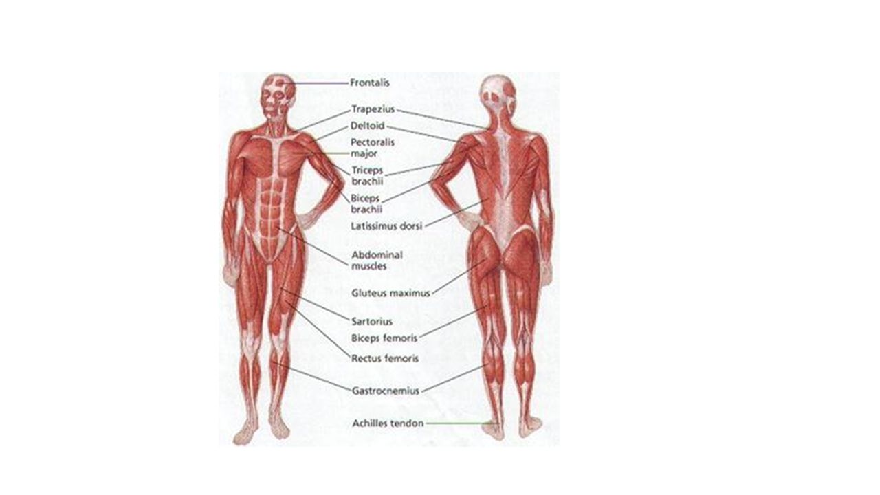
36-2 The Muscular System

18
Types of Muscle Tissue There are three different types of muscle tissue: skeletal, smooth, and cardiac

19
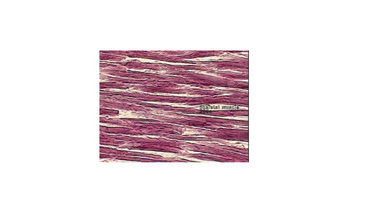
Skeletal Muscles - attached to bones, responsible for voluntary movements . Skeletal muscles look like they have alternating light and dark bands or stripes called striations. Because of this, skeletal muscle is sometimes called striated muscle.

22
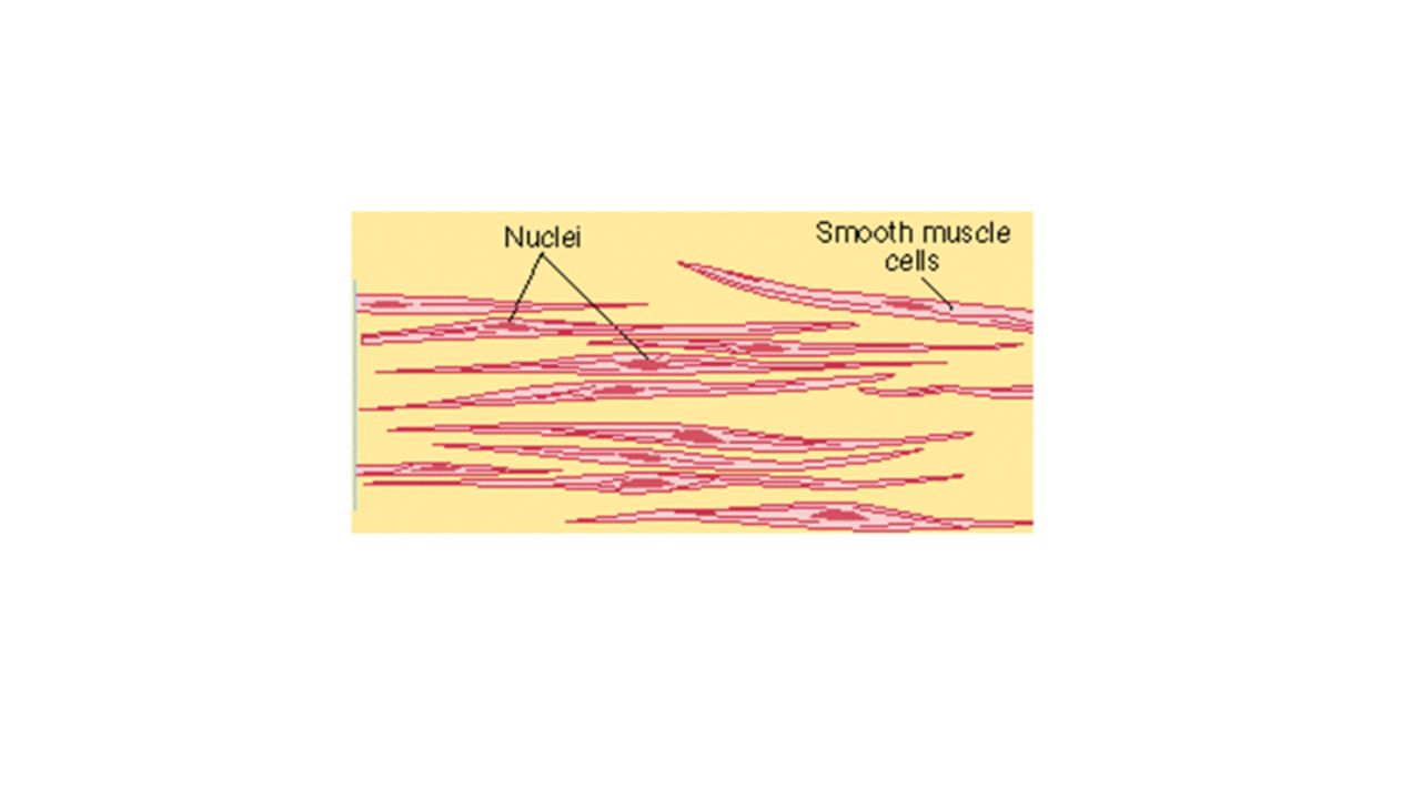
Smooth Muscles – usually not under voluntary control
Smooth Muscles – usually not under voluntary control. Smooth muscles cells are spindle-shaped, have one nucleus, and is not striated. Found in hollow structures such as the stomach, blood vessels, and the small and large intestines. Smooth muscles move food through your digestive tract, control the way blood flows through your circulatory system, and decrease the size of the pupils of your eyes in bright light.

24
Cardiac Muscle – Cardiac muscle is only found in the heart.

25
Muscle Contraction The muscle fibers in skeletal muscles are made up of smaller structures called myofibrils. Each myofibril is made up of even smaller structures called filaments. The striations in skeletal muscle cells are formed by an alternating pattern of thick and thin filaments. The thick filaments contain a protein called myosin. The thin filaments are made up mostly of a protein called actin. A muscle contracts when the thin filaments in the muscle fiber slide over the thick filaments. The energy for muscle contraction is supplied by ATP.

26
Control of Muscle Contraction
Skeletal muscles are useful only if they contract in a controlled way. Neuromuscular junction – the point of contact between a motor neuron and a skeletal muscle cell. Acetylcholine – a neurotransmitter released by vesicles in the axon terminals of the motor neurons.

27
When a muscle contracts, acetylcholine produces an impulses which causes the release of calcium ions within the muscle fiber. From the time a nerve impulse reaches a muscle cell, it is only a few milliseconds before these events occur and the muscle cell contracts. A muscle cell remains contracted until the release of acetylcholine stops and an enzyme produced at the axon terminal destroys any remaining acetylcholine. Then the cell pumps calcium ions back into storage, the cross-bridges stop forming and the contraction ends.

28
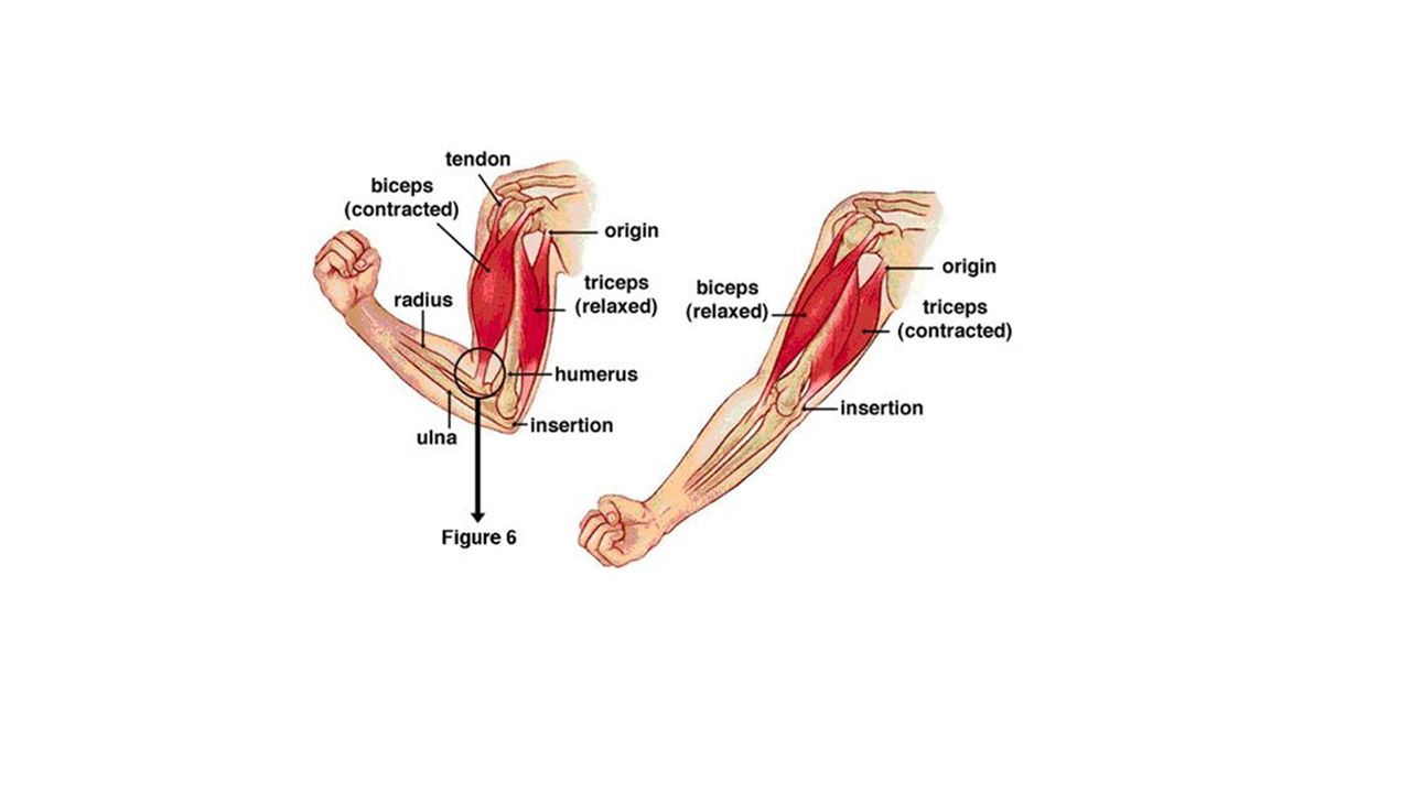
How Muscles and Bones Interact
Skeletal muscles are joined to bones by tough connective tissues call tendons. Tendons pull on the bones and make them work like levers. Most skeletal muscles work in opposing pairs. When one muscle contracts, the other relaxes. When the biceps muscle in the upper arm contracts, it bends the elbow joint. When the triceps muscle contracts, it opens the elbow joint. A controlled movement requires contraction by both muscles, like holding a tennis racket or violin. Both the biceps and triceps contract in balance.

30
36-3 The Integumentary System

31
The skin and its related structures, like hair, nails, and glands, made up the integumentary system
The skin is the largest organ of the body

32
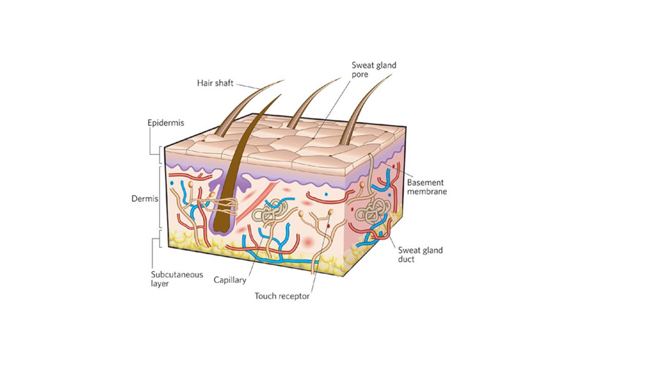
The Skin The integumentary system serves as a barrier against infection and injury, helps to regulate body temperature, removes waste products from the body, and provides protection against ultraviolet radiation from the sun. The skin is made up of two main layers: The epidermis and the dermis. Beneath the dermis is a layer of fat (hypodermis) and loose connective tissue that help insulate the body.
 and loose connective tissue that help insulate the body.")
34
Epidermis – the outer layer of the skin
The outside layer (that comes in contact with the environment) is made up of dead cells. The inner layer is made up of living cells
 is made up of dead cells. The inner layer is made up of living cells.")
35
The cells in the inner layer go through rapid cell division
The cells in the inner layer go through rapid cell division. The new cells push the old cells to the surface of the skin. The older cells become flattened and their organelles disintegrate. They being making keratin, which is a tough, fibrous protein. The keratin-producing cells die and form a tough, flexible waterproof covering of the surface of the skin. This outer layer of dead cells is shed or washed way once every four to five weeks.

36
Melanin – a dark brown pigment produced in cells called melanocytes.
Melanin protects the skin from damage by absorbing ultraviolet rays from the sun. Differences in skin color are caused by the different amount of melanin and where they are distributed.

37
Dermis – The inner layer of the skin.
The dermis lies beneath the epidermis and contains collagen fibers, blood vessels, nerve endings, glands, sensory receptors, smooth muscles, and hair follicles.

38
The skin helps to maintain homeostasis by regulating body temperature
The skin helps to maintain homeostasis by regulating body temperature. On a cold day, the blood vessels in the dermis narrow, helping to limit heat loss. On a hot day, the blood vessels widen, bringing hear from the body’s core to the skin and increasing heat loss.

39
The dermis contains two major types of glands: sweat glands and sebaceous glands. Sweat glands produce sweat when the body gets too hot. Sebaceous glands produce an oily secretion call sebum. Sebum spreads out along the surface of the skin and helps to keep the keratin- rich epidermis flexible and waterproof.

40
Skin cancer – an abnormal growth of cells in the skin which can be caused by excessive exposure to the ultraviolet radiation in sunlight. Protect yourself by wearing a hat, sunglasses, and clothes that cover your skin! And use sunscreen!

41
Hair and Nails Hair and nail is made with keratin. In other animals, keratin forms structures like bull horns, reptile scales, bird feathers, and porcupine quills

42
Hair has an important function
Hair has an important function. Hair on the head protects the scalp from ultraviolet light from the sun and keeps us warm in the cold. Hairs in the nostrils, external ear canals, and eyelashes prevent dirt and other particles from entering the body.

43
Hair follicle – cells that produce hair
Hair is a large column of cells that have filled with keratin and then died. Rapid cell growth at the base of the hair follicle causes the hair to grow longer.

44
Nails grow from the nail root, which is an area of rapidly dividing cells. Nails are also made by keratin. Nails protect the tips of the fingers and toes. Nails grow at an average rate of 3 millimeters per month, and fingernails grow four times faster than toe nails.

Similar presentations