Download presentation
Presentation is loading. Please wait.

1
Congenital and Hereditary Diseases
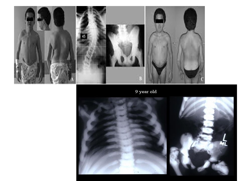
Osteogenesis Imperfecta (OI) هشاشة العظام Etiology: Mutation of two genes that encodes for α1 & α2 polypeptide chains of type I collagen (the main collagen of bone, tendon, and skin) leading to imperfect formation of osseous tissue. Signs: Present at birth but the onset may be in later developmental stages leading to limb deformities, dwarfism القزامة and some cases hearing impairment (deformation of auditory ossicle). Diagnosis: Radiograph evaluation reveals multiple fractures in various stages of healing and decrease in bone mass. X-ray film showed lower extremities (tibia and fibula) which was recognized after the child began to walk.
 هشاشة العظام. Etiology: Mutation of two genes that encodes for α1 & α2 polypeptide chains of type I collagen (the main collagen of bone, tendon, and skin) leading to imperfect formation of osseous tissue. Signs: Present at birth but the onset may be in later developmental stages leading to limb deformities, dwarfism القزامة and some cases hearing impairment (deformation of auditory ossicle). Diagnosis: Radiograph evaluation reveals multiple fractures in various stages of healing and decrease in bone mass. X-ray film showed lower extremities (tibia and fibula) which was recognized after the child began to walk.")
3
Osteopetrosis: (Marble Bone) العظم الرخامي
Etiology An increase in bone density Normal bone growth is achieved by a balance between bone formation by osteoblasts and bone resorption (break down of bone matrix) by osteoclasts. In osteopetrosis, the number of osteoclasts may be reduced, normal, or increased. Most importantly, osteoclast dysfunction mediates the pathogenesis of this disease. Signs Heavy compact bones, brittle, which requires an increase in exposure factors. Associated with Osteosclerosis. Diagnosis Radiograph shows an increase in bone density and thickness with marked reduction of the bone marrow.
 by osteoclasts. In osteopetrosis, the number of osteoclasts may be reduced, normal, or increased. Most importantly, osteoclast dysfunction mediates the pathogenesis of this disease. Signs. Heavy compact bones, brittle, which requires an increase in exposure factors. Associated with Osteosclerosis. Diagnosis. Radiograph shows an increase in bone density and thickness with marked reduction of the bone marrow.")
5
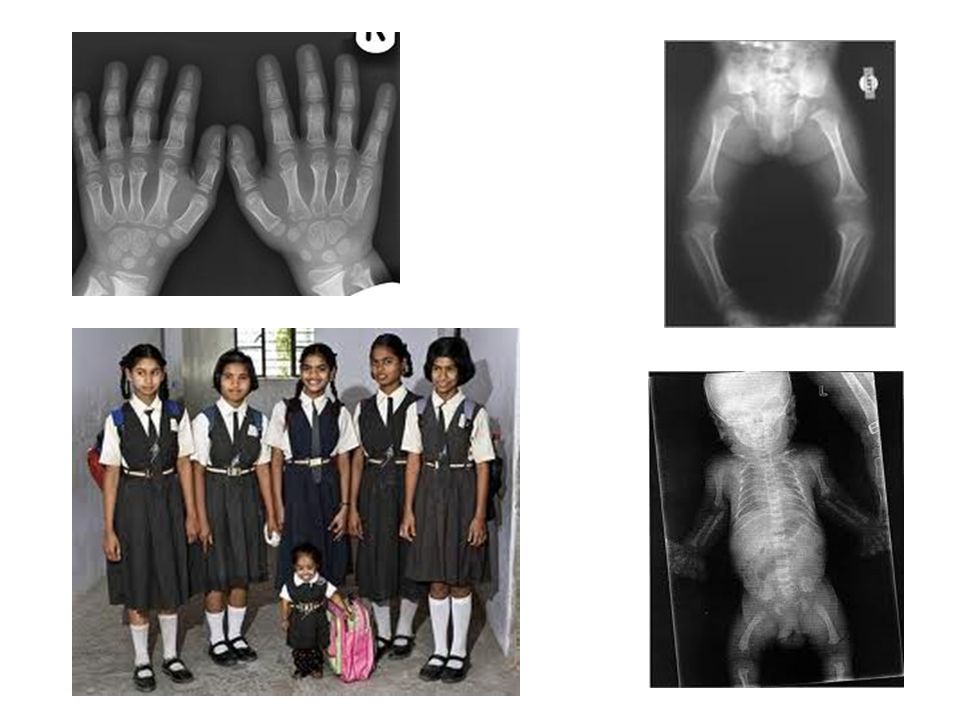
Acondroplasia Etiology
Autosomal dominant genetic disorder which causes an abnormality of cartilage formation that is a common cause of dwarfism. Signs Most common hereditary disease. Dwarfism Short limbs with normal trunk Small face with normal vault Trident handsيد خطافية Lordosis إنحناء العمود الفقري Diagnosis A skeletal survey is useful to confirm the diagnosis of achondroplasia. The skull is large, with a narrow foramen magnum, and relatively small skull base. The tubular bones are short and thick. Fibular overgrowth is present. The hand is broad with short metacarpals and phalanges, and a trident configuration.

7
Hand & Foot Malformations
Etiology Abnormal development of toes and fingers during fatal stages. Signs Syndactyly نقصان الأصابع Polydactylyزيادة الأصابع Diagnosis Webbed digitsإلتصاق الأصابع Extra digits

8
Inflammatory Diseases
Bacterial Osteomyelitis Etiology an infection (inflammation) of the bone (osteitis) or bone marrow (myelitis) caused by bacterial infection reach bone from blood or after trauma or surgery. In infants and children, the metaphysis of long bones, especially the femur and tibia, are most common affected. In adults, attacks the vertebrae, causing localized back pain and muscle spasm. Earliest changes of osteomyelitis are usually not evident on plain radiographs until about 10 days after the onset of symptoms, radionuclide (gallium) bone scanning is the most valuable imaging modality for early diagnosis.
 of the bone (osteitis) or bone marrow (myelitis) caused by bacterial infection reach bone from blood or after trauma or surgery. In infants and children, the metaphysis of long bones, especially the femur and tibia, are most common affected. In adults, attacks the vertebrae, causing localized back pain and muscle spasm. Earliest changes of osteomyelitis are usually not evident on plain radiographs until about 10 days after the onset of symptoms, radionuclide (gallium) bone scanning is the most valuable imaging modality for early diagnosis.")
9
Radiographic appearance
Lytic abscess cavities, the cortex may become so dense.

10
Vertebral osteomyelitis
loss of the sharp cortical outline.

11
Osteoarthritis Osteoarthritis is the commonest
Fig 2.B Osteoarthritis is the commonest form of arthritis. It is due to degenerative changes resulting from wearبلي and tear تمزق of the articular cartilage. (Fig.2. A and B) The common site of osteoarthritis: • Metatarsophalangeal joint of the big toe, Wrist joint, Hip and knee joint (frequently affected). • Ankle (infrequently affected). (Fig.2. C and D) Fig 2.A Fig 2.C Fig 2.D
 The common site of. osteoarthritis: • Metatarsophalangeal joint of the. big toe, Wrist joint, Hip and knee. joint (frequently affected). • Ankle (infrequently affected). (Fig.2. C and D) Fig 2.A. Fig 2.C. Fig 2.D.")
12
Radiographic changes seen in osteoarthritis: (Fig.3 and 4)
2 Radiographic changes seen in osteoarthritis: (Fig.3 and 4) 1-Narrowing of the joint space due to loss of the articular cartilage (1). 2- Reactive sclerosis of the articular surfaces of the bone ends (2). 3- Formation of osteophytes at the articular margins (small (bone) (3). 4- Development of cysts in the sub articular regions (4). 1 3 4
 1-Narrowing of the joint space due. to loss of the articular cartilage (1). 2- Reactive sclerosis of the. articular surfaces of the bone. ends (2). 3- Formation of osteophytes at the. articular margins (small (bone) (3). 4- Development of cysts in the. sub articular regions (4)")
13
Clinical manifestations of OA may include:
• Joint pain. • Tenderness. • Stiffness. • Creaking. • Locking of joints. • and sometimes local inflammation. • Disease of elder patients. • Loss of articular joint cartilage. • Narrowing of joint space. • Bony sclerosis around joint. • Usually secondary to weight-bearing, chronic wear and tear.

14
Rheumatoid arthritis:
• Rheumatoid arthritis is a polyarthritis caused by inflammatory overgrowth of synovium known as pannus. Features: 1- Periarticular soft tissue swelling and osteoporosis are the earliest change. 2-Joint space narrowing due to destruction of the articular cartilage by pannus. 3-Further destruction leads to small bony erosion which occurs initially at the joint Margins (Fig6).
.")
15
These erosions are often seen first
at: 1. Metatarso or metacarpophalangeal joints. 2. Proximal interphalangeal joints. 3. On the styloid process of the ulna. 4. Similar changes are seen in the large joints. (Fig.7). • Later extensive erosions may disrupt the joint surfaces. Ulnar deviation is usually present at this stage. • With much severed destruction the condition is referred to as arthritis multilans (Fig .8)
. • Later extensive erosions may. disrupt the joint surfaces. Ulnar. deviation is usually present at this. stage. • With much severed destruction the. condition is referred to as arthritis. multilans (Fig .8)")
16
Gout: • Gout is due to the deposition of urate crystal in the joint and in the adjacent bone. • Gout most commonly affects the metatarsophalangeal joint of the big toe. • Signs of gout: 1- Soft tissue swelling (is the earliest change) 2- Erosions (Later stage). • Unlike RA may be at a distance from the articular cortex (Fig.9) 3- There is usually no osteoporosis. 4- Localized soft tissue lumps (Fig. 9)
 2- Erosions (Later stage). • Unlike RA may be at a distance. from the articular cortex (Fig.9) 3- There is usually no osteoporosis. 4- Localized soft tissue lumps. (Fig. 9)")
17
ضبابي appearance of cranium
Paget’s Disease Fluffyزغبي radiodense cotton wool appearance Note hazy ضبابي appearance of cranium Paget’s is an osteitis deformans of unknown cause characterized by excessive and abnormal bone remodelling. Complications associated with Paget's disease include fractures, skeletal deformities, neoplasms and joint malformations. Lateral radiograph of the skull demonstrates a thickened calvarium and fluffy radiodense regions which is referred to as a "cotton-wool" appearance.

18
Paget's disease of bone is a chronic disorder that can result in enlarged and misshapen bones. Paget's is caused by the excessive breakdown and formation of bone, followed by disorganized bone remodelling. This causes affected bone to weaken, resulting in pain, misshapen bones, fractures and arthritis in the joints near the affected bones. Often Paget's disease is localized to only a few bones in the body. The pelvis, femur, and lower lumbar vertebrae are the most commonly affected bones. Paget's disease of right side. Man of 80 years age.

Similar presentations


>")


 is a common, degenerative disease, which is characterized by local degeneration of joint cartilage and new bone formation.>")