Download presentation
Presentation is loading. Please wait.

1
Haematopoiesis Lab 1

2
Bone Marrow Examination
Cellularity: (normal, decreased, increased) Evaluation of cell lines: • Erythropoiesis • Thrombopoiesis (Megakaryopoiesis) • Myelopoiesis Parasites, neoplastic cells
 Evaluation of cell lines: • Erythropoiesis • Thrombopoiesis (Megakaryopoiesis) • Myelopoiesis. Parasites, neoplastic cells.")
3
Bone Marrow Examination
The common indications are: Persistent neutropenia Persistent leukocytosis Unexplained thrombocytopenia Persistent thrombocytosis Poorly regenerative anaemia Abnormal blood cell morphology Unexplained presence of immature cells in blood

4
Cellularity The cellularity of bone marrow is estimated by examining the proportion of cells versus fat present in particles. If the particles are composed of more than 75% cells, the marrow is interpreted as hypercellular, and if the particles are composed of more than 75% fat, the marrow is interpreted as hypocellular. The overall cellularity of bone marrow decreases with age. Histological section of canine bone marrow. Pink bony trabeculae are present in the lower left corner, lower right corner, and top, and surround the haematopoietic cells and fat. The fat is the round to oval, clear areas. The erythrocytic and granulocytic precursor cells are the many small, round purple structures. The larger, densely staining purple structures distributed throughout the marrow space are megakaryocytes.

5
Cellularity: Normal Bone marrow particles filled with cells Two Megakaryocytes If 10 to 20 Megakaryocytes are seen per field using the IOx objective, the numbers are likely increased

6
Cellularity: Decreased
Hypoplasia: almost no nucleated cells, empty bm-particles

7
Cellularity: Decreased
Myelofibrosis: bm-particles containing connective tissue, and stromal cells

8
Cellularity: Increased
Increased cellularity can indicate increased cell production (regenerative response) or neoplasia
 or neoplasia.")
9
Other considerations Meyloid:Erythroid (M:E) ratio Stainable Iron
Usually between 0.7 to 1.7 Lower ratio is usually in large animals high M:E ratio could indicate the presence of either increased granulocytic cells or decreased erythroid cells Stainable Iron Hemosiderin appears gray to black It may be seen within macrophages or as free material It is decreased or absent in iron-deficient animals, and may be increased in association with all anaemias Bone marrow iron increases in some animals with age

10
EVALUATION OF CELL LINES: ERYTHROPOIESIS
Morphologic changes that occur as cells of the erythroid series undergo maturation include diminution in size, decrease in N:C ratio, progressive nuclear condensation, and the appearance of red cytoplasmic colour as hemoglobin is synthesized and accumulates within the cytoplasm.

11
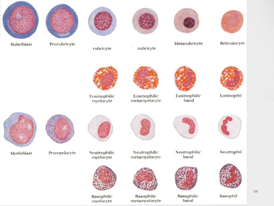
ERYTHROPOIESIS Rubriblast. Is relatively large cells with high N:C ratio and intensely basophilic cytoplasm, resulting from the presence of many polyribosomes. The nucleus of the rubriblast is usually nearly perfectly round and the chromatin is finely granular, containing one or two pale-blue to medium-blue nucleoli. Prorubricyte. Is a large round cell with a round nucleus with a coarsely granular chromatin pattern. This cell typically lacks a nucleolus. There is a small amount of deep blue cytoplasm with often a prominent perinuclear clear zone. Rubricyte. is a round cell with a round, centrally located nucleus; it is smaller than the prorubricyte. The coarsely granular chromatin is more condensed compared with the earlier stages of development, and irregular clear areas are present between the chromatin clumps. Later rubricytes stain more red (contain more haemoglobin).
.")
12
ERYTHROPOIESIS Metarubricyte. Is smaller than the rubricyte. The nucleus is round to oval, usually slightly eccentrically located, and has very condensed chromatin. There are small to moderate amounts of blue to reddish-blue cytoplasm. The metarubricytes, with more-reddish cytoplasm, contain more haemoglobin. Reticulocyte. Does not have a nucleus, and cytoplasm is reddish-blue. As cells mature, they become less blue and more red due to increased amounts of haemoglobin. Erythrocyte. does not have a nucleus, and the cytoplasm is reddish to reddish-orange. The central pallor present here is due to the biconcave discoid shape of the cells.

13
EVALUATION OF CELL LINES: MEGAKARYOPOIESIS

14
Megakaryopoiesis Megakaryoblast, promegakaryocyte, and megakaryocyte
Megakaryoblasts have a single nucleus and deeply basophilic cytoplasm. This cell type is not recognised in most normal aspirate smears, because it occurs in small numbers and is difficult to differentiate from other blast cells. Promegakaryocytes, which have two or four nuclei and deeply basophilic cytoplasm, are easily recognised. These cells are much larger than leukocytes or nucleated erythroid precursor cells. Subsequent nuclear reduplications result in progressively larger basophilic megakaryocytes. Nuclei in basophilic megakaryocytes are joined into a lobulated mass, making it difficult to count the number of nuclear reduplications that have occurred. Megakaryocytes are gigantic and vary from 50 to 200 um in diameter, with larger cells having greater nuclear ploidy. Megakaryoblast, promegakaryocyte, and megakaryocyte Megakaryocyte

15
Osteoblast / Osteoclast
An example of other cells found in a bone marrow slide and that should be differentiated from a megakaryocyte. In osteoclast nuclei are clearly separate, in contrast to the fused nuclear material present in platelet precursors.

16
EVALUATION OF CELL LINES: MYELOPOIESIS
The early granulocytic precursors have oval to indented nuclei and blue cytoplasm. The larger immature forms have small, pink cytoplasmic granules. The cytoplasm becomes less blue as the cells mature.

17
MYELOPOIESIS

18
MYELOPOIESIS The myeloblast is a large, round to oval cell with a round to oval nucleus with a finely stippled chromatin pattern and usually prominent nucleolus or multiple nucleoli. There is a small to moderate amount of blue cytoplasm and no prominent cytoplasmic granules. The promyelocyte is a large, round to oval cell with a round to oval nucleus. The nuclear chromatin pattern is finely granular. The moderate amount of cytoplasm is blue and contains multiple, pink to purple granules, which are primary granules. The neutrophilic myelocyte is a round cell that is smaller than the myeloblast and the progranulocyte. The nucleus is round to oval and may contain a single indentation. The chromatin pattern is finely to moderately granular. Nucleoli are not present. The moderate amounts of blue cytoplasm contain multiple secondary granules, also called specific granules. These secondary granules are pink for the neutrophilic lineage and difficult to see. The secondary granules for the eosinophilic and basophilic lineages are generally reddish and purple respectively.

19
MYELOPOIESIS The neutrophilic metamyelocyte is a round cell with a kidney-shaped nucleus. The chromatin is moderately granular and more condensed than that of the myelocyte. The moderate amounts of blue cytoplasm contain secondary granules, which are difficult to see. The secondary granules of the eosinophilic and basophilic metamyelocyte are generally reddish and purple respectively. The band neutrophil is a round cell with a horseshoe-shaped nucleus. The cytoplasm is blue to light blue and contains secondary granules. These granules are difficult to see in the band neutrophil. The secondary granules of the band eosinophil and basophil are generally reddish and purple respectively. The segmented neutrophil is a small round cell with a single nucleus, which has multiple segmentations. The nuclear chromatin is very condensed. There is a moderate amount of light blue to pink cytoplasm.

21
Erythroid or granulocyte precursors!
As a general rule, erythroid precursors are smaller, have more nearly spherical nuclei with more condensed nuclear chromatin Erythroid precursors have darker cytoplasm than do granulocyte precursors at similar maturation stages. Consequently, smaller and darker cells, observed by scanning marrow smears at low power, are usually erythroid precursors (unless lymphocytes are increased in numbers), and the larger, paler cells are usually granulocyte precursors.
, and the larger, paler cells are usually granulocyte precursors.")
22
Other cells of the bone marrow
Lymphocytes Monocytic series (monoblast, promonocyte, and monocyte) Macrophages Mast cells Plasma cells Basket cells The cytoplasm of macrophages generally contains vacuoles and phagocytised material such as nuclear debris, hemosiderin, and erythrocytes and leukocytes (rarely). Hemosiderin in macrophages appears gray to black. The term "basket cell" has been used to refer to free nuclei in which the chromatin is dispersed in a lacelike manner.
 Macrophages. Mast cells. Plasma cells. Basket cells. The cytoplasm of macrophages generally contains vacuoles and phagocytised material such as nuclear debris, hemosiderin, and erythrocytes and leukocytes (rarely). Hemosiderin in macrophages appears gray to black. The term basket cell has been used to refer to free nuclei in which the chromatin is dispersed in a lacelike manner.")
Similar presentations


 Course code: MLHE-201 Supervisor: Prof. Dr Magda Sultan Date : 31 / 10/2013 Outcome : The student will understand : -The process.>")


