Download presentation
Presentation is loading. Please wait.

1
Unit 3: Biological Bases of Behavior Module #5: The Brain
AP Psychology

2
TECHNIQUES TO LEARN ABOUT STRUCTURE & FUNCTION
Clinical Observation (Case Study) – Observing specific brain diseases and injuries. Over 150 years ago people were studying patients with brain damage and linked loss of structure with loss of function. Essentially, losing brain tissue caused brain damage.
 – Observing specific brain diseases and injuries. Over 150 years ago people were studying patients with brain damage and linked loss of structure with loss of function. Essentially, losing brain tissue caused brain damage.")
3
Who is Phineas Gage?

4
Phineas Gage Phineas Gage was a level-headed, calm foreman of a railroad crew in 1848. An explosion shot an iron rod through his head, severing the connections between his limbic system and his frontal cortex. Gage became hostile, impulsive, and unable to control his emotions or his obscene language. Autopsy revealed that the relationship between frontal lobes and control of emotional behavior.

5
Phineas Gage Clip

6
Lesions Precise destruction of brain tissue.
Enabled more systematic study of the loss of function resulting from surgical removal, cutting of neural connections, or destruction by chemical applications.

7
Measuring brain function
Electroencephalogram (EEG): an amplified recording of the waves of electrical activity that sweep across the brain’s surface. These waves are measured by electrodes placed on the scalp.
: an amplified recording of the waves of electrical activity that sweep across the brain’s surface. These waves are measured by electrodes placed on the scalp.")
8
Measuring brain function
The amplified tracings are called evoked potentials. Scientists introduce a stimulus and study the brain waves to learn about brain activity.

9
Measuring brain function
Positron Emission Tomography (PET) scan: a visual display of brain activity that detects where a radioactive form of glucose (sugar) goes while the brain performs a given task.
 scan: a visual display of brain activity that detects where a radioactive form of glucose (sugar) goes while the brain performs a given task.")
10
Measuring brain function
Active neurons hog the glucose (the brain’s chemical fuel), and the PET scan tracks where in the brain the radioactive glucose goes.
, and the PET scan tracks where in the brain the radioactive glucose goes.")
11
Measuring brain function
Researchers can have participants think about certain topics or do activities to see where the glucose goes (thereby showing what part of the brain is active during that activity).
.")
12
Brain Imaging Magnetic Resonance Imaging (MRI): a technique that uses magnetic fields and radio waves to produce computer-generated images that distinguish among different types of soft tissue; allows us to see structures within the brain.
: a technique that uses magnetic fields and radio waves to produce computer-generated images that distinguish among different types of soft tissue; allows us to see structures within the brain.")
13
Brain Imaging A head is put into a strong magnetic field to produce the image. Healthy Individual Schizophrenic

14
Measuring brain function
Functional Magnetic Resonance Imaging (fMRI): a technique for revealing blood flow and, therefore, brain activity by comparing successive MRI scans. MRI scans show brain anatomy; fMRI scans show brain functions.
: a technique for revealing blood flow and, therefore, brain activity by comparing successive MRI scans. MRI scans show brain anatomy; fMRI scans show brain functions.")
15
Measuring brain function
Researchers compare images taken less than a second apart, they can see which parts of the brain “light up” with increased blood flow.

16
Brain Imaging Computerized Axial Tomography (CAT or CT): two-dimensional x-ray slices that are passed through various angles of the brain, arranged to show the extent of a lesion.
: two-dimensional x-ray slices that are passed through various angles of the brain, arranged to show the extent of a lesion.")
17
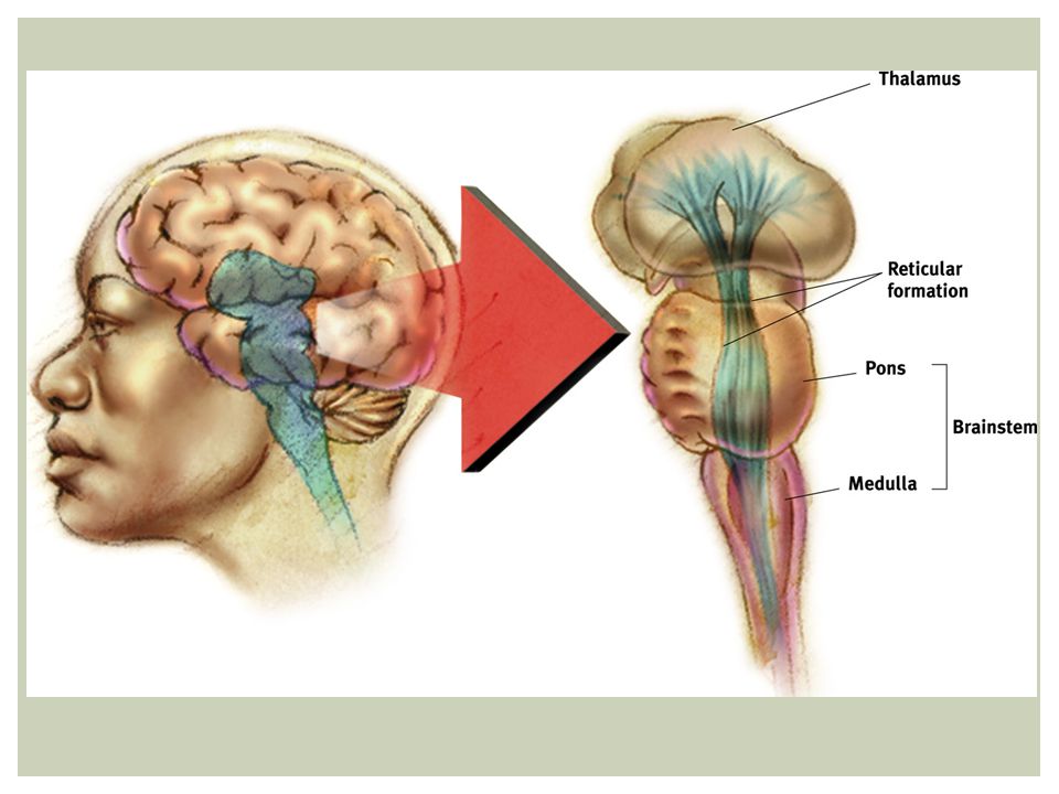
The BRAIN Brainstem – oldest part of the brain. Connects the brain with the spinal cord. Responsible for automatic survival function.

18
Waterboy Clip

19
Structure of Brain: Brainstem
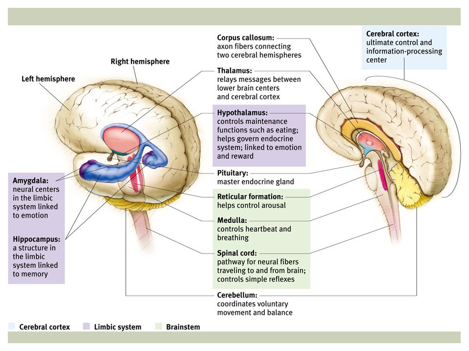
Medulla: where most fibers cross above the brain stem, resulting in contralateral (opposite side) control. regulates heart rate, blood flow, breathing, digestion, vomiting.
 control. regulates heart rate, blood flow, breathing, digestion, vomiting.")
20
Structure of Brain: Brainstem
Reticular Formation: a nerve network in the brainstem (pons) that plays an important role in controlling arousal (awakening).
 that plays an important role in controlling arousal (awakening).")
21
Structure of Brain: Brainstem
Pons: right above the medulla, helps coordinate movement, and is the bridge between cerebral hemispheres and both medulla and cerebellum.

22
Structure of Brain Thalamus: relay “station” for the brain. It receives information from all of the senses (except smell) and routes it to the brain regions that deal with those senses. Located at the top of the brain stem.
 and routes it to the brain regions that deal with those senses. Located at the top of the brain stem.")
24
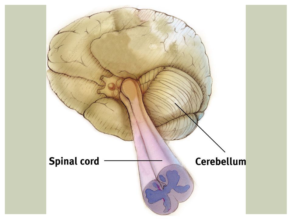
Structure of Brain Cerebellum: “little brain”-coordinates motor function integrating motion and positional information from the inner ear and muscles. helps maintain balance.

26
The Limbic System Responsible for emotions like fear and anger, and basic need for food and sex.

27
The LIMBIC SYSTEM Hippocampus: Enables formation of new long-term memories.

28
The Limbic System Amygdala: influences anger and fear. Coordinates fight-or-flight response. Removal will result in loss of anger. Stimulation will cause aggression.

29
The Limbic System Hypothalamus: controls autonomic functions such as body temperature and heart rate via control of sympathetic and parasympathetic centers in the medulla. Sets appetite drives (e.g. thirst, hunger, sexual desire) and behavior.
 and behavior.")
30
The limbic system Hypothalamus: Integrates with endocrine system by secretion of hormones that regulate hormones from the pituitary. It’s the reward center because it’s responsible for pleasure.

31
An electrode is connected to the hypothalamus, which causes the rat to cross the painful electric grid to press the pedal to send electrical impulses to its pleasure center (reward).
.")
33
Structure of Brain Basal Ganglia: links the thalamus with the motor cortex and other motor areas. regulates initiation of movements, balance, eye movements, and posture. Involved in reward/punishment learning and focus.

34
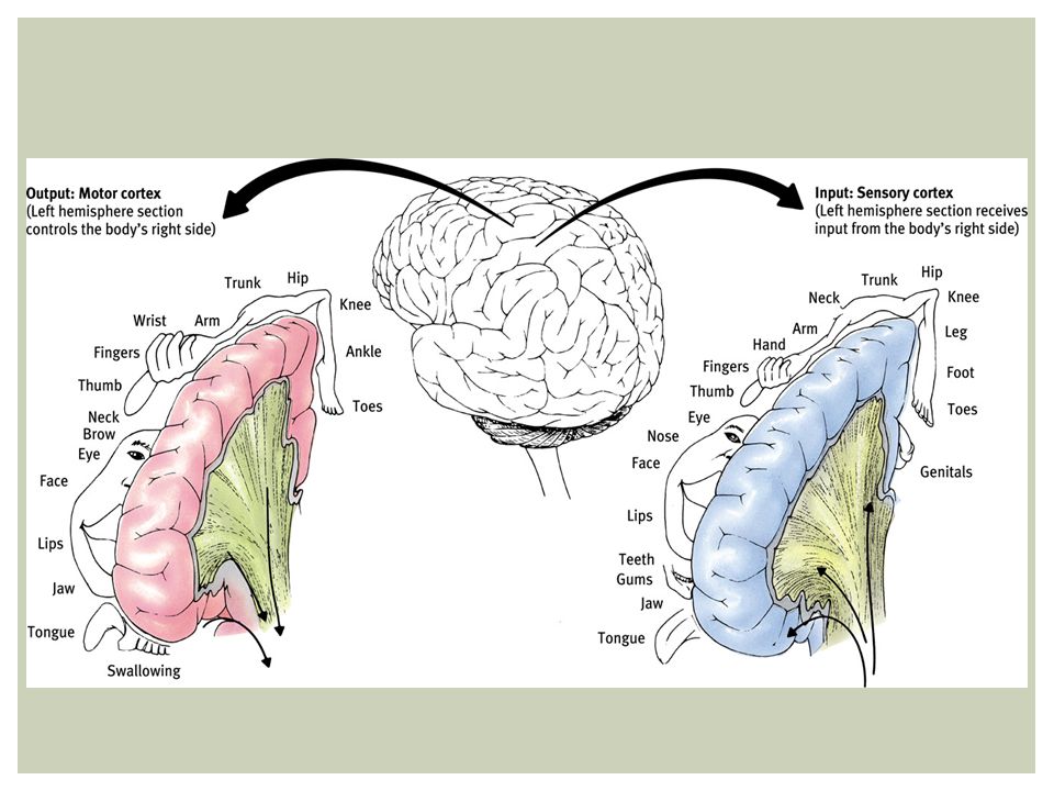
Structure of Brain Cerebral Cortex: receives and processes sensory information and directs movement. Center for higher order process such as thinking, planning, judgment.

35
Cerebral Cortex

36
Structure of Brain Frontal lobe: The part of the cerebral cortex right behind the forehead. Involved in speaking and muscle movements.

37
Structure of Brain Frontal lobe:
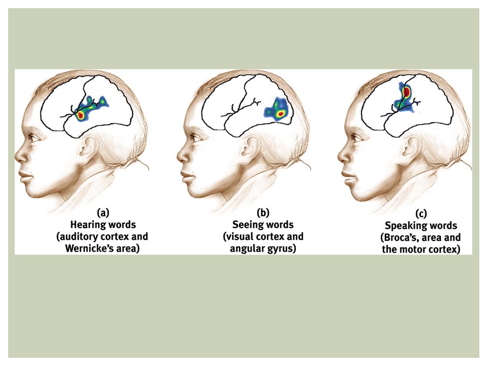
Includes Broca’s area: in left frontal lobe controls production of speech. Interpret and control emotional behaviors, make decisions, carry out plan.

38
Structure of Brain Temporal lobe: center for hearing.

39
Structure of Brain Temporal Lobe:
Includes Wernicke’s area: in left temporal lobe, plays role in understanding language and making meaningful sentences.

40
Structure of Brain Temporal Lobe:
Right temporal lobe important for understanding music/tonality. Sound from both ears is processed mostly contralaterally.

41
Structure of Brain Parietal Lobe: Responsible for cognition (thinking), information processing, pain and touch sensation, speech, and visual perception.
, information processing, pain and touch sensation, speech, and visual perception.")
42
Structure of Brain Occipital Lobe: Receives information from the eyes and processes it. Responsible for visual perception and color recognition.

43
Structure of Brain Parietal lobes Frontal lobes Temporal lobes
Occipital lobes

44
Parietal Lobe Frontal Lobe Corpus Callosum Occipital Lobe Pituitary Temporal Lobe Cerebellum Pons Medulla Spinal Cord

45
Broca’s area It was concluded that damage to this so-called Broca’s area caused a loss of ability to speak, known as expressive aphasia.

46
Wernicke’s area Carl Wernicke found another brain area involved with understanding language in the left temporal lobe. Destruction of Wernicke’s area results in loss of ability to comprehend written and spoken language, known as receptive aphasia.

47
Broca’s area Paul Broca (1861) did an autopsy on a patient named Tan, who couldn’t speak even though there was no physical damage and he could understand language. Tan’s brain showed loss of tissue in part of the frontal lobe of the left central cerebral hemisphere (as did several other similar cases).
 did an autopsy on a patient named Tan, who couldn’t speak even though there was no physical damage and he could understand language. Tan’s brain showed loss of tissue in part of the frontal lobe of the left central cerebral hemisphere (as did several other similar cases).")
48
Review!!! EEG – Wave cap; studies brain waves PET – Inject glucose, track where it goes. Shows brain activity. MRI – Uses magnetic fields to produce an image of soft tissue. fMRI – Track blood flow through brain. CAT – 2D x-ray of slice of brain.

49
Review!!! Medulla – Heart rate, breathing, blood flow Reticular Formation – Arousal (awakening), located inside of pons Pons – movement Thalamus – Relay station; receives info from body (except smell) and sends to different parts of the brain.
 and sends to different parts of the brain.")
50
Review!!! Cerebellum – Motor function, balance Hippocampus – Memories
Amygdala – Emotions (anger & fear) Hypothalamus – autonomic functions (heart rate, body temp). Regulates hormones. Reward center. Cerebral Cortex – Receives & processes sensory info & movement. Basal Ganglia – motor functions, movement, balance, eye movement.
 Hypothalamus – autonomic functions (heart rate, body temp). Regulates hormones. Reward center. Cerebral Cortex – Receives & processes sensory info & movement. Basal Ganglia – motor functions, movement, balance, eye movement.")
51
Review!!! Frontal – Speaking & muscle movement; Broca’s area (speech) Temporal – hearing, music, & sound; Wernicke’s area (understanding language & making sentences) Parietal – thinking, cognition, info processing, sensation, pain, & touch Occipital – Visual perception & color recognition
 Parietal – thinking, cognition, info processing, sensation, pain, & touch. Occipital – Visual perception & color recognition.")
52
Reading Aloud

53
Reading Aloud Depending on the damaged area different forms of aphasia occurs. Aphasia: impairment of language Damage to: A. Angular gyrus – can speak and understand but not read. B. Wernicke’s area – can’t understand words C. Broca’s area – disrupts speaking

55
Hemispheres Demonstration
Rotate your right hand in a circle. Now rotate your right foot in the same direction. Now rotate your right foot in the opposite direction, without changing the direction of your hand. Difficulties arise because you are asking your left hemisphere to do two things.

56
Hemispheres demonstration
Rotate your right hand in a circle. Now rotate your LEFT foot in the same direction. Now rotate your LEFT foot in the opposite direction, without changing the direction of your hand. Easier because your left hemisphere controls your right hand and your right hemisphere controls your left foot.

57
The Brain The last evolutionary development of the brain is localization of functions on different sides of your brain.

59
LOCALIZATION AND LATERALIZATION OF THE BRAIN’S FUNCTION
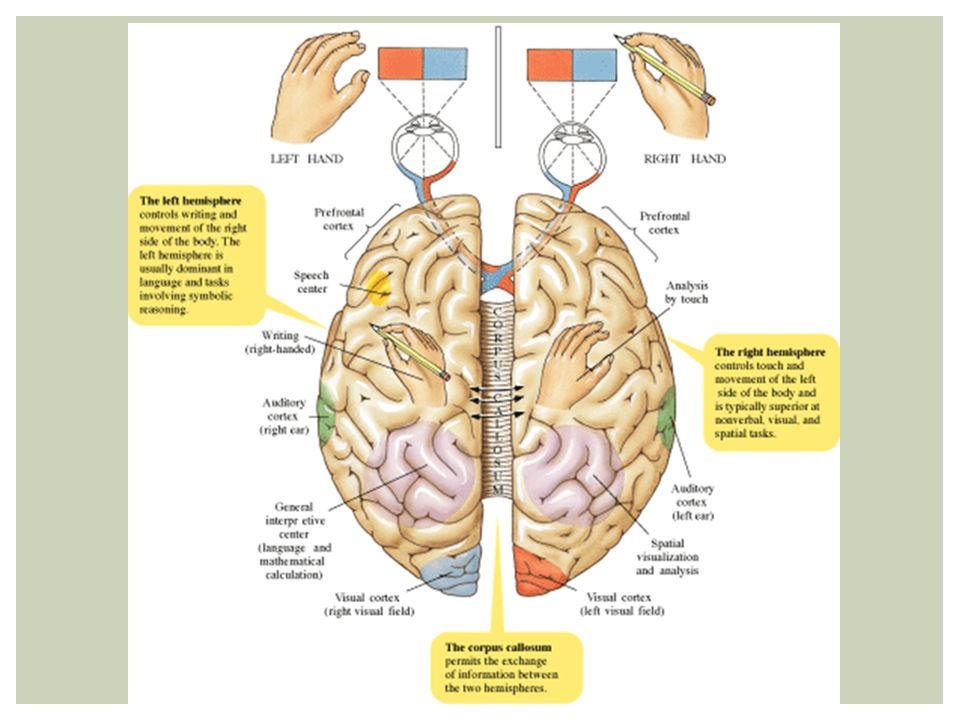
The brain is contralateral. Contralateral: right/left hemisphere controls other side of body. The right side of the brain controls the left side of the body and vice-versa.

60
Right hemisphere: nonverbal spatial, musical, and holistic functions
identifying faces recognizing emotional facial expressions controls the left side of the body

61
Left hemisphere: verbal functions mathematical functions
analytical functions language controls the right side of the body

62
LOCALIZATION AND LATERALIZATION OF THE BRAIN’S FUNCTION
Association areas: regions of the cerebral cortex that do not have specific sensory or motor functions, but are involved in higher mental functions, such as thinking, planning, remembering, and communicating.

64
Structure of Brain Plasticity: when one region of the brain is damaged, the brain can reorganize to take over its function.

65
Our Divided brain Corpus Callosum: the wide band of axon fibers connecting the two hemispheres of the brain. Split Brain – cutting the corpus callosum, resulting in two separate hemispheres.

66
Our Divided brain What we see:

67
Our Divided brain Cutting the corpus callosum alleviates the symptoms of epilepsy. Split Brain results in both hemispheres operating independently of each other and can follow two separate orders simultaneously. Eg. Drawing two pictures with both hands at the same time.

68
Split Brain Clip

69
Right Brain vs. Left Brain???

Similar presentations


>")


