Download presentation
Presentation is loading. Please wait.

1
CT SHOULDER AXIAL

2
CT SHOULDER AXIAL

3
SHOULDER AXIAL

4
CT SHOULDER AXIAL

5
CT SHOULDER AXIAL

6
CT SHOULDER AXIAL

7
CT SHOULDER AXIAL

8
CT SHOULDER AXIAL

9
CT SHOULDER AXIAL

10
SHOULDER AXIAL

11
SHOULDER AXIAL

12
CT SHOULDER AXIAL

13
CT SHOULDER AXIAL

14
CT SHOULDER AXIAL

15
CT SHOULDER AXIAL

16
CT SHOULDER AXIAL

17
CT WRIST AXIAL

18
CT WRIST AXIAL

19
CT WRIST AXIAL

20
CT WRIST AXIAL

21
CT WRIST AXIAL

22
CT WRIST AXIAL

23
CT WRIST AXIAL

24
CT WRIST AXIAL

25
CT WRIST AXIAL

26
CT WRIST AXIAL

27
CT WRIST AXIAL

28
CT WRIST AXIAL

29
CT WRIST AXIAL

30
CT WRIST AXIAL

31
CT WRIST AXIAL

32
CT WRIST AXIAL

33
MR ELBOW AXIAL

34
MR ELBOW AXIAL

35
MR ELBOW AXIAL

36
MR ELBOW AXIAL

37
MR ELBOW AXIAL

38
MR ELBOW AXIAL

39
MR ELBOW AXIAL

40
MR ELBOW AXIAL

41
MR ELBOW AXIAL

42
MR ELBOW AXIAL

43
MR ELBOW AXIAL

44
MR ELBOW AXIAL

45
MR ELBOW AXIAL

46
MR ELBOW AXIAL

47
MR ELBOW AXIAL

48
MR ELBOW AXIAL

49
MR ELBOW AXIAL

50
MR ELBOW AXIAL

51
CT ANKLE AXIAL

52
CT ANKLE AXIAL

53
CT ANKLE AXIAL

54
CT ANKLE AXIAL

55
CT ANKLE AXIAL

56
CT ANKLE AXIAL

57
CT ANKLE AXIAL

58
CT ANKLE AXIAL

59
CT ANKLE AXIAL

60
CT ANKLE AXIAL

61
CT ANKLE AXIAL

62
CT ANKLE AXIAL

63
CT ANKLE AXIAL

64
CT ANKLE AXIAL

65
MR HIP AXIAL

66
MR HIP AXIAL

67
MR HIP AXIAL

68
MR HIP AXIAL

69
MR HIP AXIAL

70
MR HIP AXIAL

71
MR HIP AXIAL

72
MR HIP AXIAL

73
MR HIP AXIAL

74
MR HIP AXIAL

75
MR HIP AXIAL

76
MR HIP AXIAL

77
MR HIP AXIAL

78
MR HIP AXIAL

79
MR HIP AXIAL

80
MR HIP AXIAL

81
MR HIP AXIAL

82
MR HIP AXIAL

83
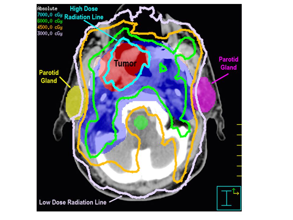
Planning Radiation Therapy
When planning radiation therapy of brain tumours from MR scans of the head, it is necessary to apply to most effective dose of radiation to the tumour, yet cause least damage to the surrounding tissue. This is a complex task demanding 3D visualization. An interactive system is being developed to produce 3D representations of the head, by combining the information from a series of cross-sectional images. Segmentation techniques are used to semi-automatically identify the tumour and other anatomical structures. One approach is for the clinician to select a point within an area of interest, such as the tumour. The system then locates the surrounding points which possess similar characteristics, within a given range of variability. This is repeated for other regions, and other images, to produce a 3D model. Given this model, an optimal treatment plan can be generated.

84
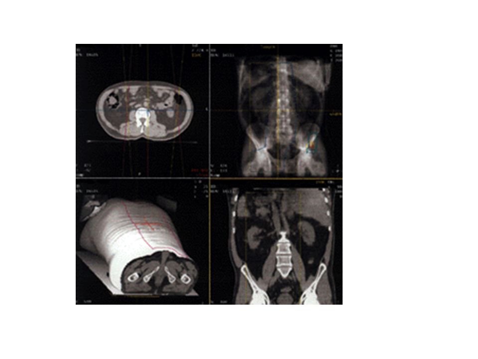
Merging Medical Images
Different types of medical image yield different information. For example, MR scans provide soft tissue information, yet nothing regarding bones. The converse is true of computed tomography (CT) scans. When planning surgery, information from both CT and MR scans may be needed. Machine vision techniques can be used to collate information from the different scans, thus producing a single 3D image showing the relationship between single 3D image showing the relationship between important features. This has been achieved, for example, by interactively labelling matching features in the original images. Bone is identified in the CT scan by locating areas with grey levels above a certain threshold value. Identifying tissue structures in the MR scan requires more sophisticated segmentation techniques. This process is now being developed to use known anatomical relationships to automatically match 3D models derived from different medical images
 scans. When planning surgery, information from both CT and MR scans may be needed. Machine vision techniques can be used to collate information from the different scans, thus producing a single 3D image showing the relationship between single 3D image showing the relationship between important features. This has been achieved, for example, by interactively labelling matching features in the original images. Bone is identified in the CT scan by locating areas with grey levels above a certain threshold value. Identifying tissue structures in the MR scan requires more sophisticated segmentation techniques. This process is now being developed to use known anatomical relationships to automatically match 3D models derived from different medical images.")
87
CT SCANNER

88
CT SCANNER

89
MRI SCANNER

90
What is PET/CT? PET/CT is a new imaging tool that combines two scan techniques in one exam - a PET scan and a CT scan. PET/CT is mainly used for diagnosis, staging or restaging malignant disease and metastases and evaluation of treatment response. It may also be used to differentiate dementia verses Alzheimer's disease. The two procedures together provide information about the location, nature of and the extent of the lesion. In other words, it answers questions like : Where is the tumor, how big is it, is it malignant, benign or due to inflammatory change, and has the cancer spread?

91
Fusion imaging

92
Nuclear Scanners

95
SCAPULA

97
HUMERUS The humerus is the arm bone (most folks call it the UPPER arm bone). It has action at the shoulder joint and at the elbow joints (two at the elbow).
. It has action at the shoulder joint and at the elbow joints (two at the elbow).")
98
RADIUS/ULNA The forearm is the region of the ulna and radius. At the proximal (in proximity to the body) is the elbow. At the distal end (distant from the body) is the wrist.
 is the elbow. At the distal end (distant from the body) is the wrist.")
99
ELBOW

102
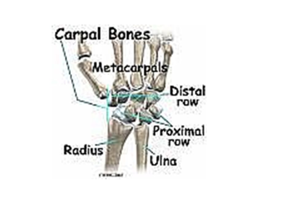
HAND ANATOMY The human hand contains 27 bones divided into three groups: the carpal bones in the wrist, the metacarpal bones forming the knuckles, and the phalanges, or finger bones.

105
FEMUR The thigh is the region of the femur. At the proximal (in proximity to the body) is the hip. At the distal end (distant from the body) is the knee.
 is the hip. At the distal end (distant from the body) is the knee.")
106
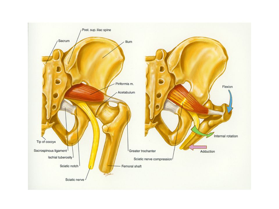
HIP

107
HIP The knee is a region and the knee is a joint. As a joint it is made up of uh... how many bones? Mmm... well, mmm, that depends if you want to count the fibula in on the action. Yeah, let's do that because it anchors the lateral ligament complex. The femur, and tibia are obvious. Add the patella and the fibula, that's four.

108
TIBIA/FIBULA The shin is the region of the tibia. At the proximal (in proximity to the body) is the knee. At the distal end (distant from the body) is the ankle.
 is the knee. At the distal end (distant from the body) is the ankle.")
109
ANKLE The ankle is a region and the ankle is a joint, but the joint isn't the same region as the region. Huh? What folks call the ankle (singular) is actually two distinctly different joints. What they call ankle motion is actually motion of both those joints.
 is actually two distinctly different joints. What they call ankle motion is actually motion of both those joints.")
110
FOOT 1 Calcaneus 2 Talus 3 Navicular 4 Medial cuneiform 5 Intermediate cuneiform 6 Lateral cuneiform 7 Cuboid 8 First metatarsal 9 Second metatarsal 10 Third metatarsal 11 Fourth metatarsal 12 Fifth metatarsal 13 Proximal phalanx of great toe 14 Distal phalanx of great toe 15 Proximal phalanx of second toe 16 Middle phalanx of second toe 17 Distal phalanx of second toe

Similar presentations


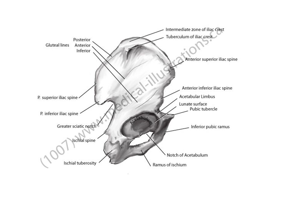


 Coxae have 3 distinct parts: – Ilium – Ischium – Pubis.>")


