Download presentation
Presentation is loading. Please wait.

1
Introduction to Dentures
Lecturer Hatem Dousouky Ahmad

2
Terminology Prosthetics Prosthesis Prosthodontics Dentulous Edentulous
Partially edentulous Complete Denture Partial Denture Immediate denture

3
“A prosthesis that replaces the lost natural dentition and associated
Complete Denture Definitions : “A prosthesis that replaces the lost natural dentition and associated structures of the mandible and / or the maxilla “ . Component Parts : Denture Base Denture Flange Denture Border (Edge) Denture Surfaces : Fitting Surface Polished Surface Occlusal Surface
 Denture Surfaces : Fitting Surface. Polished Surface. Occlusal Surface.")
4
Complete denture consists of

5
Restoration of mastication.
Objectives in complete denture construction: Restoration of mastication. Restoration of facial dimensions and contours. Restoration of speech. Restoration of the remaining natural tissues. Satisfaction and comfort of the patient.

6
Anatomical Landmarks In Relation to Complete Denture:
Extraoral landmarks Intraoral landmarks

7
Changes The Happened After Teeth Loss :
Face :

8
Extra Oral Landmarks Inter pupillary line Ala Tragus Line
Naso – Labial sulcus Philtrum Labiomental sulcus Angle of the mouth Modiolus Vermillion border

10
Landmarks of Importance in Occlusal Plane Orientation
Interpupillary line Ala tragus line

11
Landmarks restored by proper lip support
Naso – Labial sulcus Becomes deeper with age and with loss of teeth Philtrum becomes flat with loss of teeth Modiolus moves inwards and downwards Vermillion border becomes reduced in size

12
Landmarks restored by Proper Vertical Dimension
Vermillion border Angle of the mouth becomes inflammed angular cheilitis

14
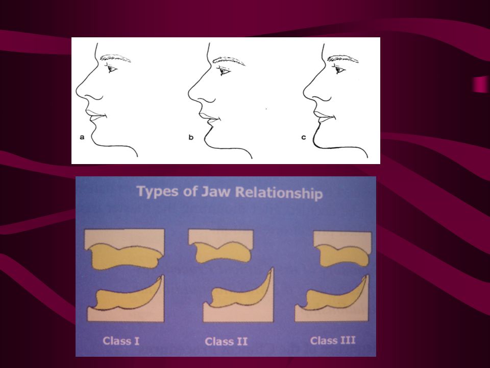
The Labiomental Sulcus
A Landmark helpful in determining the Jaw Relation Angle Class I Angle Class II Angle Class III

15
Intraoral Landmarks Mandible Maxilla Supporting structures
& Limiting structures Maxilla Supporting structures & Limiting structures

17
Maxillary Supporting Structures
Incisive Papilla 1 . The incisive papilla is a thick part of the mucous membrane covering the incisive foramen. 2 . It is located at the anterior end of the median palatine raphae . 3 . The nasopalatine nerves and vessels pass through the incisive foramen to supply the anterior 2 / 3 of the palate. 4 . In some cases due to the excessive bone resorption, the papilla may lie on the crest of the ridge. 5 . The incisive papilla should be relieved to avoid pressure on the incisive nerves and vessels.

18
Cont. max. supporting structures
Palatine Rugae Rugae Area 1 . It is an irregular shaped elevation of the soft tissue extending laterally from the midline in the anterior part of the hard palate. 2 . If serves as one of stress bearing area in the palate .

19
Cont. max. supporting structures
Median Palatine Raphae Median Palatine Raphae 1 . The midline of the hard palate is covered by a thin layer of mucoperiostium , that covers the median palatine suture . 2 . That suture joins the right and the left halves of the hard palate. 3 . It is usually relieved to increase denture stability by preventing its rocking .

20
Cont. max. supporting structures
Torus Palatinus When small relieved When large surgical excision

21
Cont. max. supporting structures
Fovea Palatina Fovia Palatina 1 . It is helps in the determination of the posterior border of the upper denture. 2 . The posterior border of the upper denture should be 2 mm posterior to the fovea Palatina .

22
Cont. max. supporting structures
Residual Alveolar Ridge Residual Alveolar Ridge 1 . It should be firm . 2 . since it is usually well developed, it might be considered as a primary stress bearing area.

23
Cont. max. supporting structures
Tuberosity Tuberosity 1 . It is important for retention and support of the upper denture against lateral movement. 2 . The denture should cover it .

24
Cont. max. supporting structures
Buttress Part Of Bone Buttress Part Of Bone 1 . It is formed of the lower portion of the zygomatic process of the maxilla (the area above the first molar teeth) . 2 . It provides excellent resistance to the vertical forces(Support).
 It provides excellent resistance to the vertical forces(Support).")
25
Maxillary Limiting Structures
Labial Frenum Labial Frenum Labial Frenum It must be relieved in the denture by making a V-shape notch in the labial flange opposite to its position .

26
Cont. Maxillary Limiting Structures
Labial Vestibule Labial Vestibule 1 . It Is the reflection of the mucosa of the lip to the mucosa of the alveolar process in the labial vestibule. 2 . The denture in this area is in relation to the orbicularis oris and the superior incisive muscles . 3 . These muscles limit the thickness and the length of the labial flange of the denture.

27
Cont. Maxillary Limiting Structures
Buccal Frenum Buccal Frenum 1 . It is a fold of mucous membrane (tendon of the buccinator muscle) varies in size in number and in position . 2 . A notch is made in the denture flange opposite to its position to facilitate its functional movements.
 varies in size in number and in position A notch is made in the denture flange opposite to its position to. facilitate its functional movements.")
28
Cont. Maxillary Limiting Structures
Buccal Vestibule Buccal Vestibule 1 . The denture in this area is related to buccinator muscle. 2 . Buccal flanges must extend in the buccal vestibule . 3 . Due to the horizontal direction of the fibers of this muscle contraction of this muscle will not displace the denture.

29
Cont. Maxillary Limiting Structures
Hamular Notch Hamular Notch 1 . It is one of the important landmarks for determination of the posterior limit of the upper denture . 2 . A straight line from hamular notch on one side to the other on the other side determine the posterior limit of the upper denture

30
Cont. Maxillary Limiting Structures
Vibrating Line ( Ah Line) Vibrating Line ( Ah Line) Postdam area 1 . It separate the movable part from the immovable part of the soft palate. 2 . This line is 2mm posterior to the fovea palatine . 3 . This line determines the posterior end of the upper denture.
 Vibrating Line. ( Ah Line) Postdam area. 1 . It separate the movable part from the immovable part of the soft palate. 2 . This line is 2mm posterior to the fovea palatine This line determines the posterior end of the upper denture.")
31
Cont. Maxillary Limiting Structures
Curvature of the soft palate I Class I gentle curvature Class II medium curvature Class III abrupt curvature II III

34
Denture should be notched
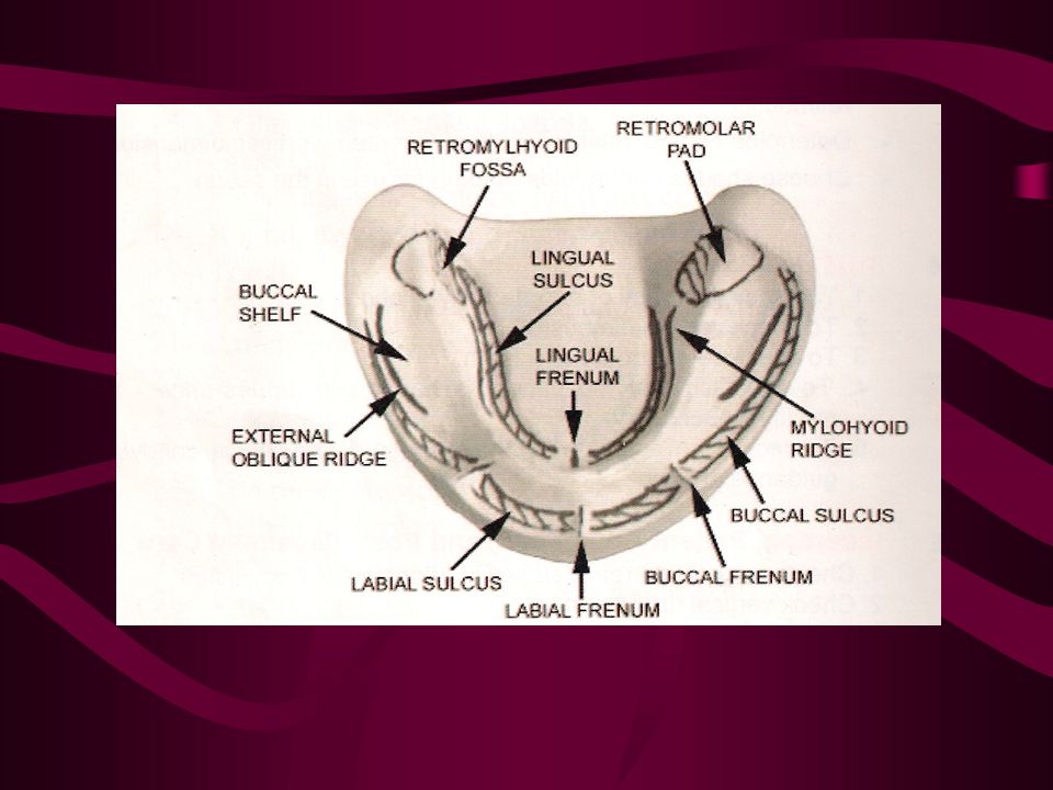
Mandibular Limiting Structures Labial Frenum Denture should be notched opposite to it. Labial Frenum

35
Cont. Mandibular Limiting Structures
Labial Vestibule Labial Vestibule Limits the denture flange thickness and length.

36
Cont. Mandibular Limiting Structures
Buccal Frenum Buccal Frenum 1 . It is a fold of mucous membrane in the premolar area, movement of the lip and the cheek move the frenum . 2 . A notch is made in the lower denture to accommodate the frenum.

37
Cont. Mandibular Limiting Structures
Buccal Vestibule Buccal Vestibule 1 . The denture in this area is related to the buccinator muscle . 2 . Its contraction does not displace the lower denture so flanges of the lower denture must extend in the buccal vestibule.

38
Cont. Mandibular Limiting Structures
Masseter muscle influencing area Masseteric notch Distobuccal area

39
Cont. Mandibular Limiting Structures
Posterior end of retromolar pad Posterior end of retromolar pad It constitutes the posterior limit of the lower denture at which postdamming can be performed.

40
Cont. Mandibular Limiting Structures
Palatoglossal arch Distolingual area Denture overextension in this area will cause sore throat.

41
Cont. Mandibular Limiting Structures
Lingual Pouch Lingual Pouch Lingual pouch More posteriorly the lingual flanges are related to the lingual pouch with its boundaries which are : Posteriorly : The palatoglosssus muscle . Anteriorly : The Mylohyoid muscle. Medially : The tongue . Laterally : The medial aspect of the mandible.

42
Cont. Mandibular Limiting Structures
Mylohyoid muscle influencing area (internal oblique ridge) Mylohyoid muscle influencing area Mylohyoid muscle influencing area
 Mylohyoid muscle. influencing area. Mylohyoid muscle. influencing area.")
43
Cont. Mandibular Limiting Structures
Sublingual salivary gland area Sublingual salivary gland area Sublingual salivary gland area The lingual flanges of the lower denture should not extend in this area because with excessive resorption of the mandible the gland may bulge superiorly above the body of the mandible.

44
Cont. Mandibular Limiting Structures
Lingual Frenum Lingual Frenum 1 . More anteriorly a fold mucous membrane attach the mucosa of the tongue tip to mucosa of the floor of the mouth 2 . It moves with the movement of the tongue so a notch is made to accommodate the frenum.

45
Michael H. Hart (born April 28, 1932 in New York City)
Graduate of the Bronx High School of Science, received his undergraduate degree at Cornell University in mathematics and later earned a Ph.D. in astrophysics at Princeton University. He also holds graduate degrees in physics, astronomy, and computer science, as well as a law degree. He was a research scientist at NASA before leaving to be a professor of physics at Trinity University in San Antonio, Texas.

48
منذ سنين وأنت تفخر بأنك مسلم... فماذا فعلت ليفخر الإسلام بك؟؟
عن أبي هريرة رضي الله عنه أن رسول الله صلى الله عليه وسلم وقف على أناس جلوس فقال ( ألا أخبركم بخيركم من شركم ) قال : فسكتوا ، فقال ذلك ثلاث مرات ، فقال رجل : بلى يا رسول الله أخبرنا بخيرنا من شرنا ، قال ( خيركم من يرجى خيره ويؤمن شره وشركم من لا يرجى خيره ولا يؤمن شره ) رواه الترمذي. أحب العباد إلى الله تعالى أنفعهم لعياله
 قال : فسكتوا ، فقال ذلك ثلاث مرات ، فقال رجل : بلى يا رسول الله أخبرنا بخيرنا من شرنا ، قال ( خيركم من يرجى خيره ويؤمن شره وشركم من لا يرجى خيره ولا يؤمن شره ) رواه الترمذي. أحب العباد إلى الله تعالى أنفعهم لعياله.")
49
Mandibular Supporting Structures
Residual Ridge (Fibrous band of connective tissue) Residual Ridge 1. It covers the crest of the lower ridge. 2. Its mobility may cause pressure symptoms under the lower denture. 3 . Also can affect denture stability .
 Residual Ridge. 1. It covers the crest of the lower ridge. 2. Its mobility may cause pressure symptoms under the lower denture. 3 . Also can affect denture stability .")
50
External oblique ridge
Cont. Mandibular Supporting Structures External oblique ridge External obique ridge

51
Cont. Mandibular Supporting Structures
Buccal Shelf Of Bone Buccal Shelf Of Bone 1 . The area that lies between the crest of the residual ridge and the external oblique ridge. 2 . It is the primary stress bearing area in the lower arch . 3 . It forms good support for the lower denture .

52
Cont. Mandibular Supporting Structures
Retromolar pad Retromolar pad 1 . It is a pear shaped area of mucous membrane at the posterior end of the mandibular ridge and anterior to the pterygomandibular raphae . 2 . It consists of mucous glands , temporal tendon , fibers of the buccinators and superior constrictor muscle . 3 . Lower denture should cover this area for retention and to cover the buccal shelf of bone (Primary stress bearing area) it act as valve seal area for the lower denture.
 it act as valve seal. area for the lower denture.")
53
Cont. Mandibular Supporting Structures
Torus mandibularis When small relieved When large surgical excision Torus mandibularis

55
Cont. Mandibular Supporting Structures
Internal oblique ridge If sharp, it should be surgically reduced. Internal oblique ridge

56
Cont. Mandibular Supporting Structures
Mental foramen When the ridge is resorbed, it appears on its crest . Since it covers nerves, it should be relieved.

57
Cont. Mandibular Supporting Structures
Genial tubercles When the ridge resorbed, it appears on its crest . It is relieved or better surgically reduced

59
THANK YOU

Similar presentations


 B.M.C.>")


>")


