Download presentation
Presentation is loading. Please wait.

1
Chest Radiography Interpretation
Dr. Raghu Ram Uppalapati

2
Lung Anatomy Trachea Carina Right and Left Pulmonary Bronchi
Secondary Bronchi Tertiary Bronchi Bronchioles Alveolar Duct Alveoli

3
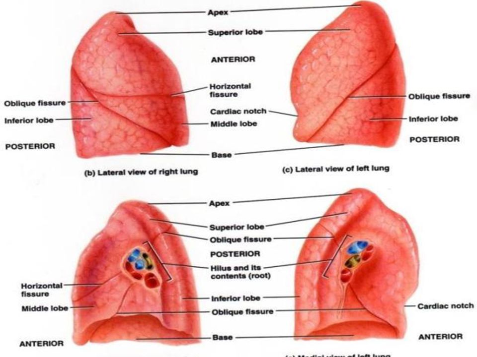
Lung Anatomy Right Lung Left Lung Superior lobe Middle lobe
Inferior lobe Left Lung

4
Lung Anatomy on Chest X-ray
PA View: Extensive overlap Lower lobes extend high Lateral View: Extent of lower lobes

5
Lung Anatomy on Chest X-ray
The right upper lobe (RUL) occupies the upper 1/3 of the right lung. Posteriorly, the RUL is adjacent to the first three to five ribs. Anteriorly, the RUL extends inferiorly as far as the 4th right anterior rib The right upper lobe (RUL) occupies the upper 1/3 of the right lung. Posteriorly, the RUL is adjacent to the first three to five ribs. Anteriorly, the RUL extends inferiorly as far as the 4th right anterior rib.
 occupies the upper 1/3 of the right lung. Posteriorly, the RUL is adjacent to the first three to five ribs. Anteriorly, the RUL extends inferiorly as far as the 4th right anterior rib. The right upper lobe (RUL) occupies the upper 1/3 of the right lung. Posteriorly, the RUL is adjacent to the first three to five ribs. Anteriorly, the RUL extends inferiorly as far as the 4th right anterior rib.")
6
Lung Anatomy on Chest X-ray
The right middle lobe is typically the smallest of the three, and appears triangular in shape, being narrowest near the hilum The right middle lobe is typically the smallest of the three, and appears triangular in shape, being narrowest near the hilum.

7
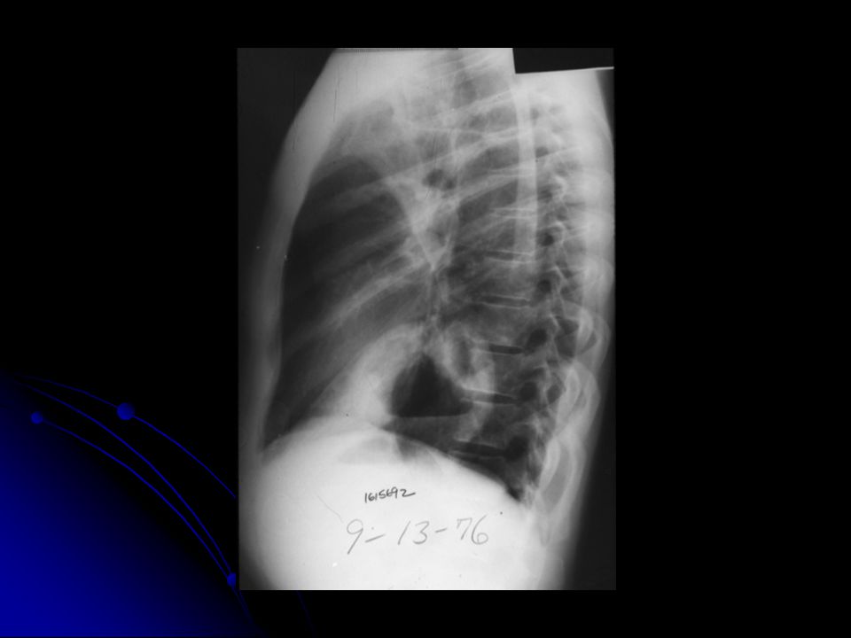
The right lower lobe is the largest of all three lobes, separated from the others by the major fissure. Posteriorly, the RLL extend as far superiorly as the 6th thoracic vertebral body, and extends inferiorly to the diaphragm. Review of the lateral plain film surprisingly shows the superior extent of the RLL. Posteriorly, the RLL extend as far superiorly as the 6th thoracic vertebral body, and extends inferiorly to the diaphragm. Review of the lateral plain film surprisingly shows the superior extent of the RLL; there is considerable overlap between the more anterosuperiorly located RUL and the RLL. Similarly, the deep posterior gutters extend considerably inferiorly; with full inspiration, the lower lobe can extend may as low as L2, becoming superimposed over the upper poles of the kidneys.

8
Lung Anatomy on Chest X-ray
These lobes can be separated from one another by two fissures. The minor fissure separates the RUL from the RML, and thus represents the visceral pleural surfaces of both of these lobes. Oriented obliquely, the major fissure extends posteriorly and superiorly approximately to the level of the fourth vertebral body. Grossly, these lobes can be separated from one another by two fissures which anatomically correspond to the visceral pleural surfaces of those lobes from which they are formed. The minor fissure separates the RUL from the RML, and thus represents the visceral pleural surfaces of both of these lobes. The minor fissure is oriented horizontally, extending ventrally from the chest wall, and extending posteriorly to meet the major fissure. Generally, the location of the minor fissure is approximately at the level of the fourth vertebral body and crosses the right sixth rib in the midaxillary line. The right major fissure is more expansive in size than the minor fissure, separating the right upper and middle lobes from the larger right lower lobe. Oriented obliquely, the major fissure extends posteriorly and superiorly approximately to the level of the fourth vertebral body. The major fissure extends anteroinferiorly, intersecting the diaphragm at the anterior cardiophrenic angle

9
The lobar architecture of the left lung is slightly different than the right.
Because there is no defined left minor fissure, there are only two lobes on the left; the left upper The lobar architecture of the left lung is slightly different than the right. Because there is no defined left minor fissure, there are only two lobes on the left; left upper

10
Lt Lower Lobes Left lower lobes and left lower lobes

11
Lung Anatomy on Chest X-ray
These two lobes are separated by a major fissure, identical to that seen on the right side, although often slightly more inferior in location. The portion of the left lung that corresponds anatomically to the right middle lobe is incorporated into the left upper lobe. These two lobes are separated by a major fissure, identical to that seen on the right side, although often slightly more inferior in location. The portion of the left lung that corresponds anatomically to the right middle lobe is incorporated into the left upper lobe. It is important to understand that in most individuals, interlobar fissures are usually not completely formed; in some individuals there may be complete absence of a fissure thus losing the demarcation between lobes on gross examination. In general, fissures are not readily identifiable on plain films, with only small portions typically visualized at best. This is because fissures which are composed of only two layers of visceral pleura, may not present a significant radiographic interface and will not produce a shadow. However, if there is fluid within the pleural space or if the visceral pleura is thickened, fissures may be seen in their entirety.

12
RUL (Right Upper Lung)
")
13
RML (Right Middle Lung)
")
14
RLL (Right Lower Lung)
")
16
LUL (Left Upper Lung)
")
17
LLL (Left Lower Lung)
")
18
Left Side Fissure LUL LLL

19
Chest Radiography: Basic Principles
A structure is rendered visible on a radiograph by the juxtaposition of two different densities

20
Silhouette Sign Loss of the expected interface normally created by juxtaposition of two structures of different density No boundary can be seen between two structures of similar density

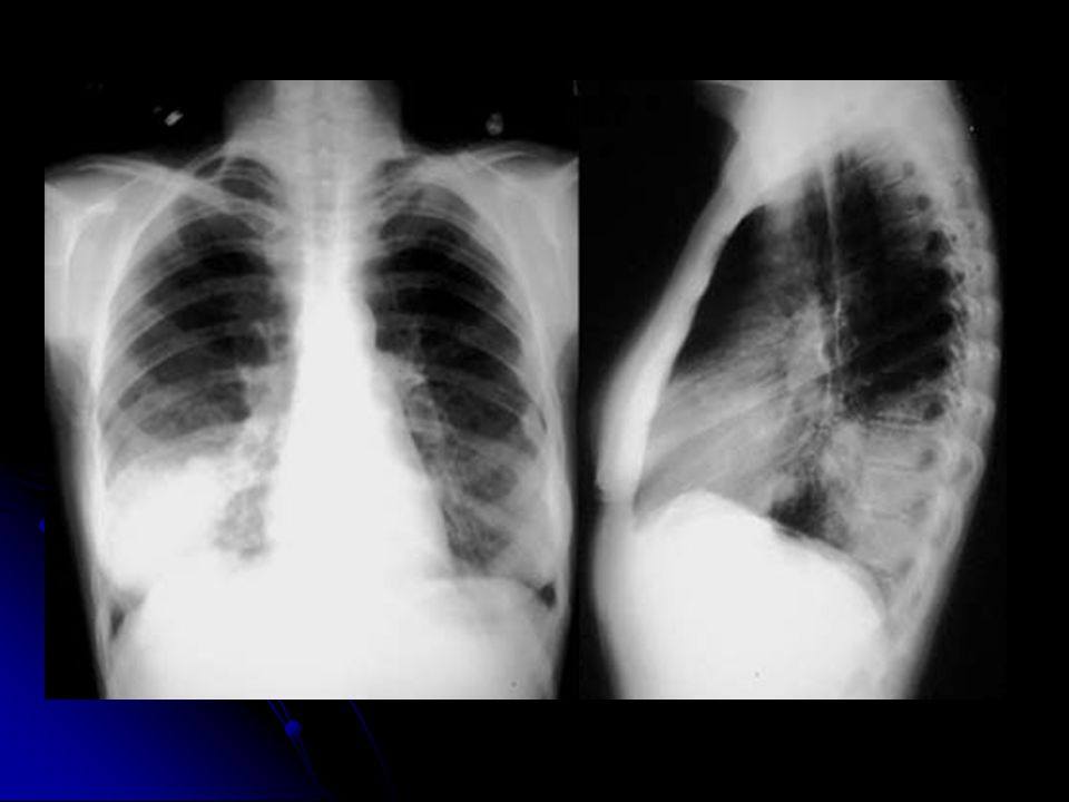
21
Right Lower Lobe Pneumonia

22
Differential X-Ray Absorption
The absence of a normal interface may indicate disease; The presence of an unexpected interface may also indicate disease The presence of interfaces can be used to localize abnormalities

23
Chest Radiographic Patterns of Disease
Air space opacity Interstitial opacity Nodules and masses Lymphadenopathy Cysts and cavities Lung volumes Pleural diseases

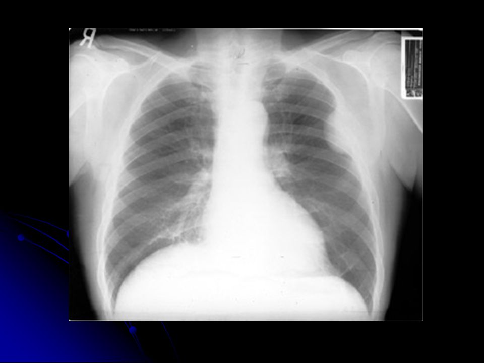
24
LUL Pneumonia

25
Air Space Opacity Components:
air bronchogram: air-filled bronchus surrounded by airless lung confluent opacity extending to pleural surfaces segmental distribution

26
Air Space Opacity: DDX Blood (hemorrhage) Pus (pneumonia)
Water (edema) hydrostatic or non-cardiogenic Cells (tumor) Protein/fat: alveolar proteinosis and lipoid pneumonia
 hydrostatic or non-cardiogenic. Cells (tumor) Protein/fat: alveolar proteinosis and lipoid pneumonia.")
27
Interstitial Opacity: Small Nodules

28
Interstitial Opacity:
Lines

29
Interstitial Opacity: Lines & Reticulation

30
Interstitial Opacity Hallmarks: small, well-defined nodules lines
interlobular septal thickening fibrosis reticulation

31
Interstitial Opacity: DDX
Idiopathic interstitial pneumonias Infections (TB, viruses) Edema Hemorrhage Non–infectious inflammatory lesions sarcoidosis Tumor
 Edema. Hemorrhage. Non–infectious inflammatory lesions. sarcoidosis. Tumor.")
32
Nodules and Masses Nodule: any pulmonary lesion represented in a radiograph by a sharply defined, discrete, nearly circular opacity mm in diameter Mass: larger than 3 cm

33
Nodules and Masses Qualifiers: single or multiple size
border definition presence or absence of calcification location

34
Well-Defined Calcification Ill-Defined Mass

35
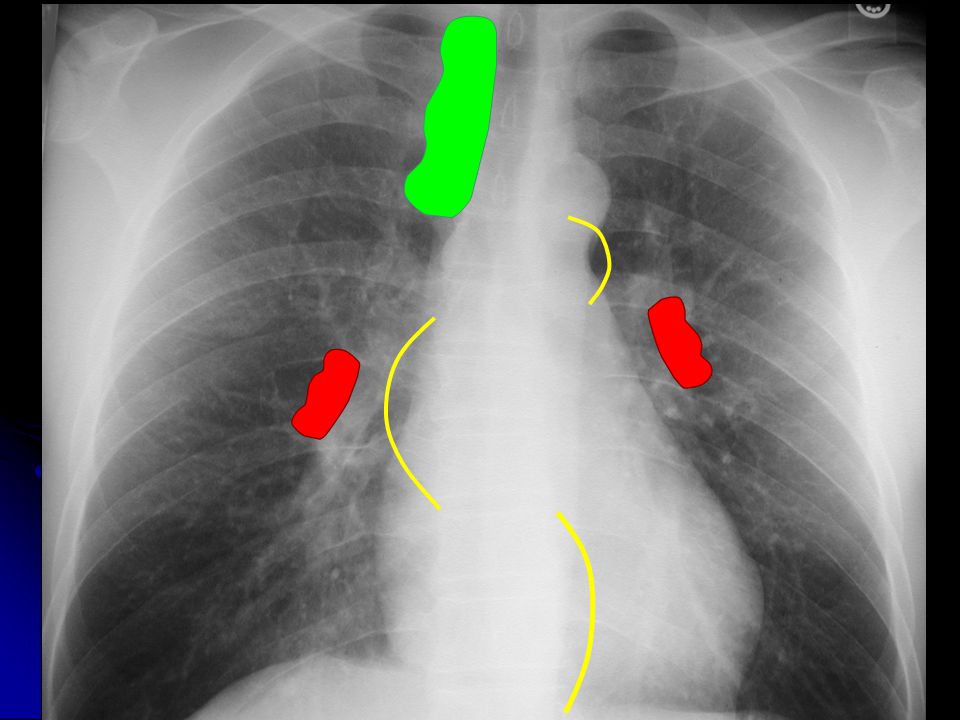
Lymphadenopathy Non-specific presentations: Specific patterns:
mediastinal widening hilar prominence Specific patterns: particular station enlargement

36
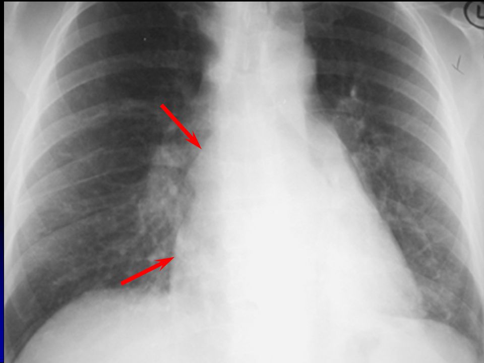
Right Paratracheal Lymphadenopathy

37
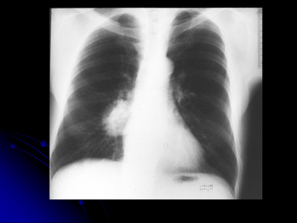
Right Hilar LAN

38
Right Hilar LAN

39
Left Hilar LAN

41
Subcarinal LAN *

42
AP Window LAN

44
Cysts & Cavities Cyst: abnormal pulmonary parenchymal space, not containing lung but filled with air and/or fluid, congenital or acquired, with a wall thickness greater than 1 mm epithelial lining often present

45
Cysts & Cavities Cavity: Abnormal pulmonary parenchymal space, not containing lung but filled with air and/or fluid, caused by tissue necrosis, with a definitive wall greater than 1 mm in thickness and comprised of inflammatory and/or neoplastic elements

46
Cysts & Cavities Characterize: wall thickness at thickest portion
inner lining presence/absence of air/fluid level number and location

47
Benign Lung Cyst : PCP Pneumatocele
Uniform wall thickness 1 mm Smooth inner lining

48
Benign Cavities : Cryptococcus max wall thickness 4 mm minimally irregular inner lining

49
Indeterminate Cavities
max wall thickness 5-15 mm mildly irregular inner lining

50
Malignant Cavities: Squamous Cell Ca
max wall thickness 16 mm Irregular inner lining

51
Pleural Disease: Basic Patterns
Effusion angle blunting to massive mobility Thickening distortion, no mobility Mass Air Calcification

52
Pleural Effusion

53
Pleural Effusion

54
Pleural Calcification

56
SOME INTERESTING X-RAYS & DISCUSSIO N

57
Chest breast implants

58
Tip of ET Pneumomediastinum

59
Potential X ray findings
wide mediastinum obliteration of aortic knob Rt mainstem shift up and right NG deviate to right pleural cap Major Vessel Injury

60
Expiration reduces lung volume, making a small pneumo easier to see

67
Irregular linear opacities are present in both lungs, especially in the periphery and the bases of the lungs. The heart is slightly enlarged, but this is not related to the pulmonary abnormalities in this case.

70
Hodgkin’s Disease

72
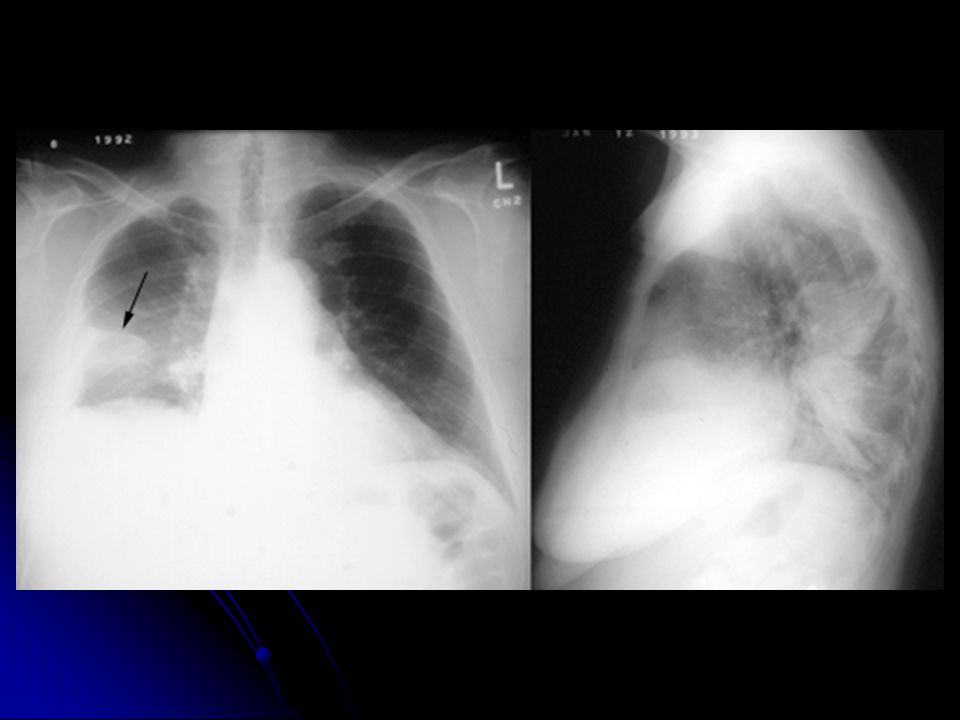
A single, 3cm relatively thin-walled cavity is noted in the left midlung. This finding is most typical of squamous cell carcinoma (SCC). One-third of SCC masses show cavitation
. One-third of SCC masses show cavitation.")
74
LUL Atelectasis: Loss of heart borders/silhouetting
LUL Atelectasis: Loss of heart borders/silhouetting. Notice over inflation on unaffected lung

76
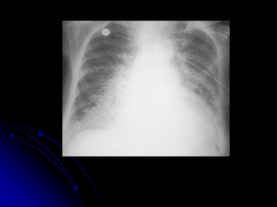
Right Middle and Left Upper Lobe Pneumonia

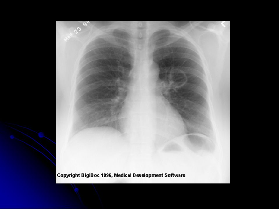
78
Pseudotumor: fluid has filled the minor fissure creating a density that resembles a tumor (arrow). Recall that fluid and soft tissue are indistinguishable on plain film. Further analysis, however, reveals a classic pleural effusion in the right pleura. Note the right lateral gutter is blunted and the right diaphram is obscurred.

80
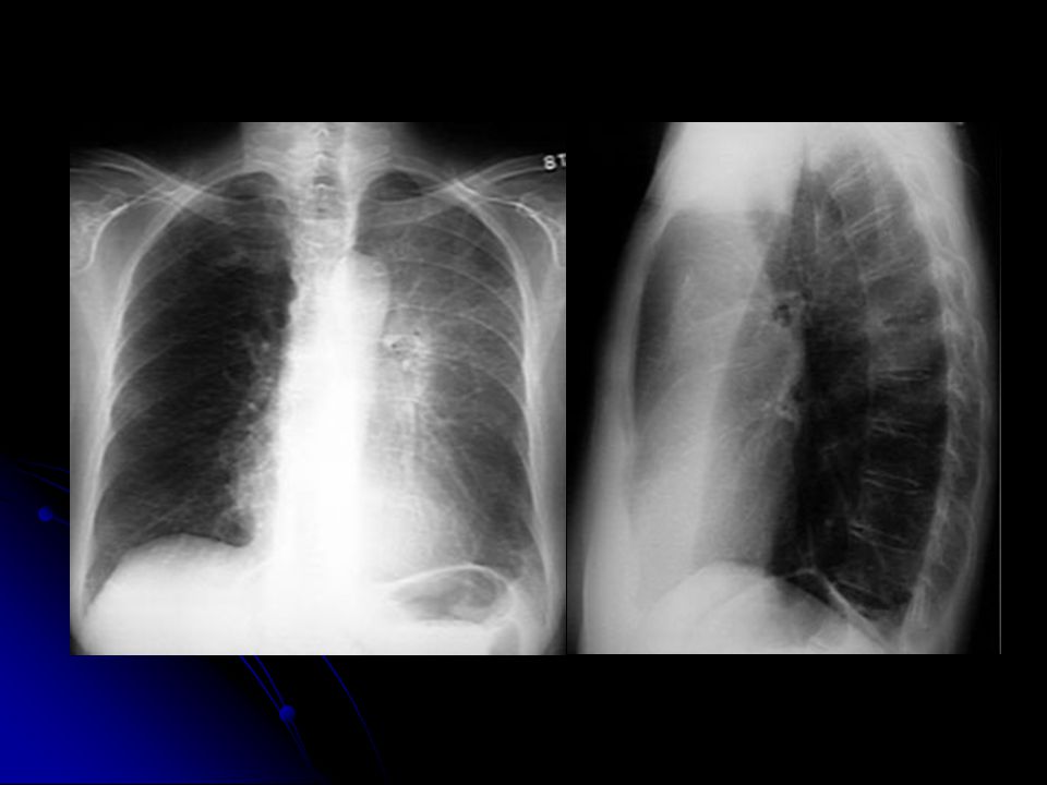
Pneumonia:a large pneumonia consolidation in the right lower lobe
Pneumonia:a large pneumonia consolidation in the right lower lobe. Knowledge of lobar and segmental anatomy is important in identifying the location of the infection

83
24 hours after diuretic therapy

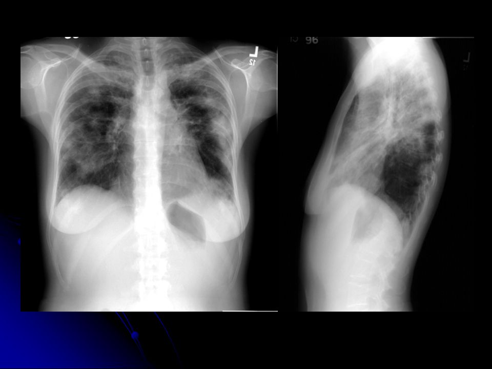
84
CHF:a great deal of accentuated interstitial markings, Curly lines, and an enlarged heart. Normally indistinct upper lobe vessels are prominent but are also masked by interstitial edema.

Similar presentations


 –Partial.>")


>")


