Download presentation
Presentation is loading. Please wait.

1
CXR of the Day!

2
Normal Chest X-Ray

4
Pleural Effusion Blunted costophrenic angles Meniscus Sign

6
Hydropneumothorax Hydrothorax + Pneumothorax Note the air-fluid level in the pleural space.

8
Air Bronchograms Diffuse increased density throughout both lungs highlighted by tubular lucencies. These are air bronchograms. They are visualized because of the alveolar filling that surrounds them. This typical alveolar-filling pattern (air space disease) suggests acute pneumonia, pulmonary hemorrhage, or pulmonary edema.
 suggests acute pneumonia, pulmonary hemorrhage, or pulmonary edema..")
10
Deep Sulcus Sign Deep sulcus sign. Portable supine radiograph in a patient status post median sternotomy. Note the increased lucency in the left upper quadrant. The highest portion of the thorax in the supine patient is the anterior cardiophrenic sulcus. This accounts for the well- defined low cardiac border (arrow) and the adjacent fat pad.
 and the adjacent fat pad..")
12
Tension pneumothorax Portable chest radiograph in a patient status-post median sternotomy and coronary artery bypass grafting. Note the large right pneumothorax displacing the mediastinum to the left and the right hemidiaphragm inferiorly. These findings indicate the presence of a tension pneumothorax on the right requiring immediate chest tube placement.

14
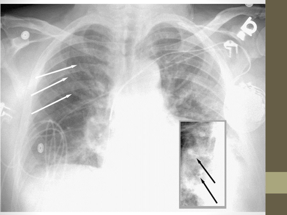
Moderate pulmonary edema Cephalization of the blood flow is visible (white arrows). The blood vessels to the apex of the lung are enlarged and similar in size to the blood vessels to the base of the lungs. The inset displays peribronchial cuffing (black arrows) the inset is from the right hilum of the same film but enhanced to make the peribronchial cuffing easier to see.
 the inset is from the right hilum of the same film but enhanced to make the peribronchial cuffing easier to see..")
16
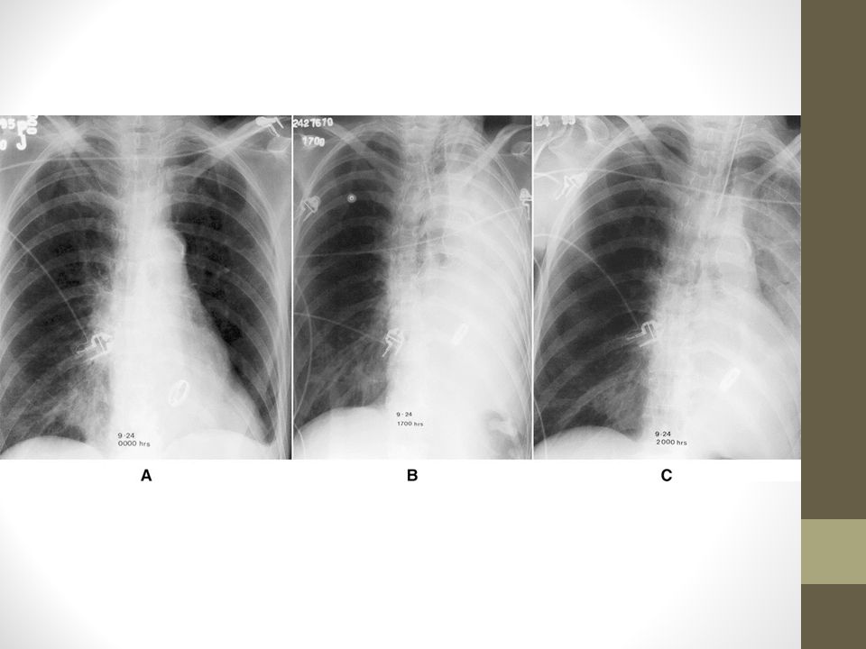
Three portable chest films obtained within a 20-hour time span. A, Good aeration of both lungs. B, Film obtained 17 hours later shows complete opacification of the left hemithorax. Bronchoscopy performed after this film revealed a mucus plug in the left main bronchus. It was removed at bronchoscopy. C, Partial reexpansion.

18
Bullous Emphysema Marked pulmonary hyperinflation is worse on the right. The asymmetrical hyperinflation is producing mediastinal shift to the left. There is flattening of the diaphragm, prominence of the clear spaces, and large areas in the upper lung zones that are devoid of any vascular markings. The walls of these bullous air spaces are well visualized.

20
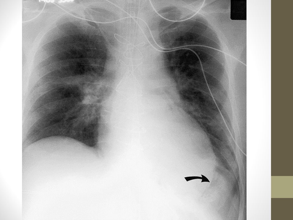
Portable supine chest film demonstrates malposition of an endotracheal tube in the right mainstem bronchus (arrow).
.")
21
Severe pulmonary edema Both lungs are opacified in a bat's wing distribution. The hilar vessels are not visible due to the edema in the lung tissue surrounding these vessels. Peribronchial cuffing can be seen at the black arrows.

23
Interstitial lung disease Posteroanterior view of the chest in a patient complaining of shortness of breath. The lung volumes are diminished. Several small cystic lucencies are visualized between the increased basilar interstitial markings representing honeycombing. The diagnosis is scleroderma lung.

Similar presentations


 Cardiomyopathy L R Shunts.>")

 –Partial.>")


>")


