Download presentation
Presentation is loading. Please wait.

1
CHAPTER 6: SKELETAL MUSCLES – PART 2
- Most skeletal muscles attach to different bones at each end having it span at least one joint - When a muscle contracts, the bone at one end usually remains still while the bone at the other end moves. ORIGIN – stationary attachment of a muscle to a bone, etc. BELLY – thick mid-region of skeletal muscle INSERTION – mobile attachment of a muscle to a bone, etc.

2
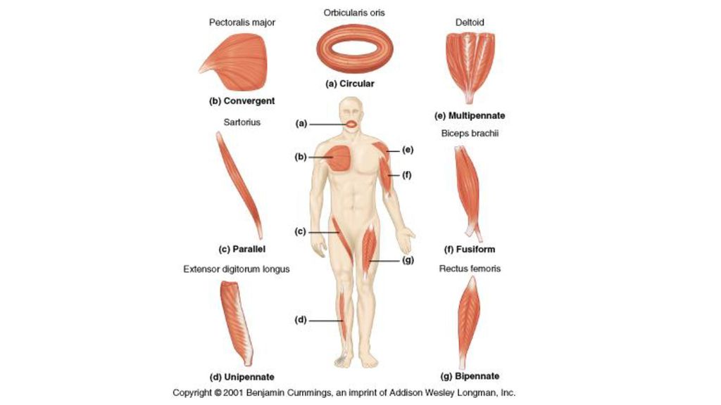
CLASSIFICATION OF MUSCLES ACCORDING TO FASCICLE ORIENTATION.
Fascicles are the “grain” visible in each muscle type. A. FUSIFORM: muscles thick in middle and tapered on ends. Ex. biceps brachii (arm), gastrocnemuius (calf). B. PARALLEL MUSCLES: long strap-like, uniform width and parallel fascicles. They usually span great distance, usually weaker than fusiform. Ex. rectus abdominis (abdomen), sartorius (inner thigh), zygomaticucs major (face). C. CONVERGENT MUSCLES: fan shaped, broad at origin and narrower at insertion. Relatively strong. Ex. pectoralis major (chest) D. PENNATE MUSCLES: (3 types) – feather shaped. Fascicles insert obliquely on tendon that runs length of muscle, like shaft of feather. 1. unipennate – all fascicles approach tendon from one side Ex. palmar interosseous muscles (hand), semimembranosus (thigh), 2. bipennate – fascicles approach tendon from both sides Ex. rectus femoris (thigh) 3. multipennate – shaped like bunch of feathers with quills converging on a single point. Ex. deltoid (shoulder) E. CIRCULAR MUSCLES (sphincters) – rings around body openings. Ex. obicularis oculi (eyelids), urethral and anal sphincters
, gastrocnemuius (calf). B. PARALLEL MUSCLES: long strap-like, uniform width and parallel fascicles. They usually span great distance, usually weaker than fusiform. Ex. rectus abdominis (abdomen), sartorius (inner thigh), zygomaticucs major (face). C. CONVERGENT MUSCLES: fan shaped, broad at origin and narrower at insertion. Relatively strong. Ex. pectoralis major (chest) D. PENNATE MUSCLES: (3 types) – feather shaped. Fascicles insert obliquely on tendon that runs length of muscle, like shaft of feather. 1. unipennate – all fascicles approach tendon from one side Ex. palmar interosseous muscles (hand), semimembranosus (thigh), 2. bipennate – fascicles approach tendon from both sides Ex. rectus femoris (thigh) 3. multipennate – shaped like bunch of feathers with quills converging on a single point. Ex. deltoid (shoulder) E. CIRCULAR MUSCLES (sphincters) – rings around body openings. Ex. obicularis oculi (eyelids), urethral and anal sphincters.")
4
TYPES OF MUSCLES (based on movement)
Muscles can’t push, they can only pull when contracted. Most actions are 2 or more muscles working together or against each other. Muscles are arranged in such a way that whatever one group does, another group can undo. Prime mover – muscle with the major responsibility for a certain movement. Antagonist – muscle that opposes or reverses a prime mover. When a primer is active, the antagonist is stretched and relaxed. Ex. bicep (primer), tricep (antagonist).
, tricep (antagonist).")
5
Synergist – muscle that aids a prime mover in a movement and helps prevent rotation, they help to stabilize joints and helps to cause unwanted movement that could cause injury. Ex. making a fist possible without bending the wrist because of synergistic muscles in the wrist that keeps it stabilized. Fixator – stabilizes the origin of a prime mover (specialized synergists) – prevents a bone from movement Ex. postural muscles that stabilize the vertebral column, muscles that anchor the scapula to the thorax.
 – prevents a bone from movement Ex. postural muscles that stabilize the vertebral column, muscles that anchor the scapula to the thorax.")
6
NAMING SKELETAL MUSCLES
Criteria: (some may be combined when naming a muscle) 1. DIRECTION OF MUSCLE FIBERS – rectus (straight) – fibers run parallel to midline of body; transverse or oblique (slanted) slanted to midline of body Ex. rectus abdominis (stomach) 2. RELATIVE SIZE OF MUSCLE – maximus (largest), minimus (smallest), longus (long), brevis (short) Ex. gluteus maximus 3. LOCATION OF THE MUSCLE – named for bone or body region with which they are associated. Ex. temporalis muscles cover the temporal bone and frontalis muscles overlie the frontal bones of the skull 4. NUMBER OF ORIGINS – bicep, tricep, quadriceps forms part of a muscle name – this means the muscle has 2, 3, or 4 origins respectively. Ex. bicep has 2 origins or “heads”; quadriceps muscles of thigh have 4. 5. LOCATION OF THE MUSCLES ORIGIN AND INSERTION – named for attachment sites (origin called first); Ex. sternocleidomastoid – muscle of neck with dual origin on sternum, clavicle, and inserts on mastoid process of temporal bone. 6. SHAPE OF THE MUSCLE – Ex. deltoid (triangular), right and left trapezius muscles (shape forms trapezoid) 7. ACTION OF THE MUSCLE – flexor, extensor, adductor, abductor. Ex. adductor longus – located on thigh, brings about thigh adduction
 1. DIRECTION OF MUSCLE FIBERS – rectus (straight) – fibers run parallel to midline of body; transverse or oblique (slanted) slanted to midline of body Ex. rectus abdominis (stomach) 2. RELATIVE SIZE OF MUSCLE – maximus (largest), minimus (smallest), longus (long), brevis (short) Ex. gluteus maximus. 3. LOCATION OF THE MUSCLE – named for bone or body region with which they are associated. Ex. temporalis muscles cover the temporal bone and frontalis muscles overlie the frontal bones of the skull. 4. NUMBER OF ORIGINS – bicep, tricep, quadriceps forms part of a muscle name – this means the muscle has 2, 3, or 4 origins respectively. Ex. bicep has 2 origins or heads ; quadriceps muscles of thigh have LOCATION OF THE MUSCLES ORIGIN AND INSERTION – named for attachment sites (origin called first); Ex. sternocleidomastoid – muscle of neck with dual origin on sternum, clavicle, and inserts on mastoid process of temporal bone. 6. SHAPE OF THE MUSCLE – Ex. deltoid (triangular), right and left trapezius muscles (shape forms trapezoid) 7. ACTION OF THE MUSCLE – flexor, extensor, adductor, abductor. Ex. adductor longus – located on thigh, brings about thigh adduction")
7
GROSS ANATOMY OF SKELETAL MUSCLES
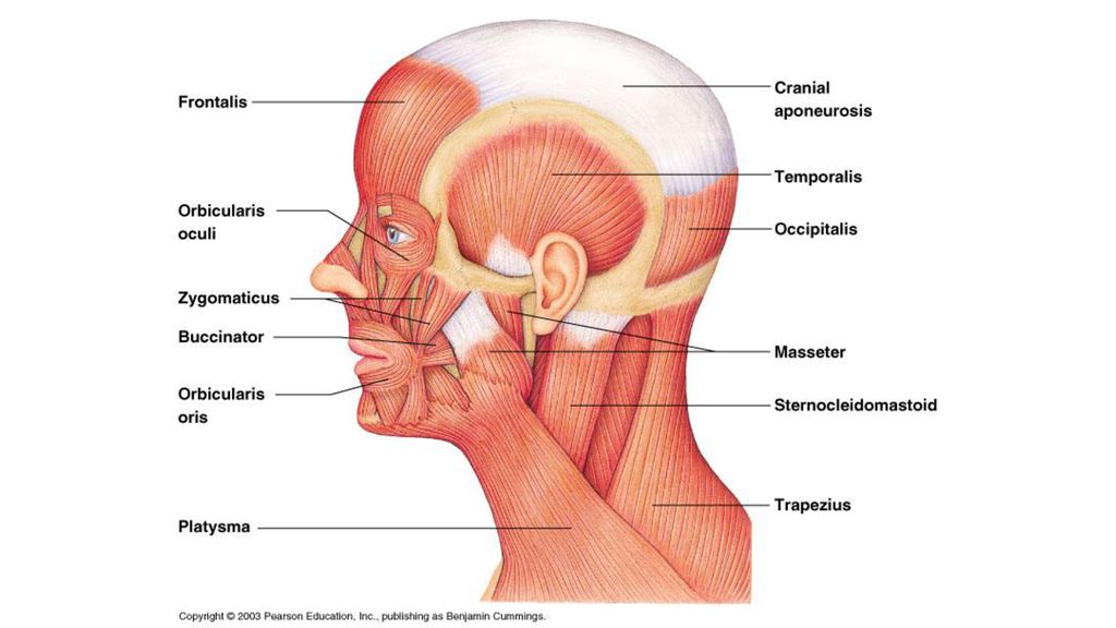
There are over 600 muscles in the body. We will study only the most important muscles. HEAD AND NECK MUSCLES 2 large categories: facial muscles and chewing muscles FACIAL MUSCLES (unique because they are inserted into soft tissues such as other muscles or skin) When these muscles pull on the face, they cause us to smile faintly, grin widely, frown, pout, deliver a kiss, etc. FRONTALIS ORBICULARIS OCULI ORBICULARIS ORIS BUCCINATOR ZYGOMATICUS OCCIPITALIS (CRANIAL APONEUROSIS)
 When these muscles pull on the face, they cause us to smile faintly, grin widely, frown, pout, deliver a kiss, etc. FRONTALIS. ORBICULARIS OCULI. ORBICULARIS ORIS. BUCCINATOR. ZYGOMATICUS. OCCIPITALIS. (CRANIAL APONEUROSIS)")
9
CHEWING MUSCLES (begin the breakdown of food for the body)
MASSETER TEMPORALIS NECK MUSCLES (move the head and shoulder girdle, small and straplike) PLATYSMA STERNOCLEIDOMASTOID
 PLATYSMA. STERNOCLEIDOMASTOID.")
10
ANTERIOR MUSCLES OF THE TRUNK
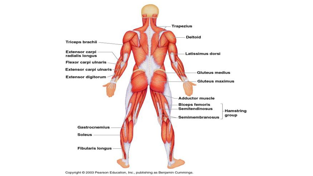
TRUNK MUSCLES (3 TYPES) those that move the vertebral column (most of which are posterior anti-gravity muscles) anterior thorax muscles, which move the ribs, head, arms muscles of the abdominal wall, which help to move the vertebral column, and most importantly, form the muscular “natural girdle” of the abdominal body wall. ANTERIOR MUSCLES OF THE TRUNK PECTORALIS MAJOR, INTERCOSTAL MUSCLES (EXTERNAL/INTERNAL), MUSCLES OF THE ABDOMINAL GIRDLE, RECTUS ABDOMINUS, EXTERNAL OBLIQUE, INTERNAL OBLIQUE, TRANSVERSE ABDOMINIS, SERRATUS ANTERIOR, TERES MAJOR, TERES MINOR (APONEUROSIS) POSTERIOR MUSCLES OF THE TRUNK TRAPEZIUS LATISSIMUS DORSI DELTOID ERECTOR SPINAE - ILIOCOSTALIS - LONGISSIMUS - SPINALIS
 those that move the vertebral column (most of which are posterior anti-gravity muscles) anterior thorax muscles, which move the ribs, head, arms. muscles of the abdominal wall, which help to move the vertebral column, and most importantly, form the muscular natural girdle of the abdominal body wall. ANTERIOR MUSCLES OF THE TRUNK. PECTORALIS MAJOR, INTERCOSTAL MUSCLES (EXTERNAL/INTERNAL), MUSCLES OF THE ABDOMINAL GIRDLE, RECTUS ABDOMINUS, EXTERNAL OBLIQUE, INTERNAL OBLIQUE, TRANSVERSE ABDOMINIS, SERRATUS ANTERIOR, TERES MAJOR, TERES MINOR (APONEUROSIS) POSTERIOR MUSCLES OF THE TRUNK. TRAPEZIUS. LATISSIMUS DORSI. DELTOID. ERECTOR SPINAE. - ILIOCOSTALIS. - LONGISSIMUS. - SPINALIS.")
11
External intercostal muscles
Internal intercostal muscles

12
Serratus Anterior

13
Teres Minor Teres Major

14
MUSCLES OF THE UPPER LIMBS (3 groups)
a. muscles that arise from shoulder girdle and cross shoulder joint to insert into humerus. PECTORALIS MAJOR LATISSIMUS DORSI DELTOID b. muscles that cause movement at the elbow joint. They enclose the humerus and insert on the forearm bones. BICEPS BRACHII BRACHIALIS BRACHIORADIALIS TRICEPS BRACHII c. muscles of the forearm, which insert on the hand bones and cause their movement. These muscles are thin and spindle-shaped, there are many of them. The forearm muscle names reflect their activity. FLEXOR CARPI RADIALIS (anterior aspect) – flexion of wrist FLEXOR DIGITORUM (anterior aspect) – flexion of fingers FLEXOR CARPI ULNARIS – flexes wrist/ adducts hand/ stabilizes wrist EXTENSOR CARPI RADIALIS LONGUS (lateral and posterior aspect) – extends wrist EXTENSOR DIGITORUM (lateral and posterior aspect) – extends fingers EXTENSOR CARPI ULNARIS – extends wrist, abducts hand
 – flexion of wrist. FLEXOR DIGITORUM (anterior aspect) – flexion of fingers. FLEXOR CARPI ULNARIS – flexes wrist/ adducts hand/ stabilizes wrist. EXTENSOR CARPI RADIALIS LONGUS (lateral and posterior aspect) – extends wrist. EXTENSOR DIGITORUM (lateral and posterior aspect) – extends fingers. EXTENSOR CARPI ULNARIS – extends wrist, abducts hand.")
15
MUSCLES OF THE HUMERUS THAT ACT ON THE FOREARM
***** ALL ANTERIOR ARM MUSCLES CAUSE ELBOW FLEXION******** In order of decreasing strength: BRACHIALIS BICEPS BRACHII BRACHIORADIALIS TRICEPS BRACHII – only muscle fleshing out the posterior humerus.

17
Flexor digitorum

18
MUSCLES OF THE LOWER LIMBS
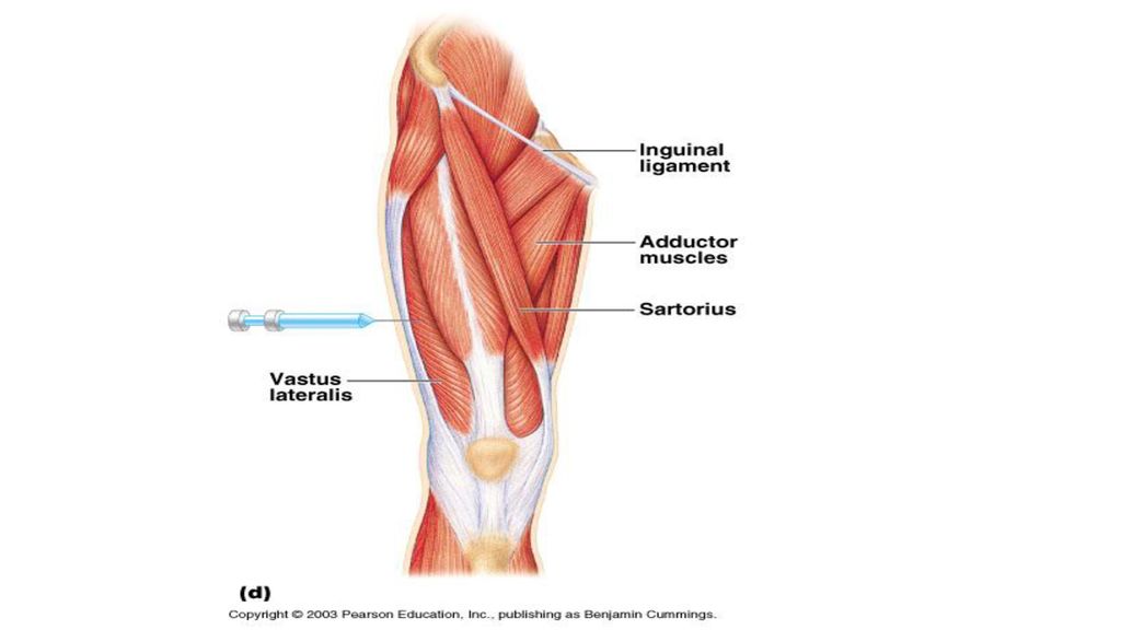
MUSCLES CAUSING MOVEMENT AT THE HIP JOINT GLUTEUS MAXIMUS, GLUTEUS MEDIUS ILIOPSOAS (2 MUSCLES) ILIACUS PSOAS MAJOR ADDUCTOR MUSCLES (3 MUSCLES) ADDUCTOR LONGUS – adducts, flexes, rotates thigh ADDUCTOR BREAVIS – same as longus) ADDUCTOR MAGNUS – adducts, flexes thigh PECTINEUS – adducts, flexes, rotates thigh laterally GRACILIS MUSCLES CAUSING MOVEMENT AT THE KNEE JOINT HAMSTRING GROUP BICEPS FEMORIS, SEMIMEMBRANOSUS, SEMITENDINOSUS SARTORIUS QUADRICEPS GROUP RECTUS FEMORIS, VASTUS MEDIALIS,VASTUS INTERMEDIUS (not on picture as in layer of muscle below vastus medialis/lateralis), VASTUS LATERALIS
 ILIACUS. PSOAS MAJOR. ADDUCTOR MUSCLES (3 MUSCLES) ADDUCTOR LONGUS – adducts, flexes, rotates thigh. ADDUCTOR BREAVIS – same as longus) ADDUCTOR MAGNUS – adducts, flexes thigh. PECTINEUS – adducts, flexes, rotates thigh laterally. GRACILIS. MUSCLES CAUSING MOVEMENT AT THE KNEE JOINT. HAMSTRING GROUP. BICEPS FEMORIS, SEMIMEMBRANOSUS, SEMITENDINOSUS. SARTORIUS. QUADRICEPS GROUP. RECTUS FEMORIS, VASTUS MEDIALIS,VASTUS INTERMEDIUS (not on picture as in layer of muscle below vastus medialis/lateralis), VASTUS LATERALIS.")
19
Pectineus Gracilis

22
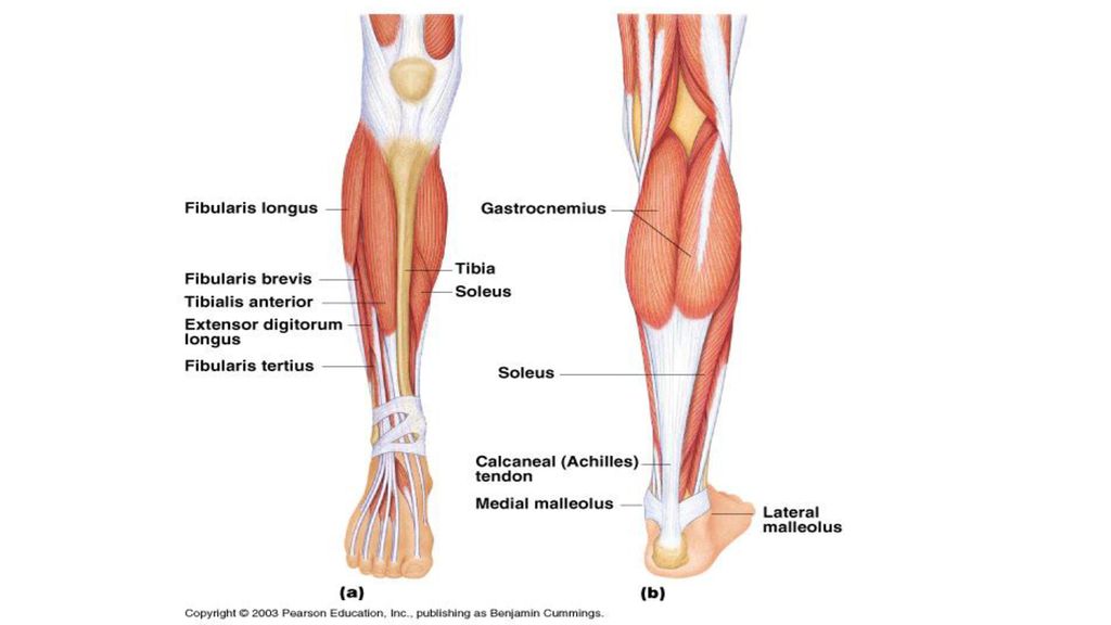
MUSCLES CAUSING MOVEMENT AT THE ANKLE AND FOOT
TIBIALIS ANTERIOR EXTENSOR DIGITORUM LONGUS FIBULARIS MUSCLES LONGUS BREVIS TERTIUS GASTROCNEMIUS SOLEUS FIBULARIS TERTIUS (CALCANEAL “ACHILLES” TENDON)
")
Similar presentations


 A muscle that provides the major force for producing a specific movement.>")


 evaluationsStudent Evaluation of Course and Instructor (SECI) evaluations Please do.>")


