Download presentation
Presentation is loading. Please wait.

1
Part I

2
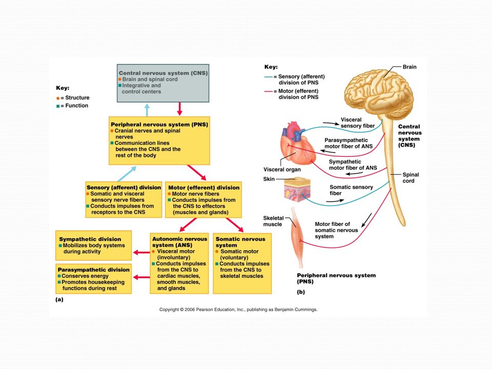
Main Divisions 1. Central Nervous System - brain and spinal cord 2. Peripheral Nervous System - outside of central a. Sensory (afferent) b. Motor (efferent)
 b. Motor (efferent).")
3
Somatic Division – controls voluntary body movements (sensory and motor) Visceral Division – controls involuntary body movement (sensory and motor) Autonomic nervous system – motor division, controls involuntary movement a. Sympathetic b. Parasympathetic

5
Cells of the Nervous System Two main types, neurons and neuroglia Neurons conduct electrical nerve impulses and release neurotransmitters 1. Longevity 2. Amitotic 3. High metabolic rate Neuroglia are supportive, protect neurons and keep them healthy

6
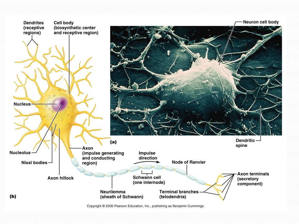
Neuron Structure 1. Cell body – in CNS, in gray matter; in PNS, in ganglia - Has nucleus with nucleoli - Has Nissl substance (rER) 2. Cell processes - Dendrites – receptive part of neuron, detect stimuli from environment - Axon – conducts impulses away from cell body
 2. Cell processes - Dendrites – receptive part of neuron, detect stimuli from environment - Axon – conducts impulses away from cell body.")
7
Axon Hillock – where axon leaves cell body Collaterals – branches of axon Telodendria – smallest branches, each ends in an axon terminal Axon terminal – where neurotransmitter is released (at synapse) Synapse –meeting of axon with another neuron, muscle, or gland
 Synapse –meeting of axon with another neuron, muscle, or gland")
9
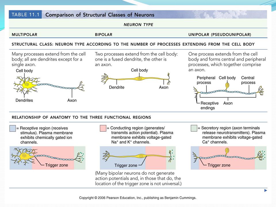
Structural classes of neurons 1. Unipolar – one process, found in PNS 2. Bipolar – 2 processes, associated with special senses 3. Multipolar – more than 2 processes (only 1 axon), make up more than 99% of neurons
, make up more than 99% of neurons.")
11
Functional Classification of Neurons Sensory (afferent) – carry impulses to CNS from skin, muscles, glands, or internal organs Unipolar, cell bodies found in ganglia outside CNS Motor (efferent) – carry impulses away from CNS to effector organs Multipolar, cell bodies in CNS Interneurons – lie between motor and sensory neurons, more than 99% of neurons Multipolar, found in CNS
 – carry impulses to CNS from skin, muscles, glands, or internal organs Unipolar, cell bodies found in ganglia outside CNS Motor (efferent) – carry impulses away from CNS to effector organs Multipolar, cell bodies in CNS Interneurons – lie between motor and sensory neurons, more than 99% of neurons Multipolar, found in CNS")
12
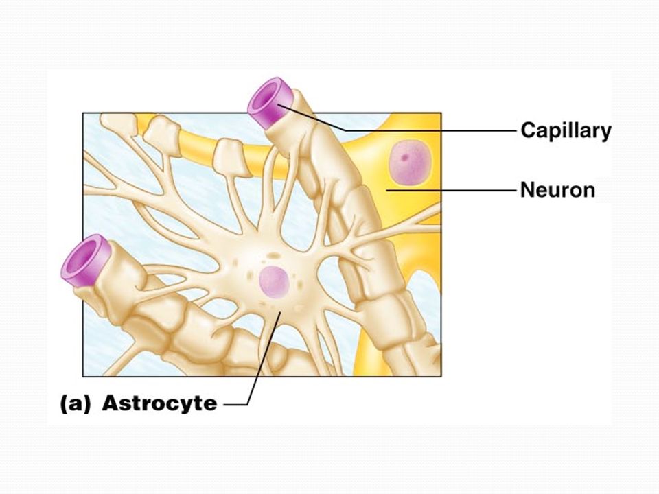
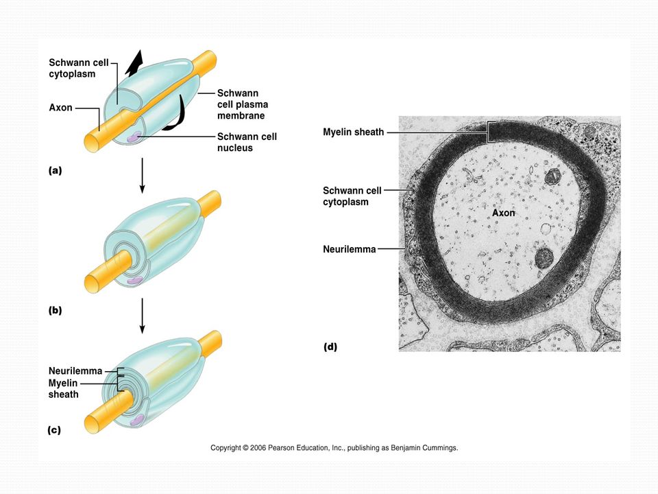
Neuroglia Central Nervous System 1. Ependymal cells – line choroid plexus, circulate CSF 2. Astrocytes – nutrient exchange between neurons and capillaries 3. Oligodendrocytes – make myelin sheaths of CNS 4. Microglia - phagocytes Peripheral Nervous System 1. Satellite cells – surround cell bodies of neurons in ganglia 2. Schwann cells – associated with all PNS axons, can form myelin sheaths around axons.

14
Microglia and Ependymal Cells

15
Figure 11.3d, e

16
Myelinated and Unmyelinated Axons Myelination – when Schwann cells in PNS wrap tightly around axon, axon is “myelinated” Myelin = lipoprotein found in plasma membrane Neurilemma = displaced cytoplasm of Schwann cell Node of Ranvier = gap between Schwann cells, site of saltatory conduction Saltatory conduction = nerve signal “jumps” from node to node Myelinated neurons conduct nerve impulses much faster than unmyelinated!!!

18
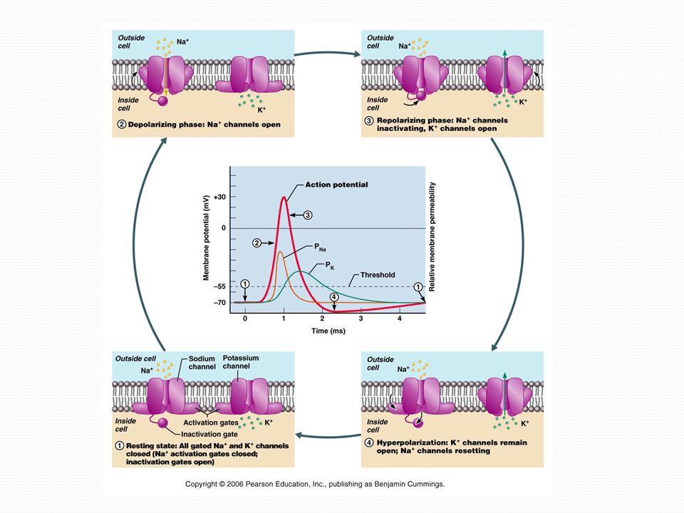
Nerve Signals Nerves conduct impulses using action potentials; long distance signals Depolarization must reach threshold level Action potentials are unstoppable Graded potentials – small changes in cell polarity; will die out as distance from origin increases; short distance signals

19
Graded Potentials

21
Synapse Presynaptic neuron – conducts impulse to synapse Postsynaptic neuron – conducts impulse away from synapse Neurotransmitter – chemical released at synapse; binds to receptors on postsynaptic membrane; some are excitatory, some are inhibitory

22
Synaptic Events

23
The Brain

24
Gray matter – inner core of CNS, can be on outer edge as well, contains cell bodies and support cells White matter – surrounds gray matter, contains myelinated fibers

25
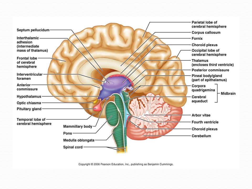
Ventricles 1. Lateral ventricles – inside cerebral hemispheres, separated by septum pellucidum; contain choroid plexus, which makes CSF 2. Third ventricle – meets with lateral ventricles through interventricular foramen 3. Fourth ventricle – meets with third ventricle by cerebral aqueduct; connects to subarachnoid space

26
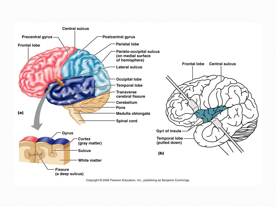
Cerebral Hemispheres Most of brain mass Have three basic regions: cortex, white matter, and basal nuclei Lobes of brain – frontal, parietal, occipital, temporal, and insula Gyrus (gyri) – ridge of tissue Sulcus (sulci) – shallow groove Fissures – deeper grooves Longitudinal fissure – separates hemispheres Transverse cerebral fissure – separates hemispheres from cerebellum
 – ridge of tissue Sulcus (sulci) – shallow groove Fissures – deeper grooves Longitudinal fissure – separates hemispheres Transverse cerebral fissure – separates hemispheres from cerebellum")
27
Cerebral Hemispheres Central sulcus – separates frontal and parietal lobes Precentral and postcentral gyri – ridges anterior and posterior to central sulcus Lateral sulcus – separates temporal lobe from parietal and frontal lobes

29
Cerebral Cortex Enables sensation, communication, memory, understanding, and voluntary movements Gray matter Three functional areas 1. Motor areas – control movement 2. Sensory areas – awareness of sensation 3. Association areas – give meaning to info we receive, can store in memory, and lets us decide on actions to take

30
Cerebral Cortex Hemispheres are contralateral to body Each hemisphere has specialization Left hemisphere – controls language, math, and logic Right hemisphere – controls visual-spatial skills, emotion, and artistic skills

31
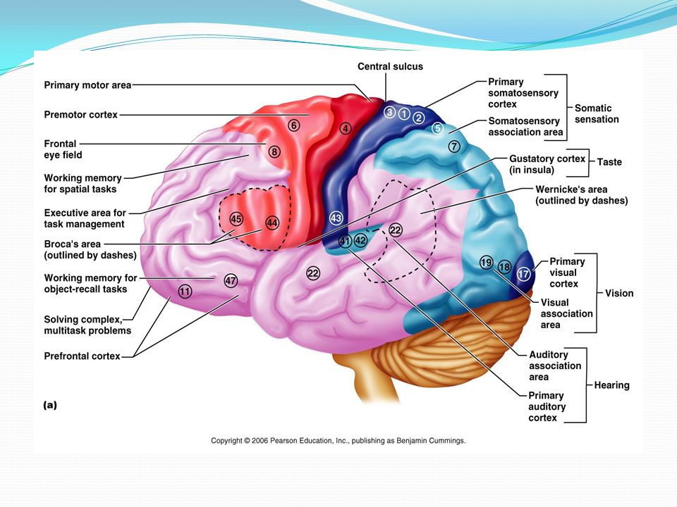
Motor Areas 1. Primary motor cortex – precentral gyrus (frontal); pyramidal cells let us control skeletal muscles, form pyramidal tracts (spinal cord) 2. Premotor cortex – in front of precentral gyrus (frontal); muscles act together to perform patterned movements 3. Broca’s area – anteroinferior to premotor cortex (frontal); activates muscles involved in speech 4. Frontal eye field – controls voluntary eye movements
; pyramidal cells let us control skeletal muscles, form pyramidal tracts (spinal cord) 2. Premotor cortex – in front of precentral gyrus (frontal); muscles act together to perform patterned movements 3. Broca’s area – anteroinferior to premotor cortex (frontal); activates muscles involved in speech 4. Frontal eye field – controls voluntary eye movements.")
32
Sensory and Association Areas 1. Primary somatosensory cortex – postcentral gyrus (parietal); receives sensory information from skin and muscles 2. Somatosensory association cortex – posterior to somatosensory cortex (parietal); integrates input from somatosensory cortex so we can comprehend sensations 3. Primary visual cortex – (occipital); receives visual information from retina
; receives sensory information from skin and muscles 2. Somatosensory association cortex – posterior to somatosensory cortex (parietal); integrates input from somatosensory cortex so we can comprehend sensations 3. Primary visual cortex – (occipital); receives visual information from retina.")
33
Sensory and Association Areas (cont.) 4. Visual association area – (occipital); interprets sensory information from visual cortex 5. Primary auditory cortex – (temporal); receives information from inner ear 6. Auditory association area – (temporal); interprets sensory information from primary auditory cortex 7. Wernicke’s area – (temporal); lets us understand spoken words
; interprets sensory information from visual cortex 5. Primary auditory cortex – (temporal); receives information from inner ear 6. Auditory association area – (temporal); interprets sensory information from primary auditory cortex 7. Wernicke’s area – (temporal); lets us understand spoken words.")
34
Sensory and Association Areas (cont.) 8. Prefrontal cortex - involved with intellect, cognition, recall, personality, judgement, and conscience

36
Consists of myelinated fibers and their tracts Commissures – connect the two hemispheres Association fibers – connect different parts of the same hemisphere Projection fibers – connect the hemispheres to lower CNS Cerebral White Matter

37
Fiber Tracts in White Matter

38
Basal Nuclei Play a role in motor control, attention, and cognition Inhibit unnecessary movements Include caudate nucleus, putamen, and globus pallidus (the putamen and globus together are called “lentiform nucleus”)
")
39
Basal Nuclei

40
Diencephalon 1. Thalamus – Relays sensory information to other parts of cerebral cortex. Mediates sensation, motor activities, cortical arousal, learning, and memory. 2. Epithalamus – roof of 3 rd ventricle; pineal gland extends from it, secretes melatonin (sleep cycle)
.")
41
Diencephalon (cont.) 3. Hypothalamus – below thalamus; connects to pituitary gland (infundibulum) a. Regulates autonomic nervous system b. Emotional responses c. Body temperature regulation d. Regulates food and water intake e. Controls pituitary gland f. Produces ADH and oxytocin
 a. Regulates autonomic nervous system b. Emotional responses c. Body temperature regulation d. Regulates food and water intake e. Controls pituitary gland f. Produces ADH and oxytocin.")
42
Brain Stem Midbrain – under diencephalon - Cerebral peduncles contain pyramidal tracts - Corpora quadrigemina (4), 2 superior colliculi coordinate head and eye movement; 2 inferior colliculi relay sound, also control reflexive reactions to sound - Substantia nigra – associated with basal ganglia
, 2 superior colliculi coordinate head and eye movement; 2 inferior colliculi relay sound, also control reflexive reactions to sound - Substantia nigra – associated with basal ganglia")
43
Brain Stem Pons Between midbrain and medulla Acts as a bridge between cerebellum/spinal cord and cerebrum Contains nuclei of 3 cranial nerves: trigeminal, abducens, and facial

44
Brain Stem Medulla oblongata Most inferior brain stem Has 2 pyramids, part of pyramidal tracts Decussation of pyramids Functions Regulates respiration and circulation Relay somatosensory information to thalamus and cerebellum Contains nuclei of 5 cranial nerves: hypoglossal, vestibulocochlear, glossopharyngeal, vagus, and accessory

46
Cerebellum Behind pons and medulla Allows for smooth, coordinated skeletal muscle movement, balance, and posture 2 hemispheres separated by vermis Gyri of cerebellum called folia White matter called arbor vitae 3 pairs of cerebellar peduncles send relays from cerebellum to pons, midbrain, and medulla No direct communication with cortex, all goes through thalamus

47
Meninges 3 layers of protective connective tissue 1. Dura mater – outer layer, connects to scalp 2. Arachnoid mater –middle layer, separated from dura by subdural space; underneath it is subarachnoid space, filled with CSF 3. Pia mater – inner layer, contains many blood vessels, directly on brain surface

48
Meninges Figure 12.24a

Similar presentations


 CNS = Brain + spinal cord Surface anatomy includes.>")
 changes in the environment (light, sound, smell, taste, touch, heat) Detect (sense) changes in the.>")


 both within.>")


 Brain and Spinal Cord Peripheral Nervous.>")
 CNS –brain –spinal cord.>")