Download presentation
Presentation is loading. Please wait.

1
The Skeletal System

2
Objectives Describe the structure & functions of the skeletal system. Describe the structure & functions of the skeletal system. Identify 4 types of bones. Identify 4 types of bones. Learn the names of major bones. Learn the names of major bones. Learn the 4 types of joints Learn the 4 types of joints

3
Skeletal System Bones Bones Ligaments Ligaments Tendons Tendons Cartilages Cartilages

4
I. Functions of the Skeletal System 1. Support bones of the legs, pelvic girdle, and vertebral column support the weight of the body. bones of the legs, pelvic girdle, and vertebral column support the weight of the body. The mandible (jawbone) supports the teeth. The mandible (jawbone) supports the teeth. Other bones support various organs and tissues. Other bones support various organs and tissues.
 supports the teeth. The mandible (jawbone) supports the teeth. Other bones support various organs and tissues. Other bones support various organs and tissues..")
5
2. Protection 2. Protection The bones of skull protect the brain.

6
The ribs and sternum protectTheheartandlungs.

7
Vertebrae protect spinal cord

8
3. Movement Skeletal muscles attach to bones to move the body. Skeletal muscles attach to bones to move the body.

9
4. Minerals The bones are the storage center for: Calcium 99% Phosphorus 85% (phosphorus helps calcium keep our bones strong)
.")
10
5. Blood Cell Formation Bones manufacture the body’s blood cells Red bone marrow produces millions of blood cells each day Adipose (fat) tissue is found in yellow marrow of certain bones.
 tissue is found in yellow marrow of certain bones..")
11
II. Structure of the Skeleton 206 Bones in the Human Body

12
22 bones in skull 6 in middle ears 1 hyoid bone…. this is the only bone NOT connected to any other…do you know where its located? 26 in vertebral column 25 in thoracic cage

13
4 in pectoral girdle 60 in upper limbs 60 in lower limbs 2 in pelvic girdle 206 TOTAL 206 TOTAL

14
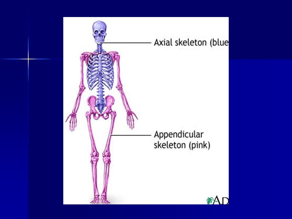
Skeletal System divided into 2 main parts Axial Skeleton – 80 bones Axial Skeleton – 80 bones – skull, vertebral column, and rib cage. Appendicular skeleton - 126 bones Appendicular skeleton - 126 bones – Bones of upper & lower limbs and the girdles (shoulder bones and hip bones)
.")
16
4 Types of Bones 1. Long Bones Much longer than they are wide. Much longer than they are wide. All bones of the limbs except for the patella (kneecap), All bones of the limbs except for the patella (kneecap), and the bones of the wrist and ankle. Consists of a shaft plus 2 expanded ends. Consists of a shaft plus 2 expanded ends.
, All bones of the limbs except for the patella (kneecap), and the bones of the wrist and ankle. Consists of a shaft plus 2 expanded ends. Consists of a shaft plus 2 expanded ends..")
17
2. Short Bones Cubed shaped bones of the wrist and the ankle. Carpal Bones

18
3. Flat Bones Thin, flattened, and usually a bit curved. Thin, flattened, and usually a bit curved. Scapulae, sternum, (shoulder blades), ribs and most bones of the skull. Scapulae, sternum, (shoulder blades), ribs and most bones of the skull. Sternum
, ribs and most bones of the skull. Scapulae, sternum, (shoulder blades), ribs and most bones of the skull. Sternum.")
19
4. Irregular Bones Have weird shapes that fit none of the 3 previous classes. Have weird shapes that fit none of the 3 previous classes. Vertebrae, hip bones, 2 skull bones (sphenoid and the ethmoid bones). Vertebrae, hip bones, 2 skull bones (sphenoid and the ethmoid bones). Sphenoid Bone
. Vertebrae, hip bones, 2 skull bones (sphenoid and the ethmoid bones). Sphenoid Bone.")
20
III. Bone Structure III. Bone Structure Bones are organs Bones are organs Composed of multiple type of tissues Composed of multiple type of tissues –Bone tissue (a.k.a. osseous tissue). –Fibrous connective tissue. –Cartilage. –Vascular tissue. –Lymphatic tissue. –Adipose tissue. –Nervous tissue.
. –Fibrous connective tissue. –Cartilage. –Vascular tissue. –Lymphatic tissue. –Adipose tissue. –Nervous tissue..")
21
All bones consist of a dense, solid outer layer known as compact bone All bones consist of a dense, solid outer layer known as compact bone and an inner layer of spongy bone – a honeycomb of flat, needle-like projections called trabeculae. and an inner layer of spongy bone – a honeycomb of flat, needle-like projections called trabeculae.

22
3 Types of Bone cells osteocyte

23
Bone Structure Bone tissue is a type of connective tissue, so it must consist of Bone cells: Bone tissue is a type of connective tissue, so it must consist of Bone cells: Osteoblasts - Bone-building cells Osteoblasts - Bone-building cells Osteocytes - Mature Bone cells maintain bone tissue Osteocytes - Mature Bone cells maintain bone tissue Osteoclasts - normal bone growth, development, maintenance, and repair. Osteoclasts - normal bone growth, development, maintenance, and repair.

24
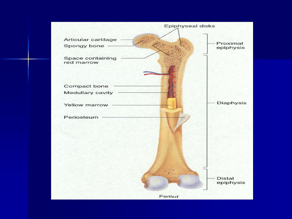
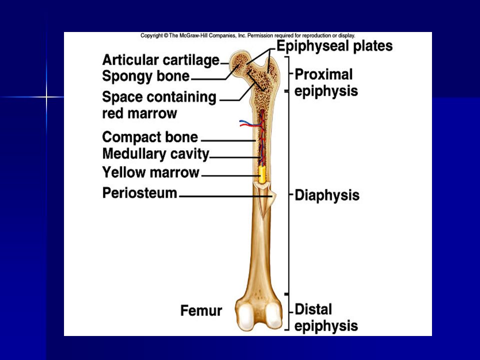
A. Long Bone Structure Shaft plus 2 expanded ends. Shaft plus 2 expanded ends. Shaft is known as the diaphysis. Shaft is known as the diaphysis. –Consists of a thick collar of compact bone surrounding a central marrow cavity –In adults, the marrow cavity contains fat or yellow bone marrow.

25
Expanded ends are epiphyses Expanded ends are epiphyses –Thin layer of compact bone covering an interior of spongy bone. –Joint surface of each epiphysis is covered w/ articular cartilage. It cushions the bone ends and reduces friction during movement.

26
Long Bone Structure The external surface of the entire bone except for the joint surfaces of the epiphyses is covered by a double- layered membrane known as the periosteum. The external surface of the entire bone except for the joint surfaces of the epiphyses is covered by a double- layered membrane known as the periosteum.

27
Long Bone Structure Periosteum is richly supplied with nerve fibers, lymphatic vessels & blood vessels.

28
Long Bone Structure Internal bone surfaces are covered with a delicate connective tissue membrane known as the endosteum. Internal bone surfaces are covered with a delicate connective tissue membrane known as the endosteum. –Covers the trabeculae of spongy bone in the marrow cavities and lines the canals that pass through compact bone. –Contains both osteoblasts and osteoclasts.

30
1. 1. Bone enclosed in periosteum, which is continuous -with tendons and ligaments -blood vessels in periosteum 2.Epiphysis- ends -spongy bone contains red marrow -compact bone, articular cartilage 3.Diaphysis- middle -compact bone -medullary cavity- contains yellow marrow (fat) -lined with endosteum
 -lined with endosteum.")
32
More structures and layers of bone

33
IV. Bones before birth - Prenatal development skeleton is mostly cartilage Cartilage cells and then osteoblasts start to deposit minerals Cartilage disk (epiphyseal disk) remains in epiphysis – growth plates Cells eventually stop dividing
 remains in epiphysis – growth plates Cells eventually stop dividing.")
34
Adults continually break down and build up bone Osteoclasts remove damaged cells and release calcium into blood Osteoblasts remove calcium from blood and build a new matrix. They become trapped osteoclasts

35
Bone Development

36
Cartilage The skeletal system is made up of not only bones but Cartilage, a strong, flexible connective tissue.

37
Cartilage Cartilage serves many functions : Lines the surfaces of joints and enables them to move smoothly Cushions joining vertebrae Supports the nose and ears Baby skeleton is mostly cartilage

38
Cartilage Cartilage cells are replaced with age by bone cells and minerals in a process call Ossification The process by which bone is formed, renewed and repaired

39
4 Types of Joints in the Body 1. Ball and Socket Joint 1. Ball and Socket Joint –Hip and Shoulder

40
2. Hinge Joint Elbow, Knee and Fingers Elbow, Knee and Fingers

41
3. Gliding Joint Small bones of Wrist Small bones of Wrist

42
4. Pivot Joint Skull and Vertebrae Skull and Vertebrae

43
Ligaments A band of A band of elastic Connective Tissue Connects a Connects a bone to a bone i.e. ACL i.e. ACL

44
Tendon Connects Connects a muscle to a bone. i.e. Quadriceps Tendon i.e. Quadriceps Tendon

45
Facts on Tendons, Muscles & Movement Tendon – fibrous cord attaches muscle to bone, when muscle contact they move the bone. Tendon – fibrous cord attaches muscle to bone, when muscle contact they move the bone.

47
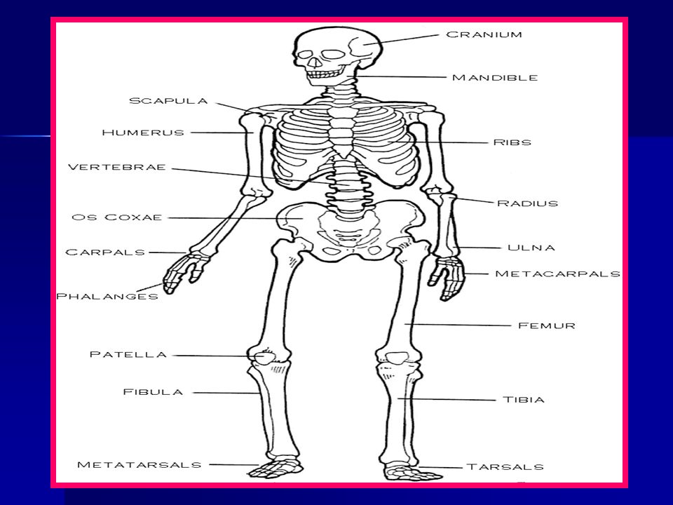
Major Bones of the Skeleton Skull Vertebrae Ribs Humerus Radius

48
Major Bones of the Skeleton Ulna Pelvis Femur Tibia Fibula

52
Theaxialskeleton

53
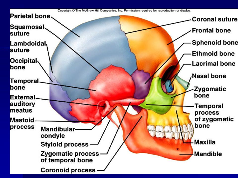
The skull Cranium - 8 sutured bones Facial bones: 13 sutured bones, 1 mandible Cranium encases brain attachments for muscles sinuses

55
Skull top view

56
Vertebral column 7 cervial vertebrae 12 thoracic vertebrae 5 lumbar vertebrae 1 sacrum (5 fused 1 coccyx (4 fused) 26 total Vertebrae vary in size and shape
 26 total Vertebrae vary in size and shape")
57
Spinal Column

58
Sacrum & Coccyx

59
Thoracic cage ribs thoracic vertebrae sternum costal cartilages True ribs are directly attached to the sternum (first seven pairs) Three false ribs are joined to the 7th rib Two pairs of floating ribs
 Three false ribs are joined to the 7th rib Two pairs of floating ribs")
61
TheAppendicularSkeleton

62
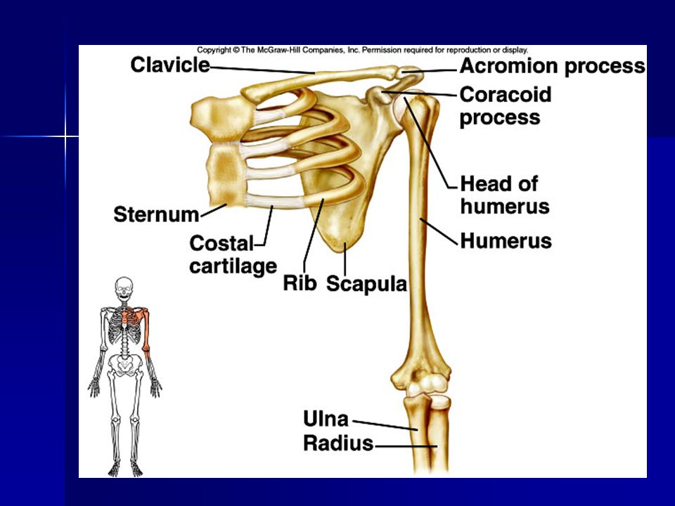
Clavicles and scapulae Help brace shoulders Attachment sites for muscles

64
Scapula Acromion Glenoid Cavity Spine Coracoid Process

65
Scapula and humerus

66
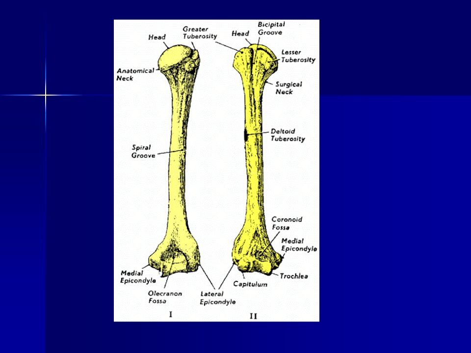
Bones of upper limb Humerus (upper arm) Radius; ulna Carpals, metacarpals, phalanges
 Radius; ulna Carpals, metacarpals, phalanges")
68
Raduis & Ulna –Wrist & elbow joints

69
Radius and Ulna

70
The Hand

71
Wrist / Carpals

72
The pelvic Girdle / Pelvis ILIUM SACRUM ACETABULUM PUBISPUBIC SYMPHISIS ISCHIUM

73
Bones of lower limb Femur Patella Tibia, fibula Tarsals, metatarslas, phalanges

74
Femur / thigh bone

75
Fibula and Tibia

76
Foot and ankle

Similar presentations


 fibers along with water and mineral salts (calcium hydroxide & calcium.>")


 hyoid bone (anchors tongue and muscles associated with swallowing) vertebral column (vertebrae and disks)>")


 Joints Cartilages Ligaments Divided into two divisions Axial skeleton –>")

