Download presentation
Presentation is loading. Please wait.

1
Odontología Digital RX SOLUCIONES DE IMAGENOLOGÍA S.A. de C.V. Guadalajara, Jal. MÉXICO TEL (33) 3499-7095 / (33) 1300-5813 radiografiadigitalschickgdl@gmail.com contacto@rxdigitalschick.com www.rxdigitalschick.com @RxDigitalDental
 / (33)")
2
1.Digital Advantage 2.Important Technical Factor of Digital Image 3.Understanding of Digital Image for Dentistry – Clinical Case 4.VATECH Products 5.Why VATECH? 6.Appendix Contents

3
Digital Technology in Dentistry

4
4 Digital Advantage Paper, paperless or hybrid – Storage Issue – Ease of data retrieval – Patient’s expectation – Patient’s confidentiality issues – Transfer speed – Portability & patient presentation

5
5 Advantages of Digital Radiography Traditional images – exposure conditions & development procedures determine the final result – brightness and contrast are fixed Digital images – overexposure and underexposure can be modify, sa ving the patient from an extra dose of radiation –↓ Image processing time –↓ radiation dosage per exposure

6
6 Advantages of Digital Radiography Environmental friendly – Eliminates the need for x-ray film, developing solutions & film d evelopment

7
7 Advantages of Digital Radiography Convenient data storage and retrieval

8
Advantages of Digital Radiography Easy data manipulation during diagnosis & treatment – Viewing digital imaging on a monitor Image can be viewed immediately for diagnosis Enlarge images for better viewing and better diagnosis Better patient communication - improves patient under standing of problem Gives the wow factor to your office

9
9 Advantages of Digital Radiography Software tools to help in diagnosis and treatment – Measuring / Drawing tools / X-ray comparisons – MORE….

10
Important Factor for Digital Image

11
–Provides a high image resolution Low resolutionHigh resolution Purpose of an X-ray Image

12
–Provides a high gradual resolution low high Purpose of an X-ray Image

13
Imaging Components Source Object Receptor Image Processing Display Post Processing Image Processing Techniques

14
Source

15
Soruce: X-Ray Tube Head Fixed-anode x-ray tube a high-voltage transformer; a lead sheath safety and protection devices enclosure An X-ray tube is a vacuum tube that produces X-rays supplied with a high voltage supply controlled by a control unit – While generating x-ray, 95% of energy is consumed in heat energy and only 5% is converted into X-ray Controlling Factors – kVp / mA / Exposure Time AC, DC and Pulsed Type X-rays Focal Spot Beam-Limiting-Device (“cone”).
.")
16
kVp / mA / Exposure Time kVp – Kilo Volts Peak Power of penetration mA – milli Amperes Dose quantity of X-ray Exposure time (secs) Increase in these factors generally provides better penetration and better quality images, while increasing radiation dosage to the patient Point to ponder: Less radiation & lower quality or higher radiation & better quality? But VATECH’S Detector provide with less radiation but better Image quality

17
Focal Spot Size The focal spot is the region of an X-ray tube from which the X-ray e manate. The sharpness of the radiographic image increases as the size of the focal spot decreases. VATECH“S”“S”“I”“I”“P”“P” Tube modelD-051SSR90/15D-051SD-054S MakerToshibaOPX (Italy)Toshiba Spot Size(mm) 0.35*0.50.5*0.50.35*0.50.5*0.5 Ug = f* b/a f = source focal-spot size. a = distance from x-ray source to front surface of material/object b = distance from the front surface of the object to the detector VATECH’S Focal Spot size is smallest in the market
Toshiba Spot Size(mm) 0.35*0.50.5* *0.50.5*0.5 Ug = f* b/a f = source focal-spot size. a = distance from x-ray source to front surface of material/object b = distance from the front surface of the object to the detector VATECH’S Focal Spot size is smallest in the market.")
18
The Receptor

19
Various type of sensors are used for different imaging pu rposes Intra Oral –CMOS, CCD Panoramic / Cephalometric –Linear CCD, CMOS –II-CCD, FPD CT –Flat Panel Detector (FPD, CMOS) –Image Intensifier CCD (II+CCD) Receptor: Sensors
 –Image Intensifier CCD (II+CCD) Receptor: Sensors")
20
CCD Charge Coupled Device Each pixel can transfer its electric charge to one or other of its neighbors. The last pixel converts the accumulated electronic charge so that the chip outputs digital bits. CMOS Complementary Metal Oxide Semiconductor Each pixel has its own charge-to-voltage conversion so that the chip outputs digital bits. Intra oral sensor, CT = CMOS (area sensor) Panoramic X-ray = CCD (linear sensor) CCD & CMOS
 Panoramic X-ray = CCD (linear sensor) CCD & CMOS.")
21
CCDCMOS Past - Particular Process - High Quality - General Process - Noise Problem Now - Fit to TDI System (Time Delay Integration) - Adapted to Panoramic X-ray - High Quality, Integrated Chips (By the advance in technology) - e.g. Canon 12Milion pixels Digital Camera (No.1 company in digital camera) - e.g. Schick : Intra-Oral Sensor CCD & CMOS
 - e.g. Schick : Intra-Oral Sensor CCD & CMOS.")
22
FPD Vs II+CCD VS

23
I. I+ CCD FPD FPD Vs IICCD

24
FPDI. I+ CCD FPD Vs II+CCD

25
Image Processing

26
Image Processing Techniques Sensor Calibration –Each sensor is made up of many pixels which have different sen sitivity and reaction even to the same X-ray signal. –Calibration has to be done to ensure that every pixel has the sa me reaction to an X-ray signal. –This requires high tech hardware and software, and is one of the main factors in producing a good digital image Image Processing Algorithms –Process by which raw data from the sensor is processed using t he various image processing algorithms to adjust gain, –offset, and various values to produce the final image

27
The Display

28
1536 pixels 2840 pixels Monitor Resolution 2048 pixels 1536 pixels Normal Monitor : 1024 x 768 Super High Resolution Monitor : 2048 x 1536 Image Resolution 2840 x 1536 pixels Display - Conclusion As you see our image resolution, it is more than the resolution of Super High Resolution Monitor. It means our image resolution is good enough.

29
Display Gray Scale –Grayscale is a range of shades of gray without apparent color –The darkest possible shade is black, which is the total absence o f transmitted or reflected light. The lightest possible shade is whit e –The result of measuring the intensity of light at each pixel in a sin gle band of the electromagnetic spectrum

30
1. Pixel Size and Image Quality 1-2. Comparison Chart of Pixel Size CompanyModelPixel Size **Pixel Size After Binning Theoretical Line Pair SO 28 ㎛ (?) PP 33 ㎛ Pano 66 / Ceph 99 ㎛ 7.6 / 5.05 lp IO 48 ㎛ 96 ㎛ 5.2 lp VATECHPaX-Series 22 / 48 ㎛ 44 / 96 ㎛ 11.4 / 5.2 lp 1-1. Factors of effecting Image Quality Sensor Sensitivity, Sensor Pixel Size, Screen Resolution, Image Processing SW, X-Ray Quality, Accuracy of Focal Layer, Patient positioning, ect. ** Pixel Size After Binning contents
 PP 33 ㎛ Pano 66 / Ceph 99 ㎛ 7.6 / 5.05 lp IO 48 ㎛ 96 ㎛ 5.2 lp VATECHPaX-Series 22 / 48 ㎛ 44 / 96 ㎛ 11.4 / 5.2 lp 1-1. Factors of effecting Image Quality Sensor Sensitivity, Sensor Pixel Size, Screen Resolution, Image Processing SW, X-Ray Quality, Accuracy of Focal Layer, Patient positioning, ect. ** Pixel Size After Binning contents.")
31
2. Sensor Resolution Real Measurement by Line Phantom 2-1. Line Pair - Unit of sensor resolution : Line Pair Per mm (lp/mm) - Theoretical formula = 1mm / (2 x Pixel Size) - e.g. Pixel Size 25 ㎛ : 1000 / (2 x 25) = 20 lp/mm 2-2. Line Phantom - The other factors are not perfect : Focal Trough, Scattering, Noise, Image processing, etc. - Manufacturers do not calculate the effect of Pixel Binning on purpose - Have no Standard Method to evaluate Line Pair by using Line Phantom (Under discussion at American Radiology Association) 2-3. Difference between theoretical Line Pair and real Line Pair contents
 - Theoretical formula = 1mm / (2 x Pixel Size) - e.g. Pixel Size 25 ㎛ : 1000 / (2 x 25) = 20 lp/mm 2-2. Line Phantom - The other factors are not perfect : Focal Trough, Scattering, Noise, Image processing, etc. - Manufacturers do not calculate the effect of Pixel Binning on purpose - Have no Standard Method to evaluate Line Pair by using Line Phantom (Under discussion at American Radiology Association) 2-3. Difference between theoretical Line Pair and real Line Pair contents.")
32
Digital image for Dentistry

33
Basic Panoramic

34
Cephalometric Image

35
Basic Dental Radiography o A radiographic image is formed by a controlled burst of X-ray radiation which penetrates oral structures at different levels, depending on varying anatomical densities, before striking the film or sensor. Teeth appear lighter because less radiation penetrates them to reach the film o The amount the X-Ray stopped (attenuated) by an object determines the radio-density of the shadows. Air Crown Root Canal Fillings Soft Tissue Bone The white or radio-opaque shadows on an image represent the various dense structures with the object which have totally stopped the X-Ray beam. The black or Radiolucent shadows represent area where the X-Ray beam has passed thru the object and has not been stopped at all. The grey shadows represent areas where X-Ray beam has been stop to a varying degreeDental caries, tooth decay, infections and other changes in the bone density, and the periodontal ligament, appear darker because X-rays readily penetrate these less dense structures Dental restorations (fillings, crowns) may appear lighter or darker, depending on the density of the material.
 by an object determines the radio-density of the shadows. Air Crown Root Canal Fillings Soft Tissue Bone The white or radio-opaque shadows on an image represent the various dense structures with the object which have totally stopped the X-Ray beam. The black or Radiolucent shadows represent area where the X-Ray beam has passed thru the object and has not been stopped at all. The grey shadows represent areas where X-Ray beam has been stop to a varying degreeDental caries, tooth decay, infections and other changes in the bone density, and the periodontal ligament, appear darker because X-rays readily penetrate these less dense structures Dental restorations (fillings, crowns) may appear lighter or darker, depending on the density of the material..")
36
Panorama

37
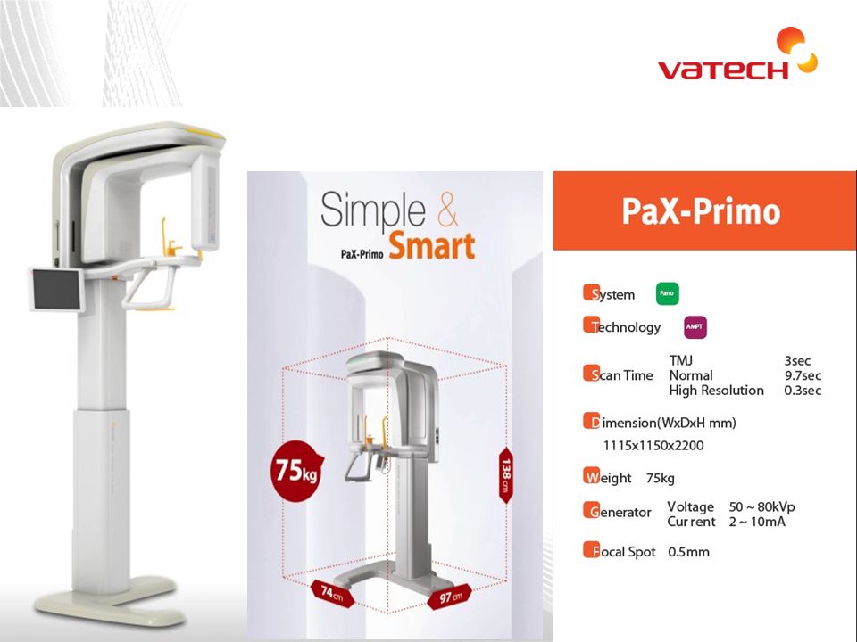
37 Introduction What is an Panorama? An Orthopantomogram (OPG) also known as “Panorex” is a panoramic scanning dental x-ray on the upper and lower Jaw. It shows a two dimensional view of half circle from ear to ear. An Orthopantomogram (OPG) also known as “Panorex” is a panoramic scanning dental x-ray on the upper and lower Jaw. It shows a two dimensional view of half circle from ear to ear. Pax primo
 also known as Panorex is a panoramic scanning dental x-ray on the upper and lower Jaw. It shows a two dimensional view of half circle from ear to ear. An Orthopantomogram (OPG) also known as Panorex is a panoramic scanning dental x-ray on the upper and lower Jaw. It shows a two dimensional view of half circle from ear to ear. Pax primo.")
38
38 2. Advantages of OPG. A large area is imagined and all the tissues within the focal through are displayed even when The patient is unable to open his mouth Image is easy to understand/useful teaching aid. Patient movement in the vertical plane distorts only the part of the image being produced that instant. overall view of the jaws allows rapid assessment of any underlying possibly unsuspected disease as well as view of the mandible heads/ TMJ. The overall view of the jaws allows rapid assessment of any underlying possibly unsuspected disease as well as view of the mandible heads/ TMJ

39
39 Clinical Importance of OPG. as part of assessment of periodontal bone support where the pocketing is greater than 6mm. as part of assessment of periodontal bone support where the pocketing is greater than 6mm. Widening of PDL space if there is pocket

40
40 Clinical Importance of OPG. assessment of wisdom teeth prior to planned surgical intervention

41
41 Clinical Importance of OPG. Fracture of all parts of mandible

42
42 2.Clinical Importance of OPG. Destructive disease of the articular surface of TMJ.

43
43 2.Clinical Importance of OPG. Vertical alveolar height as part of pre implant planning.

44
Cephalometric Radiology

45
45 Clinical application of cephalometric unit. What is a cephalometric x-ray machine A cephalometric x-ray, also simply known as a ceph, is a diagnostic radiograph used primarily for orthodontic treatment planning, and is taken during the orthodontic records appointment. Cephalometric x-rays are also used by otolaryngologists -- doctors who specialize in the treatment of ear, nose and throat (ENT) disorders such as sleep apnea -- because these x-rays provide a view of the patient's airways.orthodontic(ENT) sleep apnea A cephalometric x-ray, also simply known as a ceph, is a diagnostic radiograph used primarily for orthodontic treatment planning, and is taken during the orthodontic records appointment. Cephalometric x-rays are also used by otolaryngologists -- doctors who specialize in the treatment of ear, nose and throat (ENT) disorders such as sleep apnea -- because these x-rays provide a view of the patient's airways.orthodontic(ENT) sleep apnea
 disorders such as sleep apnea -- because these x-rays provide a view of the patient s airways.orthodontic(ENT) sleep apnea A cephalometric x-ray, also simply known as a ceph, is a diagnostic radiograph used primarily for orthodontic treatment planning, and is taken during the orthodontic records appointment. Cephalometric x-rays are also used by otolaryngologists -- doctors who specialize in the treatment of ear, nose and throat (ENT) disorders such as sleep apnea -- because these x-rays provide a view of the patient s airways.orthodontic(ENT) sleep apnea.")
46
46 Clinical application of cephalometric unit. What is cephalometric radiology? Cephalometric radiology is a standardized and reproducible form of Skull radiography used extensively in orthodontics to assess the relationship Of teeth to the jaws and jaws to the rest of facial skeleton

47
47 3.Clinical Importance of Cephalometric images. ORTHODONTICS Initial diagnosis. Treatment plan. Monitoring of Tx. process. Appraisal of treatment result.

48
48 3.Clinical Importance of Cephalometric images. Orthognathic Surgery Preoperative evaluation of skull and soft tissue. to assist in treatment planning. Post Op appraisal of the results of surgery and long term follow up studies.

49
Periopical Radiology

50
50 4.Clinical Importance of standard (periapical) images. Is an intraoral technique designed to show individual teeth And the tissues aroud the apices. Each image contains two to Four teeth and provide detailed information about the teeth And the surrounding alveolar bone. Is an intraoral technique designed to show individual teeth And the tissues aroud the apices. Each image contains two to Four teeth and provide detailed information about the teeth And the surrounding alveolar bone. What is periapical radiology?

51
51 4.Clinical Importance of standard (periapical) images. Detection of apical infection/inflammation Main indications

52
52 4.Clinical Importance of standard (periapical) images. Assessment of periodontal status Main indications

53
53 4.Clinical Importance of standard (periapical) images. Main indications Assessment of the presence of unerupted teeth Impacted 3 rd molar

54
54 4.Clinical Importance of standard (periapical) images. Main indications Assessment of root morphology before extractions Root dilacerations

55
55 4.Clinical Importance of standard (periapical) images. Main indications During Endodontics Endodontic tx

56
56 4.Clinical Importance of standard (periapical) images. Preoperative assessment and post operative appraisal of apical surgery Main indications apicoectomy

57
57 4.Clinical Importance of standard (periapical) images. Detailed evaluation of apical cyst and other lesions of alveolar bone Main indications Periapical cyst

58
58 4.Clinical Importance of standard (periapical) images. Main indications evaluation of implants post operatively. Implant after insertion

59
VATECH Products

60
Panorama Mode 1)Normal
Normal")
61
2) TMJ (Temporo Mandibular Joint) A : Temporal bone B : Condyle C : Disc
 TMJ (Temporo Mandibular Joint) A : Temporal bone B : Condyle C : Disc")
62
3) Sinus
 Sinus")
63
4) Bitewing
 Bitewing")
64
5) Orthogonal
 Orthogonal")
65
2) One Shot Ceph. 1) Scan Ceph. 7. Cephalometric Scan CephTFT Oneshot SensorLinear SensorFlat Panel Detector X-ray DoseMore than One shot typeLow QualityGoodBest PriceLow rangeHigh range

72
RX SOLUCIONES DE IMAGENOLOGÍA S.A. de C.V. Guadalajara, Jal. MÉXICO TEL (33) 3499-7095 / (33) 1300-5813 radiografiadigitalschickgdl@gmail.com contacto@rxdigitalschick.com www.rxdigitalschick.com @RxDigitalDental
 / (33)")
Similar presentations


 penetrates tissue to form a.>")
