Download presentation
Presentation is loading. Please wait.

1
Digital Imaging in Dentistry Applications and Challenges
S. Brent Dove, DDS, MS University of Texas Health Science Center San Antonio, Texas

2
“A picture is worth a thousand words” “Knowledge is valuable”
“Don’t waste it”

3
Digital Dental Radiology
Intra-oral Panoramic Cephalometric Sinus and Skull Tomography CT MRI

4
Advantages of Digital Radiology
No Darkroom No Chemical Processing Lower Cost Per Image Instant Viewing of Images Less Radiation to Patient Image Processing and Analysis Transmission of Images for Consultation

5
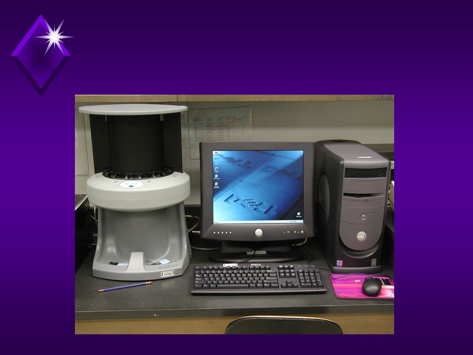
CCD/CMOS-based Sensor
X-ray Beam Scintillator Fiber Optics CCD

7
Standard Standard High & High Resolution #2 #1 #0
High Resolution = 22.5 Standard = 45 Standard & High Resolution Standard High # #1 #0

8
CCD/CMOS - 45

9
CCD/CMOS - 22

10
Storage Phosphor X-ray Beam Imaging Plate

11
Storage Phosphor Helium-Neon Laser Photoreceptor Imaging Plate

13
Storage Phosphor

14
Storage Phosphor

15
Diagnostic Accuracy Primary Dental Caries Recurrent Dental Caries
Periodontal Disease Periapical Lesions Endodontic File Length Determination

16
CCD Extra-oral Radiograpy

17
Tomography

18
Panoramic Radiography

19
Cephalometric Radiography

20
The Next Generation Three Dimensional Imaging
TACT Small Volume CT

21
TACT™ Methodology Series of images taken from different angles

22
Slices of a maxillary molar

23
Small Volume CT

24
Small Volume CT

25
Small Volume CT

26
The Future is Full of Possibilities
Optical Biopsy Laser Computed Tomography 3D Ultrasound Laser Fluorescence Computer-based FOTI

27
Computer Aided Diagnosis (CAD)
Observational Errors Misinterpretation Errors Integration of Data Errors Most Beneficial in Observational Errors

28
“CAD systems do not necessarily have to be better than the clinician, just help him not miss obvious lesions.”

29
Computer Aided Diagnosis (CAD)
PAP Cytology & Oral Brush Biopsy Mammography Chest X-ray Screening Periodontal Disease Dental Caries

30
Digital Subtraction Radiography
-

31
Neural Network Detection

32
Digital Dental Imaging
Intra-oral Radiography Panoramic Radiography Cephalometric Radiography Tomography Intra-oral Photography Extra-oral Photography Histology & Surgical Microscopy

33
Interoperability ????????

34
The DICOM Standard The Digital Imaging and Communications in Medicine (DICOM) Standard is a detailed specification that describes semantics and syntax for exchanging images and associated information. The standard applies to the operation of the interface which is used to transfer data in and out of an imaging device.
 Standard is a detailed specification that describes semantics and syntax for exchanging images and associated information. The standard applies to the operation of the interface which is used to transfer data in and out of an imaging device.")
35
DICOM Workflow DICOM Display Workstation Storage, Query/Retrieve,
Study Component LiteBox Query/Retrieve Results Management DICOM Acquisition Media Exchange Print Management Query/Retrieve, Patient & Study DICOM Archive

36
What is DICOM Digital Imaging and Communication in Medicine
Standard for communication of images and image related information between devices International in scope All biomedical imaging Voluntary standard

37
DICOM is Biomedcial Informatics
“the storage, retrieval, sharing, and optimal use of biomedical information, data, and knowledge for problem solving and decision making.” Edward Shortliffe “model formation, implementation of the model, application of implementaion to the real world, evaluation of the implementation” Titus Schleyer

38
DICOM, Informatics, Research
Digital X-ray Visible Light Clinical Trials Structured Reporting

39
The Future is Full of Possibilities

Similar presentations


 December 2004 Report. WG 22 (2004) Four (4) WG meetings in 2004 – 3 at ADA HQ in Chicago, one in Orlando in advance of ADA Annual Congress.>")


>")
>")

 -Krystal Kerney and Hui Pan.>")


