Download presentation
Presentation is loading. Please wait.

1
For: Nottingham SCRUBS 26th August 2006 Presented by: Matthew
The Chest X-Ray For: Nottingham SCRUBS 26th August 2006 Presented by: Matthew

2
Aims: Basics Best exam results Appreciate the role radiology plays
? Instil an interest in radiology

3
Contents: Densities Techniques Anatomy CXR Interpretation
Common Pathologies Questions

4
Densities The big two densities are: (1) WHITE - Bone (2) BLACK - Air
The others are: (3) DARK GREY- Fat (4) GREY- Soft tissue/water And if anything Man-made is on the film, it is: (5) BRIGHT WHITE - Man-made
 DARK GREY- Fat. (4) GREY- Soft tissue/water. And if anything Man-made is on the film, it is: (5) BRIGHT WHITE - Man-made.")
5
Techniques - Projection
P-A (relation of x-ray beam to patient)
")
6
Techniques - Projection (continued)
A-P Supine/Erect

7
Techniques - Projection (continued)
Lateral

8
Techniques - Projection (continued)
Lateral Decubitus

9
Techniques - Projection (continued)
Oblique

10
Orientation Orientation

11
Rotation

12
Rotation (continued)
")
13
Penetration

14
Inspiration/Expiration

15
Anatomy

16
Anatomy

17
Anatomy

18
Lobes Right upper lobe:

19
Lobes (continued) Right middle lobe:
 Right middle lobe:")
20
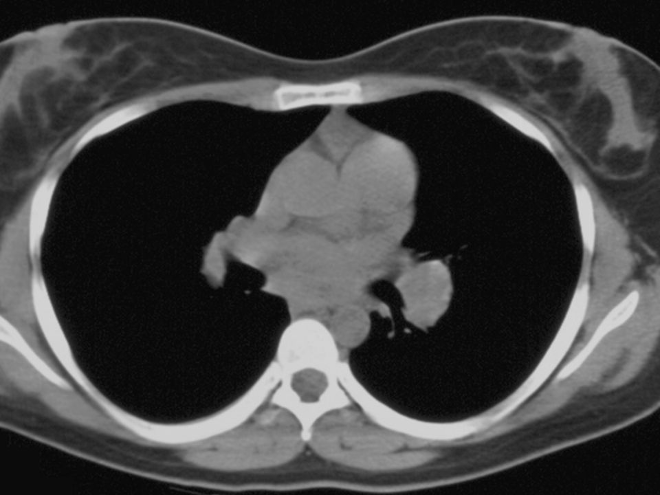
Lobes (continued) Right lower lobe:
 Right lower lobe:")
21
Lobes (continued) Left lower lobe:
 Left lower lobe:")
22
Left upper lobe with Lingula:
Lobes (continued) Left upper lobe with Lingula:
 Left upper lobe with Lingula:")
23
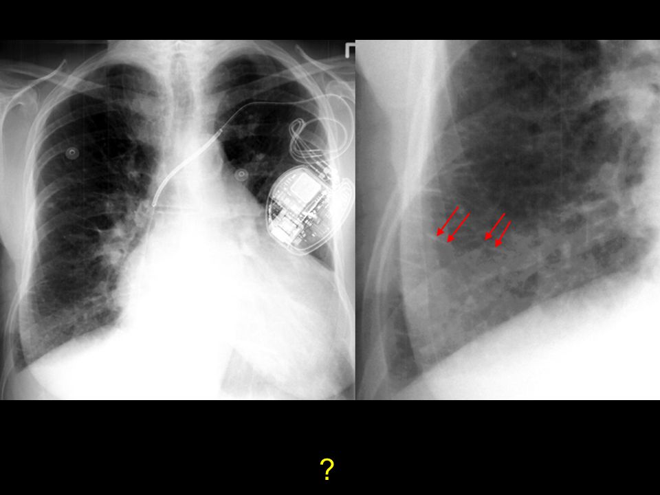
Lobes (continued) Lingula:
 Lingula:")
24
Left upper lobe - upper division:
Lobes (continued) Left upper lobe - upper division:
 Left upper lobe - upper division:")
25
Pleura Layers:

26
Heart Right border: Edge of (r) Atrium
3. Left border: (l) Ventricle + Atrium 4. Posterior border: Reft Ventricle 5. Anterior border: Right Ventricle
 Ventricle + Atrium. 4. Posterior border: Reft Ventricle. 5. Anterior border: Right Ventricle.")
27
Heart (continued)
")
28
Heart (continued)
")
29
Heart (continued) Valves
 Valves")
30
Mediastinum

31
Hilum Made of: 1. Pulmonary Art.+Veins 2. The Bronchi
Left Hilus higher (max 1-2,5 cm) Identical: size, shape, density
 Identical: size, shape, density.")
32
Hilum

33
Ribs

34
Soft tissue & bones

35
Lateral CXR

36
Lateral CXR (continued)
")
37
Lateral CXR (continued)
")
38
Lateral CXR (continued)
")
39
Lateral CXR (continued)
")
40
CXR Interpretation

41
Type Orientation Rotation Inspiration/expiration Penetration
Technical Details Type Orientation Rotation Inspiration/expiration Penetration

42
Lungs Density Symmetry Lesions

43
Heart Size:

44
Heart Size of heart Size of individual chambers of heart
Size of pulmonary vessels Evidence of stents, clips, wires and valves Outline of aorta and IVC and SVC

45
Width Contour AP window
Mediastinum: Width Contour AP window Hila: Size Location

46
Review areas: Apices Behind the heart CP angles Below the diaphragm
Soft tissues ( breast, surgical emphysema) Ribs & clavicle Vertebrae
 Ribs & clavicle. Vertebrae.")
47
Identify the lesion → localise the lesion → describe the lesion → give DD Never stop looking, carry on with your systematic approach!!

48
Pathology

49
RUL pneumonia

50
RML pneumonia

51
RLL pneumonia

52
LUL pneumonia

53
LLL pneumonia

54
Consolidation on CT

55
Hilar m l

56
The Enlarged Hila Causes: 1. Adenopathies (neoplasia, infection) 2. Primary Tumor 3. Vascular 4. Sarcoidosis

57
Multiple Masses

58
Hilar Lymphadenopathy - BL

60
Pleural Effusion

61
Pulmonary Fibrosis

62
?
63
Heart failure

64
Pneumothorax

65
RUL collapse

66
LLL collapse

67
Air under the diaphragm

68
Emphysema

69
Cervical Rib

70
Cavitating lesion

71
Hiatus hernia

72
Miliary shadowing

73
Chest Tube, NG Tube, Pulm. artery cath

74
Dextrocardia

Similar presentations