Download presentation
Presentation is loading. Please wait.

1
Before We Continue Lets Recap!

2
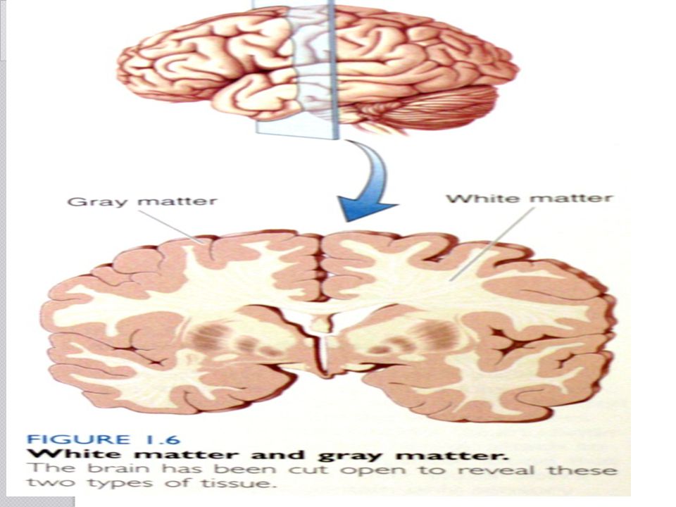
Identify the gray matter and white matter.
B A

4
The Brain C D A E F B G H I J K SfN’s Brain Facts

5
The Brain SfN’s Brain Facts

6
Dendrite, cell body & axon electroencephalograph
Review Dendrite Greek for branches of a tree. The three components of a neuron are … The functional cell that allows us to think, act, & learn. Name for the electrical impulses neurons use to send messages, The net charge of a neuron at rest. Proteins embedded into the cell membrane that allow for ions to move into or out of cells. Type of ions that depolarize the cell. How many neurons does the brain have? Name a device that measures brain waves. A neuron releases Neurotransmitters into a synapse with another axon. Assuming, this will pass stimulate to the next neuron, explain how the signal will be sent. Dendrite, cell body & axon neuron Action potential negative Protein channels positive 100 billion electroencephalograph

7
1)The Neuron receives a signal at its dendrites.
(2) This signal travels and opens specific ion channels in the axon that allow positive ions inside the cell. (3)Ion channels open sequentially, with the first open ion channels at the axon hillock, or base of the axon. 4)The Action Potential travels down the axon extremely fast, at speeds up to several hundred miles per hour. (5)Once the action potential reaches the end of the axon, it causes the release of neurotransmitters out of the neuron at the nerve terminal. The neurotransmitters diffuse a short distance and bind specific receptors on the surface of a second neuron where it will either stimulate or inhibit it from generating another action potential.
 This signal travels and opens specific ion channels in the axon that allow positive ions inside the cell. (3)Ion channels open sequentially, with the first open ion channels at the axon hillock, or base of the axon. 4)The Action Potential travels down the axon extremely fast, at speeds up to several hundred miles per hour. (5)Once the action potential reaches the end of the axon, it causes the release of neurotransmitters out of the neuron at the nerve terminal. The neurotransmitters diffuse a short distance and bind specific receptors on the surface of a second neuron where it will either stimulate or inhibit it from generating another action potential.")
8
More Review Hypothalamus true amygdala L Dopa 3 lbs
The biological clock is located in what part of the brain? True or False: The Cerebellum is crucial for motor learning. What Brain Region processes Fear and anxiety? Name the primary drug used to treat Parkinson’s Disease. To the Nearest pound, what is the weight of the human brain? The abbreviation PET stands for what brain imaging technique? Nitric oxide is the major regulator of what intracellular messenger molecule? Name the contact points where one neuron communicates with another. Hypothalamus true amygdala L Dopa 3 lbs Positron emission topography Cyclic GMP (cyclic guonosine monophosphate Synapse

9
More Questions Acetylcholine Amygdala Cerebellum Occipital Synapse
Excitatory Neurotransmitter who, when unable to bind to muscle receptors, causes myasthenia gravis. Name the part of the brain that is important for emotional learning and memory and its dysfunction is related to anxiety disorders. What part of the brain helps control movement and cognitive processes that require precise timing? Which lobe of the cerebral cortex processes visual information? The Greek word meaning to “clasp together” gives us what neuroscience term? Name the deep brain regions responsible for relaying and filtering sensory information. Name the large bundle of nerve fibers linking the left and right cerebral hemispheres. Acetylcholine Amygdala Cerebellum Occipital Synapse Thalamus Corpus Callosum

10
Limbic System Androgens Sympathetic Brocca’s Cereborspinal Fluid
The amygdala and septum are part of this group of brain structures that regulate emotion. Name the hormones, which include testosterone, that are important for male sexual development. Which branch of the autonomic nervous system mobilizes energy and resources during times of stress and arousal? This region of the brain, located in the frontal lobe of the left hemisphere, is important for the production of speech. This liquid is found within the ventricles of the brain and the central canal of the spinal cord. What is the name of the class of neurotransmitters that are chains of amino acids? Benzodiazepines (Valium) is used to increase the activity of this NT in the treatment of Huntington’s Disease. Glutamate & Asparate are both amino acid NTs though to be involved in this important function (like right now). Limbic System Androgens Sympathetic Brocca’s Cereborspinal Fluid Peptides GABA Learning & Memory
 is used to increase the activity of this NT in the treatment of Huntington’s Disease. Glutamate & Asparate are both amino acid NTs though to be involved in this important function (like right now). Limbic System. Androgens. Sympathetic. Brocca’s. Cereborspinal Fluid. Peptides. GABA. Learning & Memory.")
11
Hormone-long distance
A lack of this NT can be supplemented with L-Dopa in the treatment of Parkinson’s. An excitatory chatcolamine NT involved in the fight or flight response. Regulates Heart rate and blood pressure. NT involved in OCD and Depression. Name a difference between neurotransmitters and hormones in regard to their effective range. Which region in the brain secretes endocrine hormones? Dopamine Norepinephrine Serotinin NT-Short distance Hormone-long distance Hypothalamus

12
Brain Development (Brain Facts pages, 13-17)
")
13
Why is it important to understand development?
Congenital Defects Disease process Response to injury or external influences (drugs) Learning and memory
 Learning and memory.")
14
Background Knowledge (not in Brain Facts) Review on basic Development
Lets start at the beginning, a zygote is formed. The zygote must go through cell division

16
Embryo Developement Sperm fertilizes egg & forms a zygote

17
Zygote: Fertilized Egg
Zygote undergoes cleavage: a series of rapid cell divisions that results in the formation of many cells.

18
Zygote Blastula Cleavage
A ball of cells forms with a fluid- filled center. This hollow ball of cells is called a Blastula.

19
Zygote Blastula Cleavage

21
Blastula Gastrula Gastrulation
Gastrulation occurs when a blastula, made up of one layer, folds inward and enlarges to create a gastrula.

22
The 3 layers of the GASTRULA:
Ectoderm (outer): Skin, sensory organs, nerves Mesoderm (middle) Muscles, circulatory, reproductive, excretory systems Endoderm (inner): Digestive tract, respiratory system
: Skin, sensory organs, nerves. Mesoderm (middle) Muscles, circulatory, reproductive, excretory systems. Endoderm (inner): Digestive tract, respiratory system.")
25
video-by-yale-scientist-visualizes- fetal-development-from-conception- to-birth/?lang=en

26
Lecture Overview Summary Overview Development from embryo
Initial wiring Activity dependent fine tuning

27
How does this happen Many mechanisms of human brain development remain hidden, but Neuroscientists are beginning to uncover some of these complex steps through studies of the roundworm, fruit fly, frog, zebrafish, mouse, rat, chicken, cat and monkey. Many initial steps in brain development are similar across species, while later steps are different. By studying these similarities and differences, we can learn how the human brain develops and how brain abnormalities, such as mental retardation and other brain disorders, can be prevented or treated.

28
Summary Overview: How do cells know where to go?
Think of the Amoeba. Uses complex sensing molecules imbedded in its cell membrane to trigger chemical mechanisms that cause it to move its blobby body towards food and away from harmful substances. Neurons are also cells and, in early development, behave somewhat like Amoeba in approaching and avoiding various chemicals. But rather than the whole cell moving, neural growth involves the outreach of the cell’s connecting pathway (axon) towards its downstream partner neurons.
 towards its downstream partner neurons.")
29
Summary Overview The basic layout of visual and other maps is established during development by neurons each separately following a pattern of chemical markers to its pre-destined brain region and specific sub-areas within that region. For example, a retinal cell that responds best to red light in the upper left of the visual field will connect to cells in the brain that are tuned to the same properties and these cells, in turn, will link to other cells that use these particular properties giving rise to specific connectivity and cell receptive field properties (topographic maps).
.")
30
Summary Overview In the course of development,
detector molecules in the growing neuron interact with guide molecules to route the connection to the right general destination, sometimes over long distances as in the connection from the spinal cord to the knee. This process will get neural connections to the right general area, but aligning the millions of neurons in visual and other neural maps also involves chemical gradients, again utilizing mechanisms that are very old in evolutionary terms.

31
Summary Overview When an axon tip gets to an appropriately marked destination cell, the contact starts a process that develops rudimentary synapses. Local competition among neural axons with similar marker profiles produces some further tuning at the destination.

32
Summary Overview: Activity Dependent Tuning
In fact, the initial wiring is only approximate and leaves each neuronal axon connected to several places in the neighborhood of each of its eventual partner neurons. A second, activity dependent, mechanism is required to complete the development process. The initial chemical wiring actually produces many more connections and somewhat more neurons than are present in adult brains. The detailed tuning of neural connections is done by eliminating the extra links, as well as the strengthening functional synapses based on neural activity.

33
Lecture Overview Summary Overview Development from embryo
Neural tube development Cell division and neuronal identity Mechanisms for cell type formation and communication Initial wiring Activity dependent fine tuning Plasticity and Learning

34
Development from Embryo
The embryo has three primary layers that undergo many interactions in order to evolve into organ, bone, muscle, skin or neural tissue. The outside layer is the _________ (skin, neural tissue), the middle layer is the ___________ (skeleton, cardiac) and inner layer is the ___________ (digestion, respiratory). ectoderm mesoderm endoderm
, the middle layer is the ___________. (skeleton, cardiac) and. inner layer is the ___________. (digestion, respiratory). ectoderm. mesoderm. endoderm.")
35
Neural Tissue The skin and neural tissue arise from a single layer, known as the ectoderm in response to signals provided by an adjacent layer, known as the mesoderm. A number of molecules interact to determine whether the ectoderm becomes neural tissue or develops in another way to become skin

36
Early development Three to four weeks after conception, one of the two cell layers of the gelatin-like human embryo, now about one-tenth of an inch long, starts to thicken and build up along the middle. As this flat neural plate grows, parallel ridges, similar to the creases in a paper airplane, rise across its surface.

37
Early development Within a few days,the ridges fold in toward each other and fuse to form the hollow neural tube. The top of the tube thickens into three bulges that form the hindbrain, midbrain and forebrain. The first signs of the eyes and then the hemispheres of the brain appear later.

38
Neural Tube formation

41
In humans, during the 3rd week, this mesoderm begins to segment
In humans, during the 3rd week, this mesoderm begins to segment. The neural plate folds to form a neural groove and folds.

42
Other structures including heart, Skeleton, etc. The neural groove fuses dorsally to form a tube and "zips up” cranially and caudally and the neural crest migrates into the mesoderm.

43
BRAIN DEVELOPMENT. The human brain and nervous system begin to develop at three weeks’ gestation as the closing neural tube (left). By four weeks, major regions of the human brain can be recognized in primitive form, including the forebrain, midbrain, hindbrain, and optic vesicle (from which the eye develops). Irregular ridges, or convolutions, are clearly seen by six months.
. By four weeks, major regions of the human brain can be recognized in primitive form, including the forebrain, midbrain, hindbrain, and optic vesicle (from which the eye develops). Irregular ridges, or convolutions, are clearly seen by six months..")
45
Brain Weight

47
hindbrain midbrain forebrain
1.)The Journey of Nerve Cells Origins of the Nervous System Review The neural plate forms from which embryonic cell layer? About how long after conception does the neural plate form? When the neural plate sides rise, fold towards each other, and close, the is__________ formed. By 4 weeks, the top (anterior) of the tube thickens into 3 bulges that form the ______, _______, & _______. ectoderm About 3-4 weeks neural tube hindbrain midbrain forebrain
The Journey of Nerve Cells Origins of the Nervous System. Review. The neural plate forms from which embryonic cell layer About how long after conception does the neural plate form When the neural plate sides rise, fold towards each other, and close, the is__________ formed. By 4 weeks, the top (anterior) of the tube thickens into 3 bulges that form the ______, _______, & _______. ectoderm. About 3-4 weeks. neural tube. hindbrain midbrain forebrain.")
48
General Brain Development
1. The appropriate number of neurons migrate to their appropriate places. 2. Axons and dendrites form connections. 3. Axons recognize a target cell. 4. Connections mature and change with different activity/experiences.

49
Cellular signals from the mesoderm layer turn genes on and off.
1.)The Journey of Nerve Cells Origins of the Nervous System Induction What is the process by which a single cell line becomes many different, specialized cells? How can this process occur in the germ layers when they all possess the same genes? Differentiation Cellular signals from the mesoderm layer turn genes on and off.
The Journey of Nerve Cells Origins of the Nervous System. Induction. What is the process by which a single cell line becomes many different, specialized cells How can this process occur in the germ layers when they all possess the same genes Differentiation. Cellular signals from the mesoderm layer turn genes on and off.")
50
1.)The Journey of Nerve Cells Origins of the Nervous System
Induction Molecules released from the mesoderm can trigger cells from which layer to become nervous tissue? Specifically, what is the process called? What happens to the majority of ________ cells that do not receive the mesoderm signal molecules. ectoderm Neural induction ectoderm They become skin

51
Remember the three germ layers?
1.)The Journey of Nerve Cells Origins of the Nervous System Remember the three germ layers? Ectoderm Mesoderm Endoderm Of these three, which will develop into both the nervous system and skin cells? Skin and neural tissue arise from one layer, the ectoderm, in response to signals provided by the adjacent layer, the mesoderm.
The Journey of Nerve Cells Origins of the Nervous System. Remember the three germ layers Ectoderm. Mesoderm. Endoderm. Of these three, which will develop into both the nervous system and skin cells Skin and neural tissue arise from one layer, the ectoderm, in response to signals provided by the adjacent layer, the mesoderm.")
52
Inhibitory control may direct neural development.
Many molecules interact to determine whether the ectoderm becomes neural tissue or develops into skin. Studies in frogs show that one major mechanism depends on specific proteins that inhibit the activity of other proteins. In areas where no inhibition occurs, the tissue becomes skin. In areas where proteins secreted from the mesoderm do lead to inhibition, the tissue becomes neural.

53
No Inhibition Ectoderm SKIN
1.)The Journey of Nerve Cells Origins of the Nervous System No Inhibition Ectoderm SKIN
The Journey of Nerve Cells Origins of the Nervous System. No Inhibition. Ectoderm. SKIN.")
54
Inhibition from Mesoderm Proteins
1.)The Journey of Nerve Cells Origins of the Nervous System Inhibition from Mesoderm Proteins Ectoderm SKIN Nervous Tissue
The Journey of Nerve Cells Origins of the Nervous System. Inhibition from Mesoderm Proteins. Ectoderm. SKIN. Nervous Tissue.")
55
1.)The Journey of Nerve Cells Origins of the Nervous System
Neuron or Glial cell? Once the ectoderm is destined to become neural tissue, it has two possibilities Neuron: Signaling Cell Glial Cell: Neuron Support Cell depends on its Cellular interactions Position Environmental cues

56
SONIC HEDGEHOG Protein
1.)The Journey of Nerve Cells Origins of the Nervous System SONIC HEDGEHOG Protein Example, a key factor in spinal cord development is a secreted protein called sonic hedgehog. The protein, initially secreted from mesodermal tissue (lying beneath the developing spinal cord), marks adjacent (closest) neural cells to become a specialized class of glial cells. Cells farther away are exposed to lower concentrations of sonic hedgehog, and they become the motor neurons. An even lower concentration promotes the formation of interneurons
The Journey of Nerve Cells Origins of the Nervous System. SONIC HEDGEHOG Protein. Example, a key factor in spinal cord development is a secreted protein called sonic hedgehog. The protein, initially secreted from mesodermal tissue (lying beneath the developing spinal cord), marks adjacent (closest) neural cells to become a specialized class of glial cells. Cells farther away are exposed to lower concentrations of sonic hedgehog, and they become the motor neurons. An even lower concentration promotes the formation of interneurons.")
57
Sonic Hedgehog Protein Graded Effect
1.)The Journey of Nerve Cells Origins of the Nervous System MESODERMAL TISSUE Sonic Hedgehog Protein Graded Effect GLIAL CELLS Motor Neurons Interneurons
The Journey of Nerve Cells Origins of the Nervous System. MESODERMAL. TISSUE. Sonic Hedgehog Protein Graded Effect. GLIAL CELLS. Motor Neurons. Interneurons.")
58
MIGRATION 3-4 Weeks after conception:
Once neural induction has occurred, the next step for new neurons migration Ectoderm starts to thicken and build up along the middle. Cells continue to divide, forming a flat neural plate Parallel ridges form & rise across neural plate’s surface. Within a few days, the ridges fold in toward each other and fuse to form a hollow neural tube. The top of the tube thickens into three bulges that form the hindbrain, the midbrain, and the forebrain. At week 7, the first signs of the eyes and the brain’s hemispheres appear.

59
1.)The Journey of Nerve Cells Origins of the Nervous System
3-4 ______ weeks after conception, one of the germ layers (_________) of the human embryo starts to thicken and build up along the middle, forming the ____________ ectoderm Neural plate
 of the human embryo starts to thicken and build up along the middle, forming the ____________. ectoderm. Neural plate.")
60
1.)The Journey of Nerve Cells Origins of the Nervous System
As this flat neural plate grows, ________________ rise across its surface. The ridges fold in toward each other and fuse to form the hollow ___________ (by day 28). Parallel ridges neural tube
. Parallel ridges. neural tube.")
61
hindbrain midbrain forebrain
1.)The Journey of Nerve Cells Origins of the Nervous System The top of the tube thickens into three bulges that form the __________, __________, and __________. hindbrain midbrain forebrain Video: Neural Tube Formation
The Journey of Nerve Cells Origins of the Nervous System. The top of the tube thickens into three bulges that form the __________, __________, and __________. hindbrain. midbrain. forebrain. Video: Neural Tube Formation.")
63
1.)The Journey of Nerve Cells Origins of the Nervous System
The human brain and nervous system begin to develop at about three weeks’ gestation with the closing of the neural tube (left image). By four weeks, major regions of the human brain can be recognized in primitive form, including the forebrain, midbrain, hindbrain, and optic vesicle (from which the eye develops). This is the outline for the lecture. Irregular ridges, or convolutions, are clearly seen by six months.
. By four weeks, major regions of the human brain can be recognized in primitive form, including the forebrain, midbrain, hindbrain, and optic vesicle (from which the eye develops). This is the outline for the lecture. Irregular ridges, or convolutions, are clearly seen by six months.")
65
FIGURE 3.5 Brain Development. This schematic outline of brain development shows its relation to the ventricles. Views (a) and (c) show early development. Views (b) and (d) show later development. View (e) shows a lateral view of the left side of a semitransparent human brain with the brain stem “ghosted in.” The colors of all figures denote corresponding regions.
 and (c) show early development. Views (b) and (d) show later development. View (e) shows a lateral view of the left side of a semitransparent human brain with the brain stem ghosted in. The colors of all figures denote corresponding regions.")
67
1.)The Journey of Nerve Cells Origins of the Nervous System
Migration As neurons are produced, they move from the neural tube’s ventricular zone, or inner surface, to near the border of the marginal zone, or outer surface. After neurons stop dividing, they form an intermediate zone where they gradually accumulate as the brain develops.

68
1.)The Journey of Nerve Cells Origins of the Nervous System
The Journey of Nerve Cells Origins of the Nervous System")
69
1.)The Journey of Nerve Cells Origins of the Nervous System
Brain Facts Page 11

70
How do they know where to go?
1.)The Journey of Nerve Cells Origins of the Nervous System How do they know where to go? Neuron migration requires multiple guidance mechanisms, including the recognition of the proper path and the ability to move long distances. 90 % of migration in humans, are glia, which project radially from the intermediate zone to the cortex. Glia are providing temporary scaffolding for ushering neurons to their destination.
The Journey of Nerve Cells Origins of the Nervous System. How do they know where to go Neuron migration requires multiple guidance mechanisms, including the recognition of the proper path and the ability to move long distances. 90 % of migration in humans, are glia, which project radially from the intermediate zone to the cortex. Glia are providing temporary scaffolding for ushering neurons to their destination.")
71
How do they know where to go?
1.)The Journey of Nerve Cells Origins of the Nervous System How do they know where to go? This process of radial migration occurs in an “inside-out” manner; that is, the cells that arrive the earliest (the oldest ones) form the deepest layer of the cortex, whereas the late-arriving (the youngest) neurons form the outermost layer. In another mode, inhibitory interneurons migrate tangentially across the brain.
The Journey of Nerve Cells Origins of the Nervous System. How do they know where to go This process of radial migration occurs in an inside-out manner; that is, the cells that arrive the earliest (the oldest ones) form the deepest layer of the cortex, whereas the late-arriving (the youngest) neurons form the outermost layer. In another mode, inhibitory interneurons migrate tangentially across the brain.")
72
What if they get bad directions?
1.)The Journey of Nerve Cells Origins of the Nervous System What if they get bad directions? Environmental factors, such as alcohol, cocaine, or radiation, prevent proper neuronal migration and result in misplacement of cells, which may lead to mental retardation or epilepsy. Mutations in genes that regulate migration have been shown to cause some rare genetic forms of retardation and epilepsy in humans.
The Journey of Nerve Cells Origins of the Nervous System. What if they get bad directions Environmental factors, such as alcohol, cocaine, or radiation, prevent proper neuronal migration and result in misplacement of cells, which may lead to mental retardation or epilepsy. Mutations in genes that regulate migration have been shown to cause some rare genetic forms of retardation and epilepsy in humans.")
73
1.)The Journey of Nerve Cells Origins of the Nervous System

74
Neural Development What did you just learn?
Explain the formation of nervous tissue from to migration. Neural Development

75
Once our neural cells are where they need to be, how do their axons find their targets?
Making Connections

76
Neurons Network Near & Far
1.)The Journey of Nerve Cells: Making Connections Neurons Network Near & Far What are the three main components of neurons? Cell Body, Dendrites, and Axons The axons must grow and travel long distances to make connections. Most Axons will connect with the dendrites of other neurons Motor Neuron axons make connections with muscles (from the brain to the foot)
The Journey of Nerve Cells: Making Connections. Neurons Network Near & Far. What are the three main components of neurons Cell Body, Dendrites, and Axons. The axons must grow and travel long distances to make connections. Most Axons will connect with the dendrites of other neurons. Motor Neuron axons make connections with muscles (from the brain to the foot)")
77
Growth Cones Axons are guided on their migration by Growth Cones.
1.)The Journey of Nerve Cells: Making Connections Growth Cones Axons are guided on their migration by Growth Cones.
The Journey of Nerve Cells: Making Connections. Growth Cones. Axons are guided on their migration by Growth Cones.")
78
Growth cones Enlargements on the axon’s tip
1.)The Journey of Nerve Cells: Making Connections Growth cones Enlargements on the axon’s tip Actively explore the environment to seek destination. Growth cones have receptors for specialized ‘guidance molecules’ Some molecules lie on the cells that growth cones contact, whereas others are released from sources found near the growth cone. Signal-receptor interactions direct growth cones to move forward, stop, recoil, or change direction.
The Journey of Nerve Cells: Making Connections. Growth cones. Enlargements on the axon’s tip. Actively explore the environment to seek destination. Growth cones have receptors for specialized ‘guidance molecules’ Some molecules lie on the cells that growth cones contact, whereas others are released from sources found near the growth cone. Signal-receptor interactions direct growth cones to move forward, stop, recoil, or change direction.")
79
~ Netrin – attracts growth cones ~ Semaphorin – repulses growth cones
1.)The Journey of Nerve Cells: Making Connections Guidance Molecules Guidance molecule are proteins that direct growth cones to their proper destinations. Worms and flies have similar families of proteins that perform similar function, and provide researchers with model systems. Examples: ~ Netrin – attracts growth cones ~ Semaphorin – repulses growth cones ~ Ephrin – repulses growth cones ~ Slit – repulses growth cones
The Journey of Nerve Cells: Making Connections. Guidance Molecules. Guidance molecule are proteins that direct growth cones to their proper destinations. Worms and flies have similar families of proteins that perform similar function, and provide researchers with model systems. Examples: ~ Netrin – attracts growth cones. ~ Semaphorin – repulses growth cones. ~ Ephrin – repulses growth cones. ~ Slit – repulses growth cones.")
80
1. Brain Wiring: Spinal Cord and Nerves
Brain Facts Page 13

81
What happens once an axon reaches its target?
Are we there yet? What happens once an axon reaches its target? Axon releases specific neurotransmitter Dendrite becomes specialized to receive signal Synapse matures - connections

82
1.)The Journey of Nerve Cells Origins of the Nervous System
Which message to send? A combination of signals also determines the type of chemical messages, or neurotransmitters, that a neuron will use to communicate with other cells. Current research suggests the target cells themselves induce the genes for the NTs that will affect them. Neurons grown near cardiac cells create Ach

83
Axons Form Synapses Each neuron is connected by 1,000’s of synapses, so each one MUST be SPECIFIC Synapse forms at particular part of target Axon releases specific neurotransmitter Dendrite becomes specialized to receive signal

84
Forming Synapses Synapse matures: As behavior changes and organism matures, synapses continue to be modified and or strengthened by intermediate molecules between the axon terminal and the target dendrite or cell body. Defects in these intermediate molecules are thought to be involved in Autism and synapse degradation during aging.

85
To Recap Ectoderm Nervous tissue
Induced by molecules from Mesoderm Neural Plate forms Differentiation occurs-Glial cells or neurons Induced environmental factors, other cells, and positioning Neurons Migrate from neural tubes inner to outer layer. Nervous tissue accumulates in the intermediate zone as the brain developes. Axons begin journey to their final destinations led by Growth Cone. Axon terminals form synapses with their target cell, dendrite, or cell body

86
With the pathway made, what can we now add to make the message faster?
After the synapse is made, the myelin sheath can be wrapped around the axon to help speed up the electrical signals. This process can take years to complete.

87
Review: Name the three germ layers formed during gastrulation.
Explain how the 2 of the layers interact to form the nervous system. Provide a specific example how a specific protein effects the formation of a specific nervous system structure. Be sure to explain how orientation influences development. You are examining the neural tube looking for signs of mitosis. Where should you look? Explain why? If you follow a new cell, what will it do after shortly after its formation.

88
2. Paring Back “Only about half the neurons generated during development survive to function in the adult.” Why??

89
2. Paring Back Apoptosis - programmed cell death
Trophic factors – Nutrient molecules necessary cell survival (different trophic factors are specific to neuron type)
")
90
“The connections that are active
2. Paring Back If a cell does not receive the appropriate trophic factors from its target, it will initiate apoptosis Example: Nerve Growth Factor is necessary for the survival of sensory neurons. “The connections that are active and generating electrical currents survive, whereas those with little or no activity are lost.”

91
3. Critical Periods Definition: windows of time during development when the nervous system must obtain certain critical experiences, such as sensory, movement, or emotional input, to develop properly. These periods are characterized by high learning rates.

92
3. Critical Periods: Use it or Lose It
After a critical period, connections diminish in number and are less subject to change, but the ones that remain are stronger, more reliable, and more precise. Injury or deprivation, either sensory or social, occurring at a certain stage of postnatal life may affect one aspect of development, whereas the same injury at a different period may affect another aspect.

93
“Enriched” environment
3. Critical Periods Language Eyesight “Enriched” environment

94
Use brain development as an argument on why the driving age should be changed to 21 years old. (hint: Page 17 Brain Facts)
.")
95
Brain development in people continues into the early 20s — even the brain of an adolescent is not completely mature. One of the later aspects of brain development is the completion of myelination of the axons connecting one brain area to another. This process starts around birth and moves from the back of the brain to the front: The frontal lobes are the last to become “connected” with fast- conducting myelinated axons. Major functions of the frontal lobes are judgment, insight, and impulse control, and so the acquisition of these attributes becomes the last step in the creation of an adult human brain.

96
So at 21, is your brain set?

97
Plasticity Plasticity is the ability of the brain to modify itself and adapt to challenges of the environment Plasticity can be categorized as experience expectant or experience- dependent

98
Plasticity Experience-expectant plasticity: the integration of environmental stimuli into the normal patterns of development. Certain environmental exposures during critical periods of development are essential for healthy maturation. Example: finches need to hear adult songs before sexual maturation in order for them to learn to sing at a species appropriate level of intricacy.

99
REVIEW

Similar presentations


 & the Peripheral Nervous System.>")


