Download presentation
Presentation is loading. Please wait.

2
Dys- scope Pneathorac Tachyhema Brady-ology Cardiascopy epi-peri- Endo-sub-

3
Respiration › Unconscious exchange of air between lungs and the external environment › Breathing Two types › External Exchange of carbon dioxide CO2 and oxygen O2 between the environment and the organism › Internal Exchange of O2 and CO2 between internal body fluids (Ex: blood and individual cells)
")
4
Oxygen › Breathed into the lungs (O 2 ) › Body has a 4-6 minute supply of oxygen Carbon Dioxide › Exhaled out of the lungs › CO2 Gas exchange › Transfer of oxygen from inhaled air into the blood and the transfer of carbon dioxide from the blood into the exhaled air
 › Body has a 4-6 minute supply of oxygen Carbon Dioxide › Exhaled out of the lungs › CO2 Gas exchange › Transfer of oxygen from inhaled air into the blood and the transfer of carbon dioxide from the blood into the exhaled air")
5
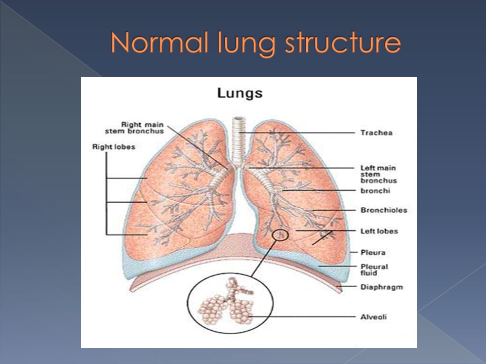
Nose/mouth › Route to take in O2 and expel CO2. mucous membranes warm and humidify air Cilia › Tiny hairs that protect the nasal passages, trachea and bronchi › Move back and forth as air is inhaled, pushing foreign particles (dust) toward the nostrils or pharynx
 toward the nostrils or pharynx.")
6
Pharynx › Also known as the Throat Larynx – also know as the Larynx – also know as the voice box Between pharynx and trachea › Contains vocal cords that vibrate against each other to cause sound › www.fauquierent.net/voice2.htm www.fauquierent.net/voice2.htm Epiglottis www.youtube.com/watch?v=QvGYvK6qScE www.youtube.com/watch?v=QvGYvK6qScE › Lid like structure at the base of the tongue that closes during swallowing so that food/drink does not enter the lungs

7
Trachea › Also known as Windpipe › Connects the pharynx to the lungs › Wrapped in cartilage for protection

8
Bronchus (plural bronchi) › The trachea divides into two main bronchi (left mainstem and right mainstem) **Main airways within the lungs Bronchioles › Branch off of the bronchi. › Do not contain cartilage › Small airways within the lungs

9
Alveolar sacs › Located at the end of the bronchioles › Look like a bunch of grapes › Surrounded by capillaries › Site of gas exchange

10
Lungs › Right lung: 3 lobes › Left lung: 2 lobes Why is the left lung smaller than the right? http://www.youtube.com/watch?v=GERsM FWYZrw http://www.youtube.com/watch?v=GERsM FWYZrw

12
Normal vs. Abnormal lung structure

13
Auscultation › Listening to sounds within the body using a stethoscope Percussion › Tapping on a surface to determine the difference in the density of the underlying tissue › “is the underlying tissue solid or filled with air? › Lungs should sound hollow when tapping over them; should NOT sound solid › http://www.youtube.com/watch?v=CvyrA8c5cY0&NR=1/ http://www.youtube.com/watch?v=CvyrA8c5cY0&NR=1/ Sputum › Mucous expelled from the respiratory tract mixed with saliva › Able to make diagnoses based on color and composition of sputum

14
Rales › Clicking, rattling and crackling noises › Caused by the opening of small airways (alveoli collapse by fluid or lack of air) › Heard during inspiration where there is fluid or pus in the alveoli › Cause: pneumonia, bronchitis Wheezing › Whistling sound caused by bronchial airways being narrowed or obstructed › Cause: asthma, bronchitis
 › Heard during inspiration where there is fluid or pus in the alveoli › Cause: pneumonia, bronchitis Wheezing › Whistling sound caused by bronchial airways being narrowed or obstructed › Cause: asthma, bronchitis")
15
Rhonchi › Coarse rattling sound (like snoring) caused by sputum in the bronchial airways › Heard during inhalation and exhalation › Cause: bronchitis › http://www.easyauscultation.com/lung-sounds- reference-guide.aspx http://www.easyauscultation.com/lung-sounds- reference-guide.aspx
 caused by sputum in the bronchial airways › Heard during inhalation and exhalation › Cause: bronchitis › reference-guide.aspx reference-guide.aspx")
16
Chest X ray (CXR) › X ray of the thoracic region of the body to look for lung problems (pathology) › Diagnose pneumonia, tuberculosis(TB,) asthma
 › X ray of the thoracic region of the body to look for lung problems (pathology) › Diagnose pneumonia, tuberculosis(TB,) asthma")
18
Hypoxemia Hypoxemia - low levels of oxygen in the blood Dyspnea Dyspnea - difficult, painful breathing Shortness of breath Shortness of breath (SOB) - difficulty “catching” your breath Intubation Intubation - placement of an endotracheal (ET) tube into the trachea of a person who is unable to breathe on their own
 - difficulty catching your breath Intubation Intubation - placement of an endotracheal (ET) tube into the trachea of a person who is unable to breathe on their own")
19
http://www.youtube.com/watch?v=441l GyW6EYs/ http://www.youtube.com/watch?v=441l GyW6EYs/

20
Upper respiratory infection (URI) › The common cold, Viral illness, highly contagious › Lasts for a week › Symptoms: runny nose, cough, fever, sore throat, congestion Would antibiotics be effective as treatment? Why/why not? Influenza › The flu, Viral illness, highly contagious › Symptoms: chills, fever, cough, sore throat, runny nose, muscle pain, fatigue › Annual vaccination is recommended for the elderly, health care workers, people with chronic diseases and pregnant women Why is a vaccine needed every year?

21
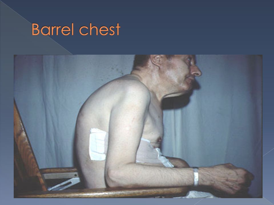
Chronic, no cure Alveoli lose elasticity and deteriorate, CO 2 becomes trapped in alveoli and they become over expanded, gas exchange is poor smoking Usually caused by heavy smoking Symptoms: dyspnea, feeling of suffocation, barrel chest, chronic cough, cyanosis Treatment: bronchodilators, oxygen, avoiding smoking, prompt treatment of infections

22
Smoker’s lung with emphysema Normal lungs

24
Chronic Obstructive Pulmonary Disease Any chronic lung disease that results in obstruction of the airways Chronic bronchitis, emphysema, asthma and tuberculosis can lead to COPD Smoking is the primary cause

25
Airflow obstruction due to bronchospasm, swelling of the bronchioles and/or bronchi, and increased mucous in the airways. › Bronchospasm: severe contraction of smooth muscle covering bronchioles/bronchi Symptoms: wheezing, cough, dyspnea, chest tightness, SOB Reversible with inhaled medications that relieve the bronchospasm

27
Bronchoscopy › A bronchoscope (camera) is inserted into the airways through the nose or mouth so the doctor may look for abnormalities within the bronchi. › http://www.youtube.com/watch?v=KqZc1JqArco http://www.youtube.com/watch?v=KqZc1JqArco Tracheostomy › An airway is created by making an incision into the trachea through the neck › Used as an emergency or a permanent fix

28
1. Place one balloon over the mouth of the bottle. Force balloon into bottle 2. Cut off the neck of second balloon about 1-1.5 inches from top 3. Secure second balloon to bottom of bottle with rubber band and tighten. 4. Pull balloon secured to b bottom of bottle down. What do you see? How does this demonstrate how the lung works?

29
You providing patient care in a clinic. A child comes in complaining of not feeling well. As part of your assessment, you check the child’s oxygen level. The value is 89. What do you consider when determining what to do for the child?

Similar presentations


 into the atmosphere Filter, moisten,>")


 › “physical.>")

