Download presentation
Presentation is loading. Please wait.

1
JOINTS

2
2 Movement at a joint occurs when a muscle contracts and its fibers pull its moveable end (insertion) towards its fixed end (origin). Types of Joint Movements

3
3 Abduction/adduction Dorsiflexion/plantar flexion Flexion/extension/hyperextension (all): © The McGraw-Hill Companies, Inc./Womack Photography Ltd. Abduction Adduction Dorsiflexion Plantar flexion Extension Flexion Copyright © The McGraw-Hill Companies, Inc. Permission required for reproduction or display.

4
© McGraw-Hill Companies / Womack Photography Ltd. Circumduction Medial rotation Lateral rotation Supination Pronation 4 Types of Joint Movements Rotation (medial rotation; lateral rotation) Circumduction Supination/pronation
 Circumduction Supination/pronation.")
5
5 Types of Joint Movements Eversion/inversion Protraction/retraction Elevation/depression InversionEversion Protraction Retraction Elevation Depression © McGraw-Hill Companies / Womack Photography Ltd.

6
6 Joints Joints (articulations) form wherever two or more bones meet. Functions of joints: 1) bind parts of the skeleton 2) allow for bone growth 3) permit skeletal parts to change shape during childbirth 4)enable movement in response to skeletal muscle contractions Joints
 bind parts of the skeleton 2) allow for bone growth 3) permit skeletal parts to change shape during childbirth 4)enable movement in response to skeletal muscle contractions Joints.")
7
7 Structural classification: (A) Fibrous joints Bones connected by dense connective tissue (many collagen fibers) Between bones in close contact (B) Cartilaginous joints Hyaline cartilage or fibrocartilage connect the bones (C) Synovial joints Most complex Allow free movement Classification of Joints
 Fibrous joints Bones connected by dense connective tissue (many collagen fibers) Between bones in close contact (B) Cartilaginous joints Hyaline cartilage or fibrocartilage connect the bones (C) Synovial joints Most complex Allow free movement Classification of Joints")
8
8 A) Fibrous joints: 1) Syndesmosis: A sheet or bundle of fibrous tissue connecting bones Slight movement Lies between tibia and fibula (interosseous membrane) Copyright © The McGraw-Hill Companies, Inc. Permission required for reproduction or display. Fibula Interosseus membrane of leg Tibia Medial malleolus Anterior tibiofibular ligament (interosseus ligament) Lateral malleolus A: Fibrous Joints
 Lateral malleolus A: Fibrous Joints.")
9
9 2) Suture: Between flat bones of the Skull Teeth-like projections of bone -thin layer of connective tissue between No movement 3) Gomphosis: Cone-shaped bony process in a socket No movement Tooth in jawbone Periodontal ligament Alveolar process of mandible Root of tooth Crown of tooth Copyright © The McGraw-Hill Companies, Inc. Permission required for reproduction or display. Parietal bone Margin of suture Sutural bones Suture Occipital bone (b)(a) (both): Courtesy of John W. Hole, Jr. A: Fibrous Joints
(a) (both): Courtesy of John W. Hole, Jr. A: Fibrous Joints.")
10
Fontanel- membranous connections between certain cranial bones in the infant which allow the skull to change shape during childbirth (the 'soft spot' on the baby's cranium). As the bones grow, the fontanels are replaced by sutures. A: Fibrous Joints

11
11 B) Cartilaginous joints: 1)Synchondrosis: Bands of hyaline cartilage unite bones Slight movement Epiphyseal plate (temporary) Between manubrium and the first rib (costal cartilages) Copyright © The McGraw-Hill Companies, Inc. Permission required for reproduction or display. Thoracic vertebra Costal cartilage Manubrium First rib B: Cartilaginous Joints

12
12 2) Symphysis: Pad of fibrocartilage between articular surfaces of bones Slight movement Pubic symphysis Joint between bodies of adjacent vertebrae (intervertebral disc) Gelatinous core Spinous process Band of fibrocartilage Pubis Fibrocartilage disc of symphysis pubis Intervertebral discs (a) (b) Body of vertebra B: Cartilaginous Joints
 Symphysis: Pad of fibrocartilage between articular surfaces of bones Slight movement Pubic symphysis Joint between bodies of adjacent vertebrae (intervertebral disc) Gelatinous core Spinous process Band of fibrocartilage Pubis Fibrocartilage disc of symphysis pubis Intervertebral discs (a) (b) Body of vertebra B: Cartilaginous Joints")
13
13 Most joints are this type All are freely movable Copyright © The McGraw-Hill Companies, Inc. Permission required for reproduction or display. Spongy bone Joint cavity filled with synovial fluid Synovial membrane Articular cartilage Joint capsule General Structure of a Synovial Joint

14
14. General Structure of a Synovial Joint: 1. Articular cartilage (hyaline cartilage) - covers articular ends of bones 2. Joint capsule - strengthened by ligaments, holds bones together 3.Synovial membrane - lines the inner layer of a joint capsule; secretes synovial fluid: -Synovial fluid has the consistency of uncooked egg white; moistens and lubricates articular surfaces, provides nutrients, and contains stem cells which may function in ligament regeneration following injury General Structure of a Synovial Joint
 - covers articular ends of bones 2. Joint capsule - strengthened by ligaments, holds bones together 3.Synovial membrane - lines the inner layer of a joint capsule; secretes synovial fluid: -Synovial fluid has the consistency of uncooked egg white; moistens and lubricates articular surfaces, provides nutrients, and contains stem cells which may function in ligament regeneration following injury General Structure of a Synovial Joint.")
15
15. Some synovial joints also have: 4.Meniscus (plural – menisci) - disc of fribrocartilage between the articular surfaces; cushion and help distribute body weight 5. Bursa (pl. Bursae) – fluid-filled sacs; also lined with synovial membrane and filled with synovial fluid. *Usually between skin and underlying bony prominences; cushion and aid the movement of tendons that glide over bony part or other tendons *Name indicates location EX: Suprapatellar bursa General Structure of a Synovial Joint
 - disc of fribrocartilage between the articular surfaces; cushion and help distribute body weight 5. Bursa (pl. Bursae) – fluid-filled sacs; also lined with synovial membrane and filled with synovial fluid. *Usually between skin and underlying bony prominences; cushion and aid the movement of tendons that glide over bony part or other tendons *Name indicates location EX: Suprapatellar bursa General Structure of a Synovial Joint.")
16
16 Six Types of Synovial Joints Hip bone Head of femur in acetabulum Femur (a) Ball-and-socket joint 1. Ball-and-socket joints: a. a spherical head articulating with the cup-shaped cavity of another bone b. very wide range of motion is possible c. examples include the hip joint, shoulder joint.

17
17 Six Types of Synovial Joints Phalanx Condylar joint Metacarpal 2. Condylar joints: a. An ovoid condyle fitting into an elliptical cavity of another bone b. Permits a variety of movements c. examples – joints between metacarpals and phalanges

18
18 Types of Synovial Joints Carpals (c) Plane joint 3. Plane joints: a. Articular surfaces are nearly flat b. permit surfaces to slide back and forth c. examples – most of the joints of the carpus and tarsus

19
19 Types of Synovial Joints Ulna (d) Hinge joint Radius Humerus. 4. Hinge joints: a. Convex surface of one bone fits into the concave surface of another bone b. Movement in one plane only (like a door on its hinges) c. examples – elbow, joints of the phalanges
 c. examples – elbow, joints of the phalanges.")
20
20 Types of Synovial Joints Axis (e) Pivot joint Dens Transverse ligament Atlas 5. Pivot joints: a. Cylindrical surface of one bone rotates within a ring of bone and ligament. b. Rotational movement c. example – atlas rotates around the dens of the axis – turn head side to side

21
21 Types of Synovial Joints Saddle joint Trapezium First metacarpal 6. Saddle joints: a. Between bones with complementary surfaces with concave and convex regions. b. Variety of movements c. example – trapezium and metacarpal 1 of the thumb

22
22

23
Knee joint - Anterior view, Right knee:

24
BONE EXPERIMENT (1) Use complete sentences. 1. Describe a cooked chicken leg bone (Demo) - hardness and flexibility. (2) 2. Background information: Describe what minerals (calcium phosphate) do for Bone and what collagen does for bone. Vinegar dissolves minerals. Baking a long time denatures collagen. (3) 3. Hypothesis: a) bone soaked in 5% acetic acid (vinegar) (4) b) bone baked for 5 hours in 250 degree F oven (4) 4. Procedure: a) Cooked bones soaked in 5% acetic acid approximately 12 weeks. (2) b) Cooked bones were baked for 5 hours in 250 degree F oven. (2) 5. Observations: a) Bone soaked in acid (2) b) Baked bone (2) 6. Discussion/ Conclusion: a) Bone soaked in vinegar (4) b) Baked bone (4)
 - hardness and flexibility. (2) 2. Background information: Describe what minerals (calcium phosphate) do for Bone and what collagen does for bone. Vinegar dissolves minerals. Baking a long time denatures collagen. (3) 3. Hypothesis: a) bone soaked in 5% acetic acid (vinegar) (4) b) bone baked for 5 hours in 250 degree F oven (4) 4. Procedure: a) Cooked bones soaked in 5% acetic acid approximately 12 weeks. (2) b) Cooked bones were baked for 5 hours in 250 degree F oven. (2) 5. Observations: a) Bone soaked in acid (2) b) Baked bone (2) 6. Discussion/ Conclusion: a) Bone soaked in vinegar (4) b) Baked bone (4).")
26
1) dorsiflexion – movement at the ankle that brings the foot closer to the shin. 2) adduction – moving a part toward the midline 3) rotation – moving a part around an axis 4) supination – turning the hand so that the palm is facing anteriorly, (or upward) 5) inversion – turning the foot so that the plantar surface (sole) faces medially 6) retraction – moving a part backward 7) elevation – raising a part 8) abduction – moving a part away from the midline 9) circumduction – moving a part so that its end follows a circular path 10) pronation – turning the hand so that the palm is facing posteriorly (or downward)
 adduction – moving a part toward the midline 3) rotation – moving a part around an axis 4) supination – turning the hand so that the palm is facing anteriorly, (or upward) 5) inversion – turning the foot so that the plantar surface (sole) faces medially 6) retraction – moving a part backward 7) elevation – raising a part 8) abduction – moving a part away from the midline 9) circumduction – moving a part so that its end follows a circular path 10) pronation – turning the hand so that the palm is facing posteriorly (or downward).")
27
Sprains

28
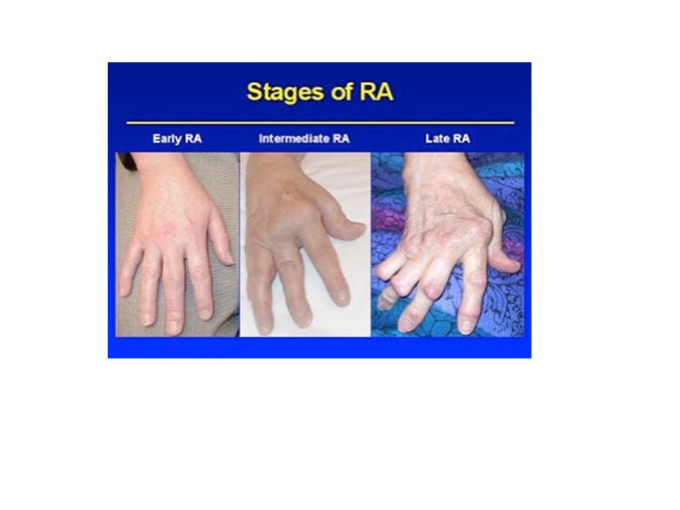
Rheumatoid Arthritis - Autoimmune Disease

30
Juvenile Arthritis

31
Osteoarthritis “Wear & Tear” Arthritis

32
Lyme Arthritis -

33
Lyme Arthritis – Bacterial Infection Borrelia burgdorferi

Similar presentations


 and type of substance.>")


 Function (range of motion)>")