Download presentation
Presentation is loading. Please wait.

1
ENT Clinical methods ICM - 1

2
LEARNING OBJECTIVES To :
Gain understanding of basic components of the ear , nose and throat examination. Learn about the basic tools that are used for the ear , nose and throat examination.

3
Patient encounter Hand hygiene & Introduce yourself to the patient
History taking: Personal data Patient complaint Present history Past history Family history Occupational history

4
Ear symptoms Pain Discharge Hearing loss Tinnitus Vertigo

5
Nasal symptoms Nasal obstruction Nasal discharge Epistaxis Headache
Sneezing Hyposmia / anosmia Nasality problem

6
Oropharyngeal symptoms
Pain / soreness Excess phlegm Dysphagia Lump feeling Dry mouth Bad odor Bad taste Snoring

7
Laryngeal symptoms Change of voice Cough Choking Stridor

8
Clinical examination Explain to the patient what you are going to do
Patient sits in a revolving chair facing doctor who also sits in a revolving chair

9
Basic instruments

10
Source of light Torch Lamp & mirror Fiber-optic light

11
Otoscope

12
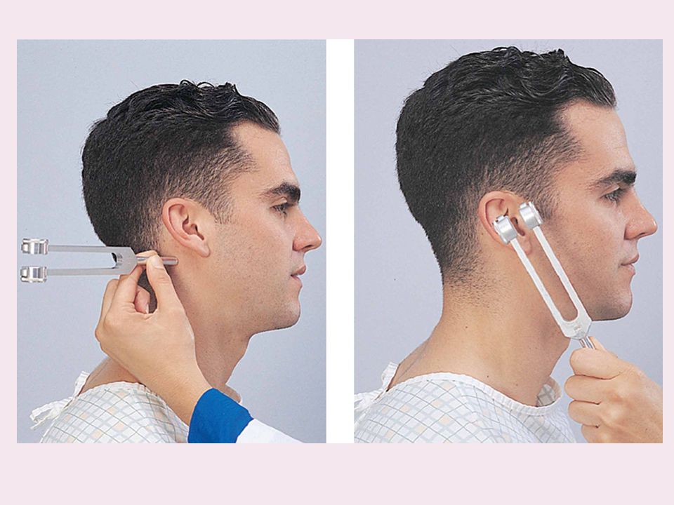
Tuning fork

13
Nasal specula

14
Tongue blades

15
Laryngeal mirror and postnasal mirror

16
Ear examination

17
Inspection Palpation Otoscopy Tests with tuning forks

18
Inspection of the auricle

19
Behind the auricle

20
Mention the finds as per the name of the area of Pinna affected ; e. g
Mention the finds as per the name of the area of Pinna affected ; e.g., mass over lobule , ulcer over tragus etc

21
Preliminary inspection of External Auditory Canal

22
Otoscopy

23
Normal tympanic membrane

24
Rinne’s Test

26
Weber test Wrap the tuning fork strongly on your palm and then press the butt of the instrument on the top of the patient's head in the midline and ask the patient where they hear the sound.

27
Normally, the sound is heard in the center of the head or equally in both ears.

28
Nose

30
Examination of the nose- inspection
Skin lesions Swelling Sinus Bruising Erythema Ulceration

31
Examination of the nose- inspection
Frontal view Side view Basal view

32
Examination of the nose- inspection
Size in relation to the rest of the face Deviation of bridge Dorsum: Convexity (hump) Concavity (saddling) of the dorsum of the nose
 Concavity (saddling) of the dorsum of the nose.")
33
Examination of the nose- inspection
Shape of the tip of nose Pointed bulbous

34
Shape of the columella and nostrils
short/ long columella narrow/wide nostrils

35
Is septum midline or is there any deviation of the nasal septum?

36
Palpation of the nose Press along the bridge of the nose with both index fingers feeling bony skeleton and skin thickness. Press on the tip of the nose with one index finger to elicit tenderness

37
Inspection and palpation
Palpation of sinuses: Frontal sinus: forehead and below eyebrow Maxillary sinus: cheek ( canine fossa) Ethmoids : in the inner canthus area
 Ethmoids : in the inner canthus area.")
38
Nasal patency Ask the patient to exhale in front a shiny surface
(a cold metal tongue depressor) Look for cloudiness due to condensation of water vapor
 Look for cloudiness due to condensation of water vapor.")
39
Anterior rhinoscopy Hold the nasal speculum in the left hand in closed fashion and introduce it gently in skin lined nasal vestibule Avoid contact with the sensitive septum and lateral nasal wall.

40
Anterior rhinoscopy Open the speculum gently in vestibule
Examine floor, medial wall and lateral wall. Look for hyperemia/ discharge/ Little's area septal deviation/ perforation/ hypertrophic turbinates/ polyps Roof of nose needs endoscopy

41
Posterior rhinoscopy

42
oro-pharyngeal Examination
Mouth & oro-pharyngeal Examination

43
Preparation If patient is wearing artificial denture, give him paper towel to get it out so that gingival mucosa can be examined .

44
Inspection-Lips Note their color, any fissures, cracking, ulceration
or any mass.

45
Inspection-gums Note color of gums (normally pink). There may be brown patches in dark races. Look for black lines (in lead poisoning) and red swollen inter-dental papillae in gingivitis.
 and red swollen inter-dental papillae in gingivitis.")
46
Inspection- oral vestibule
Make patient open his mouth. Retract cheek mucosa with tongue depressor Look for color, ulcers, white patches and nodules.

47
Inspection- oral vestibule
Look for opening of parotid duct (opposite crown of second upper molar). Do massaging of the parotid gland and note flow of saliva from Stensen's duct.
. Do massaging of the parotid gland and note flow of saliva from Stensen s duct.")
48
Inspection-Roof of oral cavity
Look for any cleft, oro-nasal fistula, high arched palate, mass bony growth, or ulcer.

49
Inspection-Tongue Ask patient to protrude his tongue out: inspect it for symmetry( a test for cranial nerve XII) Note color and texture of dorsum of tongue

50
Inspection-Tongue Note any white or reddened area, nodules or ulceration Inspect sides, undersurface of tongue

51
Inspection-Floor of the mouth
Midline frenulum Papillae of submandibular duct. Milk the gland and see the salivary flow

52
Palpation Palpate any suspicious lesions especially in smokers and alcoholic individuals above 50 years of age.

53
Pharynx – inspection Ask patient to open mouth without protruding the tongue. Use a tongue depressor to get a good exposure of posterior pharyngeal wall - not so far that you induce gagging.

54
Pharynx- inspection Inspect soft palate, anterior and posterior pillars, medial surface of tonsils. Note congestion, exudates, swelling, ulceration and tonsillar enlargement/atrophy

55
Pharynx –inspection Ask patient to say ah. Soft palate will rise which confirms intactness of vagus nerve.

56
Take-Home Points Good lighting , right tools and Thorough exam is essential .

Similar presentations


 37Palpates auricles bilaterally 38 Otoscopic examination bilaterally.>")

 MBBChir(Cantab) MRCS DOHNS MSc.>")


