Download presentation
Presentation is loading. Please wait.

1
Ears, Nose, Mouth, Throat

2
Ears

3
Summary of any symptom should include PQRSTU
P= provocative or palliative Q= quality or quantity R= region or radiation S= severity scale T= timing (onset, duration, frequency) U= understand client’s perception
 U= understand client’s perception.")
4
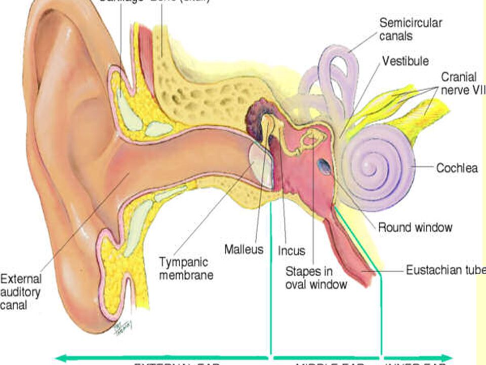
Anatomy The ear is responsible for hearing and balance
Consists of 3 regions External ear Middle ear Inner ear

5
Structure and Function
External Ear – auricle/pinna movable cartilage and skin Mastoid process= important Landmark External Auditory Canal – the opening in the external ear; cul-de-sac 2.5 to 3 cm. Long in adult and ends at the eardrum. Lined with glands that secrete cerumen

9
External Ear 2 types of cerumen
Whites and blacks – wet, sticky, and honey colored Asians and Native Americans – dry and flaky Lubricates & protects Moves to meatus with chewing & talking Outer 1/3 of canal is cartilage, inner 2/3 consists of bone covered with skin

11
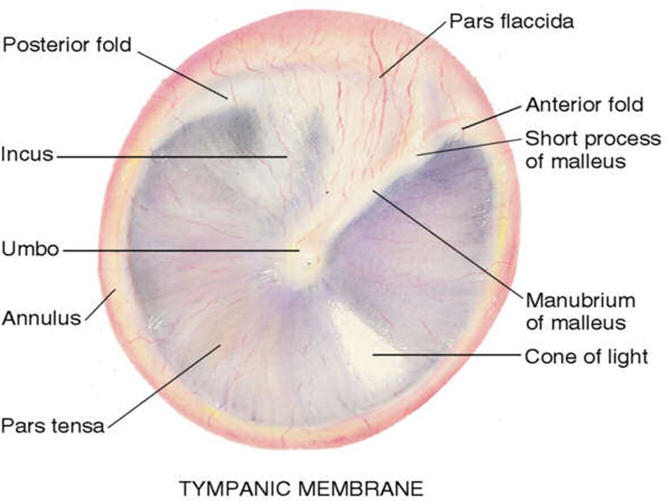
External Ear Tympanic membrane (eardrum) separates external and middle ear. Translucent membrane Pearly, gray color Cone of light reflection when using otoscope Oval and slightly concave shape, pulled in at center by malleus

13
External Ear Malleus (hammer) – one of the middle ear ossicles 3 parts
Umbo, manubrium short process, may show through the drum Lymphatic drainage of the external ear flows into Parotid, mastoid, superficial cervical nodes

14
Middle ear Tiny air–filled cavity in the temporal bone contains:
Auditory ossicles (bones) Malleus Incus Stapes Openings to Outer ear covered by tympanic membrane Inner ear = oval and round windows Eustachian tube connects middle ear to the nasopharnyx for air passage (normally closed, opens with swallowing/yawning)
 Malleus. Incus. Stapes. Openings to. Outer ear covered by tympanic membrane. Inner ear = oval and round windows. Eustachian tube connects middle ear to the nasopharnyx for air passage (normally closed, opens with swallowing/yawning)")
15
Middle ear has 3 functions
Conducts sound vibration from outer ear to inner ear Protects the inner ear by reducing the amplitude of loud sounds Eustachian tube allows equalization of air pressure on each side of the ear drum to avoid rupture ( high altitudes)
")
16
Inner Ear Contains the Bony Labyrinth which holds the sensory organs for hearing and equilibrium Vestibule Semicircular canals Cochlea (contains the central hearing apparatus)
")
17
Function of hearing 3 levels
Peripheral – ear transmits sound and converts its vibrations into electrical impulses that can be analyzed by the brain. The electrical impulses are conducted by the auditory process of cranial nerve VIII (Acoustic) to the brain stem Amplitude=loudness Frequency=pitch
 to the brain stem. Amplitude=loudness. Frequency=pitch.")
18
Sound waves cause the eardrum to vibrate
Vibrations travel via the ossicles thru the oval window, the cochlea and are scattered against the round window The basilar membrane of the cochlea contain the organ of Corti receptor hair cells that translate the vibrations to electric impulses The impulses go to the brainstem via Acoustic nerve (VIII)
")
19
Brain stem – function is binaural interaction – permits identification of sound and locating the direction of a sound in space. The acoustic nerve (Cranial nerve VIII) sends signals from each ear to both sides of the brain stem. Brainstem is sensitive to intensity & timing from the ears depending on head position
 sends signals from each ear to both sides of the brain stem. Brainstem is sensitive to intensity & timing from the ears depending on head position.")
20
Cerebral cortex – interprets the meaning of the sound and begins the appropriate response

21
Pathways of hearing Air conduction (AC)– normal pathway of hearing, the most efficient Bone conduction (BC)– bones of the skull vibrate and transmit vibrations to the inner ear and acoustic nerve
– bones of the skull vibrate and transmit vibrations to the inner ear and acoustic nerve.")
23
Hearing loss Conductive – mechanical dysfunction of the external or middle ear resulting in partial hearing loss (if ↑ amplitude to reach nerve elements in inner ear, person can hear) Causes= impacted cerumen, FB, perforated eardrum, pus/bld in the middle ear, otosclerosis
 Causes= impacted cerumen, FB, perforated eardrum, pus/bld in the middle ear, otosclerosis.")
24
Hearing loss Sensorineural ( perceptive) – pathology of the inner ear, acoustic nerve or auditory areas of the cerebral cortex. ↑ amplitude may not help Causes= Presbycusis, a nerve degeneration due to aging (50yrs) or ototoxic drugs Equilibrium – labyrinth feeds info to the brain about the body’s position in space, inflammation causes vertigo.
 – pathology of the inner ear, acoustic nerve or auditory areas of the cerebral cortex. ↑ amplitude may not help. Causes= Presbycusis, a nerve degeneration due to aging (50yrs) or ototoxic drugs. Equilibrium – labyrinth feeds info to the brain about the body’s position in space, inflammation causes vertigo.")
25
Subjective data Earaches Infections- otitis media Discharge
Hearing loss Environmental noise Tinnitus- ototoxic: ASA, Aminoglycosides (gentamicin) etc. Vertigo Self care behaviors
 etc. Vertigo. Self care behaviors.")
26
Objective data External ear = Inspect and Palpate Size and shape
Skin condition Tenderness- pinna & tragus; mastoid process External auditory meatus- cerumen

27
Inspect using Otoscope
Pull pinna up & back for adult/older child Pinna down for infant & ↓ 3yrs. Maintain hold on pinna until exam is complete. Avoid inner, bony section of canal= sensitive to pain Can angle otoscope towards nose

28
Inspect using Otoscope
External canal Color Swelling Lesions Discharge ; color and odor. Clean or change speculum before examining other ear.

30
Perform the otoscope exam prior to hearing tests.

31
The following slide show a furuncle which is an infected hair follicle

33
Tympanic membrane Color – normal is shiny, translucent, pearl-grey
Characteristics – landmarks; umbro, manubrium, and short process Position – flat, slightly pulled in at the center and flutters when person holds nose and swallows Integrity of membrane – intact? Scarring = dense white patch

34
Hearing tests Begins with the history-Conversational tone
The following tests may indicate the presence of hearing loss but not the degree.

35
Hearing tests Voice– place a finger on the tragus of one ear and while rapidly pushing it in and out of the meatus, place your head 1 –2 feet from your client’s other ear, shield your lips and whisper a 2 syllable word. Repeat on the opposite ear using another word, have the client identify the words (Used to detect high-tone loss)
")
36
Normal Response to Voice test
Correct identification of whispered words bilaterally

37
Tuning fork tests- measure hearing by AC and BC
To activate the tuning fork, hold it by the stem and strike the tines softly on the back of the hand Weber test – used when hearing is reported as better in one ear than other (bone conduction)
")
38
Normal finding for the Weber test is
Tone heard = loud bilaterally If sound lateralizes to one ear it indicates conductive or sensorineural loss.

39
Rinne test – compares bone conduction and air conduction
Normally sound is heard 2X as long by air conduction as by bone conduction Normal response ; positive Rinne Test = AC>BC Bilaterally Sound is heard longer by BC with a conductive loss.

40
Weber test Rinne test

41
Nose, Throat and Mouth

42
Nose First segment of the respiratory system
Warms, moistens and filters inhaled air Sensory organ for smell

43
External parts Bridge Tip Nares Vestibule -nares widen in to vestibule
Columella divides the nares Ala –lateral outside wing of the nose bilaterally Upper 1/3 nose is bone; rest is cartilage

44
Internal Nasal cavity, extends back over the roof of the mouth
Nasal hair, ciliated mucous membrane – red due to ↑ bld supply Septum-divides cavity into 2 passages

45
Internal Superior, middle, inferior turbinates- 3 parallel bony projections on lateral walls of each cavity Meatus- cleft underlying each turbinate. The sinuses drain into the middle, tears from the nasolacrimal duct drain into the inferior

47
Internal Olfactory receptors- in roof of the nasal cavity & upper part of septum. Merge into the olfactory nerve (I) goes to the temporal lobe of the brain
 goes to the temporal lobe of the brain.")
49
Foreign Body

51
Paranasal sinuses- air- filled pockets in the cranium Purpose
↓ wt. of the skull Serve as resonators for sound Provide mucous for the nasal cavity Sinus openings are narrow = susceptible to occlusion resulting in inflammation/sinusitis

52
Frontal sinuses Maxillary sinuses Ethnoid sinuses Sphenoid sinuses Frontal & Maxillary sinuses are accessible to examination

55
Mouth First segment of the digestive system
Airway for the respiratory system ORAL CAVITY Lips Palate Hard Soft Uvula – hangs down from the soft palate

56
Cheeks- side walls of cavity Tongue
Papillae- rough, bumpy elevations on dorsal Frenulum Taste buds Teeth – 32 permanent

58
Salivary glands Parotid- largest of the glands, located in the cheeks, front of the ear. Stenson’s duct opens in buccal mucosa Submandibular- walnut size, beneath the mandible at the angle of the jaw. Wharton’s duct either side of the frenulum Sublingual –smallest, almond shape, under tongue

60
Throat Area behind the mouth & nose
Oropharynx – separated from the mouth by a fold of tissue on each side called anterior tonsillar pillars Tonsils – lymphoid tissue behind pillars

61
Posterior pharyngeal wall located behind the tonsils
Nasopharynx continues from the oropharynx but it is above it and behind the nasal cavity. It holds the adenoids and the eustachian tube openings.

62
Subjective data Nose Discharge Frequent colds Sinus pain Trauma
Epistaxis Allergies Altered smell

63
Subjective data Mouth and Nose
Sores, lesions Sore throat Bleeding gums Toothache Hoarseness Dysphagia Altered taste

64
Smoking Alcohol intake Self care behaviors

65
Objective behavior Nose – Inspect and palpate INSPECT for:
Symmetry, deformity Inflammation Skin lesions Color If injury – palpate gently

66
Test for Patency Test for Sense of Smell – Cranial nerve I (olfactory)
")
67
Inspect nasal cavity/ septum
Deviated septum? Can see middle & inferior turbinates

68
Inspect and palpate Paranasal Sinuses
Press thumbs over frontal & maxillary sinuses Transillumination for sinus inflammation Frontal & Maxillary sinuses Darken room

71
Mouth - Inspect Use gloves, tongue depressor, light Lips Teeth Gums
Buccal mucosa –Stenson’s duct (parotid) Palate
 Palate.")
74
Throat - Inspect Tonsils Posterior pharyngeal wall
Grade size 1+ visible …………….2+ ½ way b/t tonsillar pillars and uvula …………… touching the uvula …………… touching each other Posterior pharyngeal wall Gag reflex cranial nerves IX = glossopharyngeal and X = Vagus Cranial nerve XII = hypoglossal- stick out tongue Halitosis – Due to ????

Similar presentations