Download presentation
Presentation is loading. Please wait.

1
MUSCULI MASTICATORII Muscles of mastication

2
Movement of temporomandibular joint
4 pairs of muscles attached to the mandible Movement of temporomandibular joint Arise from the bones of the neurocranium Pennate structure Fasciae Blood supply: maxillary artery Nerve supply: mandibular nerve

3
Masseter muscle

4
Thick, quadrilateral muscle
Superficial and deep portion: The Superficial Portion Origo: maxillary process of zygomatic bone and the anterior ⅔ of the lower border of the zygomatic arch Fibers pass downward and backward Insertion: tuberositas masseterica (the angle and lower ½ of the lateral surface of the ramus of the mandible)
")
5
The Deep Portion Smaller and more muscular in texture Origo: posterior ⅓ of lower border and the whole of the medial surface of the zygomatic arch Fibers pass downward and forward Insertion: the upper ½ of the lateral surface of the ramus mandible

6
Functional organization of the human masseter muscle
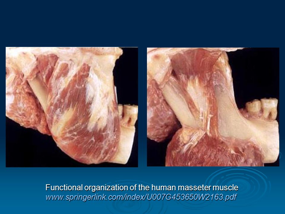
7
Function Bilateral contraction: The superficial part: elevation
propulsion The deep portion: Unilateral contraction: lateropulsion

8
The Architecture The typical pennate structure - zones of muscular and aponeurotic attachments The pennate structure allows spread the infection (submasseteric abscess) The differential activity of the muscular planes during masticatory function makes it necessary to respect the anatomic and functional individuality in the diagnosis and treatment of dysfunctional disorders of the masticatory apparatus
 The differential activity of the muscular planes during masticatory function makes it necessary to respect the anatomic and functional individuality in the diagnosis and treatment of dysfunctional disorders of the masticatory apparatus.")
9
The Masseteric Fascia Firmly connected with the muscle
From arcus zygomaticus to basis mandibulae Dorsally merge with fascia parotis (fascia parotideomasseterica)
")
10
Palpation The Superficial Portion The Deep Portion Palpace:
lékař stojí za pacientem a 4 prsty palpuje sval – pacient stiskne čelisti k sobě několikrát po sobě. Muscle palpation is a very important step in the diagnosis of TMD and myofascial pain syndromes. By means of mechanical stimuli caused by digital pressure, nociceptive neurons located in the muscular and myofascial structures are stimulated to detect and transmit pain messages to the central nerve system. The graduation of patient's response to palpation allows evaluating the severity of pain and is used to measure the efficacy of a given treatment modality in follow-up visits. Palpation should be performed with a pressure of 1.5 Kg, which is strong enough to elicit pain message in symptomatic patients, and mild enough to not cause pain in asymptomatic control subjects2,15,40. Palpation should be done bilaterally, in a relaxed position, with the tip of the finger or by pincer palpation, when no underline bone support is present. Yet, during the examination, the patient should be seated facing the orthodontist in such a way that the clinician can observe the patient's reactions. Obr. dole: vpravo: extraorální bilaterální palpace. Vlevo: extra a intraorální unilaterální palpace. The Superficial Portion The Deep Portion

11
Temporalis muscle

12
Broad, triangular muscle
Localized in the temporal fossa Origo: - the whole of the temporal fossa - the deep surface of the temporal fascia Fibers converge as they descend, and end in a tendon, which passes deep to the zygomatic arch Insertion: apex and medial surface of the coronoid process (crista temporalis) and the anterior border of the ramus of the mandible to retromolar triangle
 and the anterior border of the ramus of the mandible to retromolar triangle.")
13
The temporalis muscle consists of three parts: superficial zygomatic
Detailed description The temporalis muscle consists of three parts: superficial zygomatic deep portion The human temporalis muscle: Superficial, deep, and zygomatic ... onlinelibrary.wiley.com › ... › Journal Home › Vol 22 Issue 6

14
The Superficial Portion
Origo: temporal aponeurosis, temporal line Insertion: the coronoid process of the mandible

15
The Zygomatic Portion Origo: superior and medial zygoma and zygomatic arch Insertion: coronoid process Fuse with tendon of the superficial part of the temporalis muscle and with the deep masseter portion. Rostrally interdigitated with the portion of the deep temporalis part

16
The Deep Portion Origo: bony surface of the frontal, sphenoid, parietal and temporal bones Insertion: coronoid process and the ramus of the mandible, just caudal to the last molar (retromolar triangle) Fibers from deep part interdigitated with the buccinator and the superior pharyngeal constrictor
 Fibers from deep part interdigitated with the buccinator and the superior pharyngeal constrictor.")
18
Function Elevation (anterior part) Retropulsion (posterior part)
 Retropulsion (posterior part)")
19
The Temporal Fascia Covers the temporal muscle
Origo: superior temporal line Insertion: zygomatic arch Has two layers: 1. superficial inserted into the lateral border of the arch 2. deep inserted into the medial border of the arch Between these layers is a small quantity of fat and attachment of the superficial fibres of the temporal muscle

20
Skull

21
Palpation The Anterior Part The Posterior Part Palpace:
lékař stojí za pacientem a 4 prsty palpuje sval – pacient stiskne čelisti k sobě několikrát po sobě. Muscle palpation is a very important step in the diagnosis of TMD and myofascial pain syndromes. By means of mechanical stimuli caused by digital pressure, nociceptive neurons located in the muscular and myofascial structures are stimulated to detect and transmit pain messages to the central nerve system. The graduation of patient's response to palpation allows evaluating the severity of pain and is used to measure the efficacy of a given treatment modality in follow-up visits. Palpation should be performed with a pressure of 1.5 Kg, which is strong enough to elicit pain message in symptomatic patients, and mild enough to not cause pain in asymptomatic control subjects2,15,40. Palpation should be done bilaterally, in a relaxed position, with the tip of the finger or by pincer palpation, when no underline bone support is present. Yet, during the examination, the patient should be seated facing the orthodontist in such a way that the clinician can observe the patient's reactions. Obr. dole: vpravo: extraorální bilaterální palpace. Vlevo: extra a intraorální unilaterální palpace. The Anterior Part The Posterior Part

22
Lateral (Externus) Pterygoid Muscle
 Pterygoid Muscle")
23
Short, thick muscle, conical in form
Located in the infratemporal fossa Two heads: upper and lower The Upper/Sphenoid Head: Origo: infratemporal facies of the great wing of the sphenoid bone The Lower/Pterygoid Head: Origo: lateral surface of the lateral pterygoid plate

24
Fibres pass horizontally backward and lateralward
Insertion: fovea pterygoidea (depression in front of the neck of the condyle) - joint capsule and articular disc of the TMJ (front margin)
 - joint capsule and articular disc of the TMJ (front margin)")
25
Alternating tendinous sheets characteristic of a penniform structure

26
Function Bilateral contraction: propulsion
Unilateral contraction: lateropulsion

27
Medial (Internus) Pterygoid Muscle
 Pterygoid Muscle")
28
Thick, quadrilateral Located in the infratemporal fossa Two heads: Origo: - tuberosity of the maxilla (lateralis) - medial surface of the lateral pterygoid plate (medialis) Fibers pass downward, lateralward and backward Insertion: tuberositas pterygoidea (lower and back part of the medial surface of the ramus and angle of the mandible)
 Fibers pass downward, lateralward and backward. Insertion: tuberositas pterygoidea (lower and back part of the medial surface of the ramus and angle of the mandible)")
29
1 - lateral part 2 - medial part Inner surface of the ramus mandibulae

30
7 musculo-aponeurotic layers are making up a penniform structure
The Architecture 7 musculo-aponeurotic layers are making up a penniform structure 7 5 3 1 6 4 2

31
Function Bilateral contraction: propulsion elevation
Unilateral contraction: lateropulsion

32
The Interpterygoid Fascia
Between the medial and lateral pterygoid muscles Origo: the base of the skull Insertion: inner surface of the mandible The border is reinforced by sphenomandibular ligament Contain nerves and vessels Allow spread of infection

33
pteryg. lat. pteryg. med.

Similar presentations


>")
 Process = Prominence or extension.>")


