Download presentation
Presentation is loading. Please wait.

1
IN THE NAME OF GOD THANKS

2
FOLLOW-UP

3
It is assumed that subsequent to traumatic injury, the conduction capability of the nerve endings and/or sensory receptors is sufficiently derange to inhibit the nerve impulse from an electric or thermal stimulus. This makes the traumatized tooth vulnerable to false-negative readings from such tests.

4
Teeth that give a response at the initial examination cannot be assumed to be healthy and continue to give a response overtime. Teeth that yield no response cannot be assumed to have necrotic pulps, because they may give a response at later follow-up visits. It has been demonstrated that it may take as long as 9 months for normal blood flow to return to the coronal pulp of a traumatized fully formed tooth. As circulation is restored, the responsiveness to pulp tests returns.

5
The transition from no response to a response at a subsequent test may be considered a sign of a healing pulp. The repetitious finding of responses may be taken as a sign of a healthy pulp. The transition from a response to no response may be taken as an indication that the pulp is probably under-going degeneration, and therefore an intervention in the form of endodontic treatment might be indicated. The persistence of no response would suggest that the pulp has been irreversibly damaged, but even this is not absolute.

6
Thermal and electric pulp tests of all anterior teeth (canine to canine) of the maxillary and mandibular jaws should be performed at the time of the initial examination and carefully recorded to establish a baseline for comparison with subsequent repeated tests in later months. These tests should be repeated at 3 weeks, at 3, 6, and 12 months, and at yearly intervals following the trauma.

7
Dichlorodifluoromethane spray is a very inexpensive alternative to CO2 snow; how cold it is will give much more reliable responses than water ice.

8
The electric pulp test relies on electric impulses directly stimulating the nerves of the pulp. These tests have limited value in young teeth but are useful incases where the dentinal tubules are closed and do not allow dentinal fluid to flow in them. This situation is typical of teeth in elderly patients or in traumatized teeth that are undergoing premature sclerosis.

9
Attempts have been made to use LDF technology for pulp vitality diagnosis in traumatized teeth, because this would provide a more accurate reading of the vitality status of the pulp. Presently the cost of a laser Doppler flowmetry machine limits its use in private dental offices; these are used primarily in hospitals and teaching institutions.

11
RADIOGRAGHY

12
One radiograph will be insufficient in most dental trauma cases
One radiograph will be insufficient in most dental trauma cases. take at least four different radiographs for almost every injury. Those four include a direct 90-degree on the axis of the tooth, two with different vertical angulations, and one occlusal film .

13
An occlusal film of a luxation injury of central incisors
An occlusal film of a luxation injury of central incisors.Both were diagnosed to be laterally luxated with apical translocation.Note that the left central is completely obliterated and has a history of being luxated some years prior.

14
only resorptive defects on the mesial or distal aspects of the root can be predictably detected after some time,but the facial and palatal or lingual aspects are much harder to see. To overcome these difficulties, it is essential to take as many different horizontal angled radiographs as feasible incases of suspected root resorption. One should also carefully evaluate the adjacent bone .

15
Angled radiographs to show internal resorption
Angled radiographs to show internal resorption. radiographs from two different horizontal projections depict

16
In instances of soft-tissue laceration, it is advisable to radio-graph the injured area prior to suturing to be sure that no foreign objects have been embedded. A soft-tissue radiograph with a normal-sized film briefly exposed at reduced kilovoltage should reveal the presence of many foreign substances, including tooth fragments.

17
A B

18
In reviewing the films of traumatized teeth, special attention should be directed to the dimension of the root canal space, the degree of apical closure of the root, and the relationship of root fractures to the alveolar crest. Whereas conventional periapical films are generally useful, an occlusal or Panorex film can supplement them when the clinician is looking for bone fractures, dislocation of teeth, or the presence of foreign objects.

20
Continued root development after partial pulpotomy
Continued root development after partial pulpotomy. A, radiograph of immature tooth with a complicated crown fracture. B, At the time of placement of calcium hydroxide after partial pulpotomy. C, Follow-up radiograph confirming that the pulp maintained vitality and the root continued to develop.

21
Successful pulpotomy followed by a pulpectomy at 18 months

22
A and B, A complicated crown fracture involving the maxillary left central incisor of an 8-year-old child. C, A periapical radiograph revealed incomplete root formation. D, Postoperative film after a calcium hydroxide pulpotomy and placement of a stainless steel crown. E, A 2-year recallfilm demonstrates complete root formation. . G, Postoperative film taken after nonsurgical root canal treatment to facilitate placement of a post and core.

23
A, A radiograph of a mandibular left central incisor with a history of lateral luxation and a lateral incisor that was avulsed and demonstrates inflammatory resorption. B, Clinical view of a sinus tract that developed as a result of pulp necrosis.

25
Root Resorption

26
Types of resorptions: Internal root resorption
External root resorption

27
Internal resorption Internal resorption is characterized by resorption of the Internal aspect of the root by multinucleated giant cells adjacent to granulation tissue in the pulp.

28
Clinical manifestations :
Asymptomatic First recognized clinically through routine radiograghs. Positive or negative response to pulp sensivity testing. Pink spot.

29
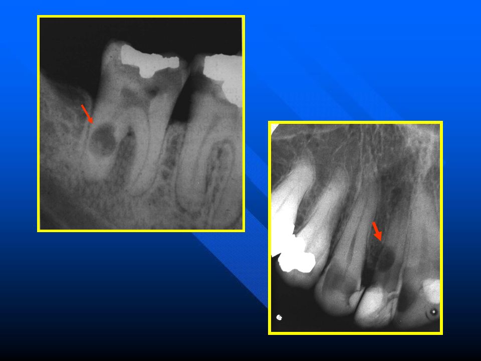
Radiographic appearance:
Fairly uniform radiolucent enlargement of the pulp canal. Original outline of the root canal is distorted. The adjacent bone is not affected with internal root resorption.

32
Treatment: root canal therapy

34
External root resorption
Surface root resorption Replacement root resorption Apical root resorption (Apical periodontitis) Inflammatory root resorption (Periradicular periodontitis) Cervical root resorption
 Inflammatory root resorption. (Periradicular periodontitis) Cervical root resorption.")
35
Surface root resorption
Caused by an injury (alone) to the external root Surface. Localized injury: spontaneous healing with cementum within 14 days. The pulp is not involved.
 to the external root Surface. Localized injury: spontaneous healing with cementum within 14 days. The pulp is not involved.")
36
Asymptomatic No visible in radiograghs

38
Replacement root resorption
Diffuse injury: Healing by osseous Replacement . Bone comes into contact with the root without an intermediate attachment apparatus (Dentoalveolar ankylosis).
.")
39
Clinical manifestations:
Lack of mobility. Metallic sound to percussion. Infraocclusion. Ultimately the tooth is lost because of loss of root support.

40
Radiographic appearance:
Lamina dura is lost between root and bone. Moth-eaten appearance Treatment: Treatment strategies involve minimizing the initial inflammation in response to the injury.

42
2 years after injury 4 years later

44
Apical root resorption
Pulp space infection in conjunction with damage to the external root surface without loss of cementum.

45
Clinical manifestations:
Asymptomatic First recognized clinically through routine radiograghs.

46
Radiographic appearance:
Resorption is observed as radiolucent area at the apex of root. Resorption is observed as radiolucent area of adjacent bone.

48
3 month later 6 month later 6

49
Inflammatory root resorption
Pulp space infection in conjunction with serious damage to the external root surface with loss of cementum results in periradicular root and bone resorption .

50
Radiographic appearance:
Resorption is observed as progressive radiolucent areas of the root and adjacent bone.

53
Treatment: Minimizing the subsequent inflammation were the focus of the emergency visit. Clinician’s attention to pulp space infection should ideally be 7-10 days after the injury. Elimination of the pulp infection.

54
3 weeks after injury 2 years later

57
Cervical root resorption
A progressive root resorption of inflammatory origin usually occurring immediately below the epithelial attachment of the tooth. It appears to be a delayed reaction after an injury. The pulp plays no role in cervical root resorption and is normal.

58
Clinical manifestations:
Asymptomatic. Often recognized clinically through routine radiograghs. The sensivity test result is within normal limits. Pink spot. Bone loss.

59
Radiographic appearance:
Initially,radiolucency near the attachment level would be seen.If the process is long standing and extensive ,the radiolucent area extend a considerable way in a coronal and apical direction.

60
Outline of the canal can be seen through the radiolucency of this resorptive defect.
Resorption is observed as radiolucent area of adjacent bone.

67
Thank you

Similar presentations


>")


. All rights reserved. Endodontics Chapter 54 Copyright 2003, Elsevier Science (USA). All rights reserved. No part.>")


