Download presentation
Presentation is loading. Please wait.

1
Knee Outline

2
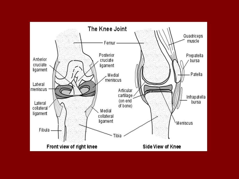
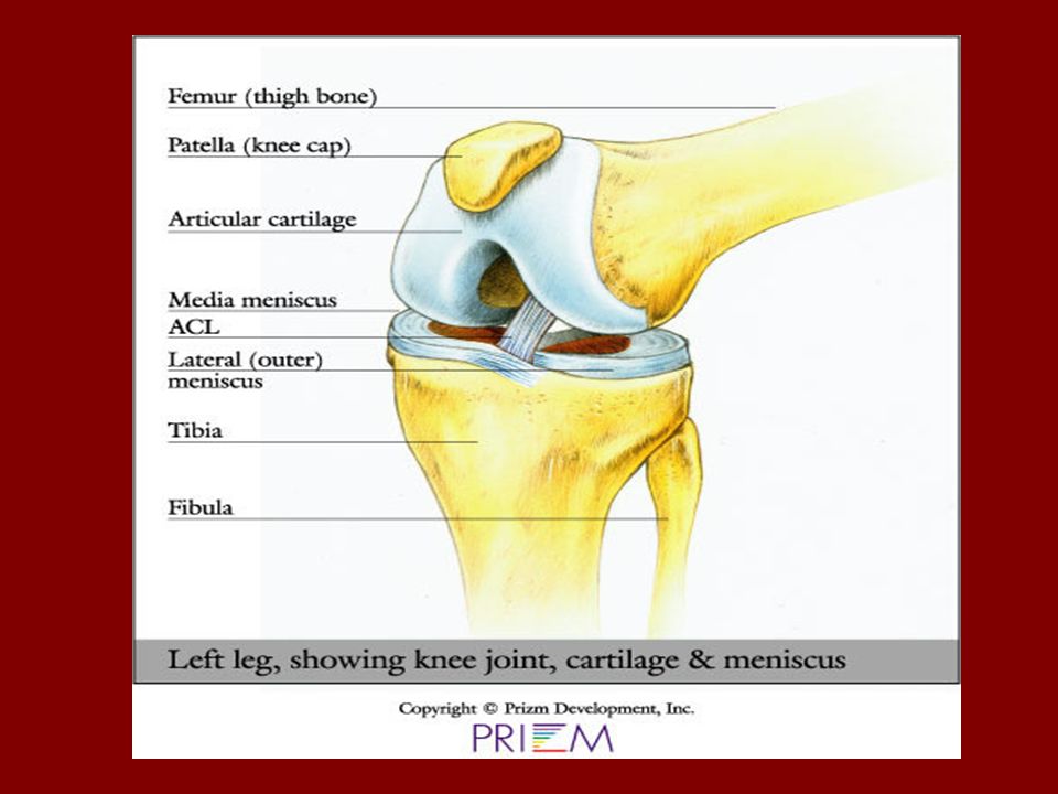
Bones of the Knee Femur- thighbone, largest and hardest bone in body
Tibia- largest lower leg bone. Distal end forms medial malleolus Fibula- smaller lower leg bone. Non-weight bearing. Distal end forms lateral malleolus. Patella- kneecap. Largest sesmoid bone.

3
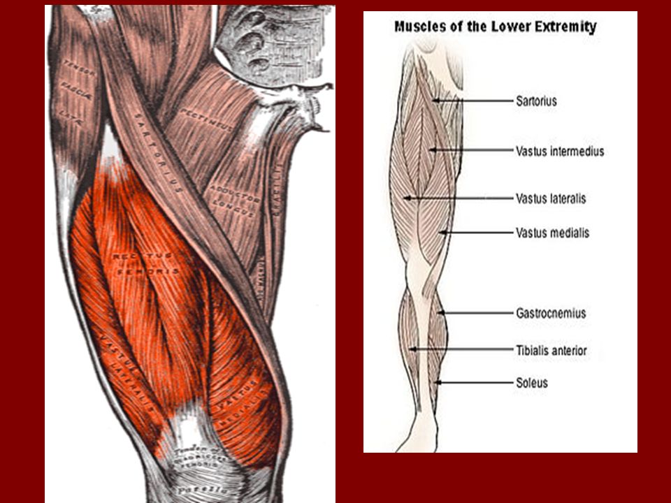
Anterior Muscles of the Knee Joint
Quadriceps Group ( 4 )- “ Body’s own Natural Knee Brace” Rectus Femoris- Longest and most anterior. Knee extension and Hip flexion. Vastus Intermedius- Deepest Quad. Muscle. Underneath Rectus Femoris, knee extension. Vastus Lateralis- On the lateral side of the thigh. Knee extension. Vastus Medialis- Most medial on thigh. Forms the bulge above medial side of the knee. Knee extension.
- Body’s own Natural Knee Brace Rectus Femoris- Longest and most anterior. Knee extension and Hip flexion. Vastus Intermedius- Deepest Quad. Muscle. Underneath Rectus Femoris, knee extension. Vastus Lateralis- On the lateral side of the thigh. Knee extension. Vastus Medialis- Most medial on thigh. Forms the bulge above medial side of the knee. Knee extension.")
5
Anterior Muscles of the Knee Joint cont.
Sartorius-Located on the medial aspect of the thigh, from center of thigh to the medial side of the knee. Gracilis- “ Groin Muscle” On the medial aspect of the thigh. Adduction of hip & flexion of knee

7
Posterior Muscles of the Knee Joint
Hamstrings Group ( 3 ) located posteriorly Semitendinosus- On the medial aspect of the thigh. Knee flexion & medial rotation. Biceps Femoris- Lies lateral to Semitendinosus. Knee flexion & lateral rotation. Semimembranosus- On top of # 1 & # 2. Knee flexion & medial rotation.
 located posteriorly. Semitendinosus- On the medial aspect of the thigh. Knee flexion & medial rotation. Biceps Femoris- Lies lateral to Semitendinosus. Knee flexion & lateral rotation. Semimembranosus- On top of # 1 & # 2. Knee flexion & medial rotation.")
8
Semitendinosus Biceps Femoris Semimembranosus

9
Distal Muscles of the Knee Joint
Popliteus-On the back of the knee from upper lateral to lower medial. Knee flexion & medial rotation of flexed leg. Gastrocnemius- “Calf Muscle” Split in two sections. Knee flexion. Plantaris- From upper lateral knee to medial malleolus. Ankle plantar flexion, & Knee flexion.

11
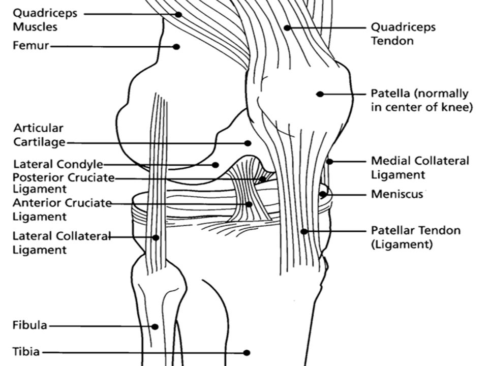
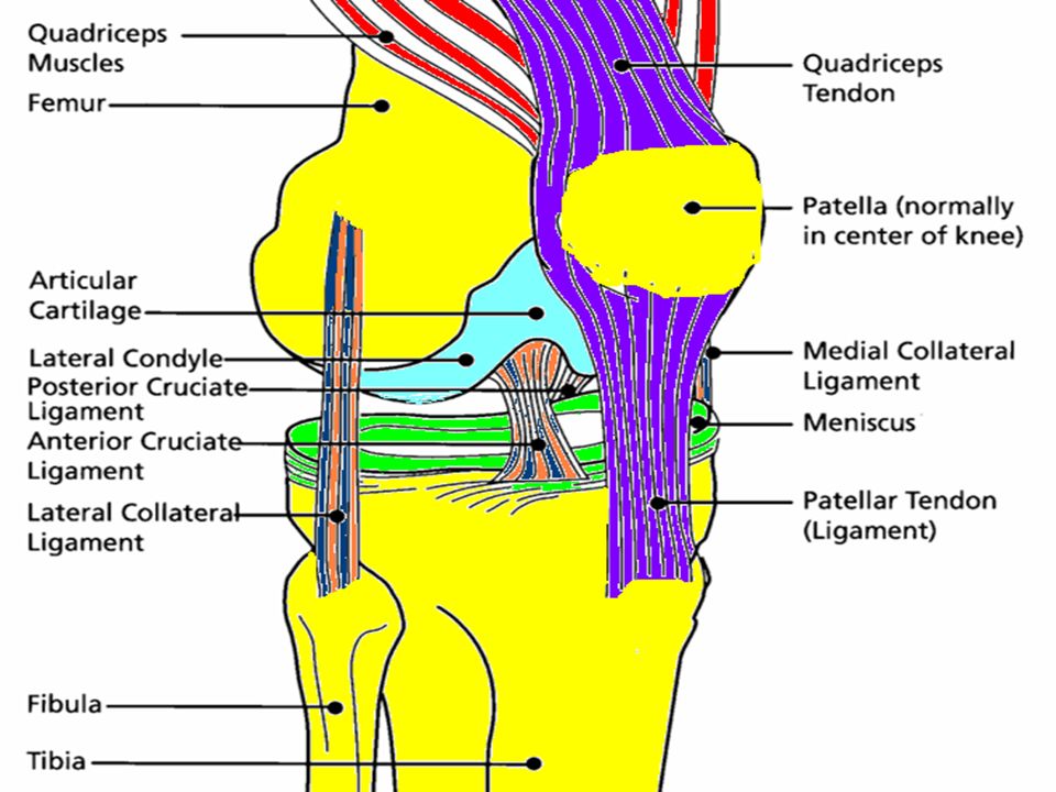
Side Ligaments of the Knee
Medial Collateral Ligament ( MCL )-on medial side of the knee, broad flat ligament. Secures femur to the tibia. Most injured ligament. Lateral Collateral Ligament ( LCL )- on lateral side. Cord-like shaped ligament. Secures femur to Fibula.
-on medial side of the knee, broad flat ligament. Secures femur to the tibia. Most injured ligament. Lateral Collateral Ligament ( LCL )- on lateral side. Cord-like shaped ligament. Secures femur to Fibula.")
13
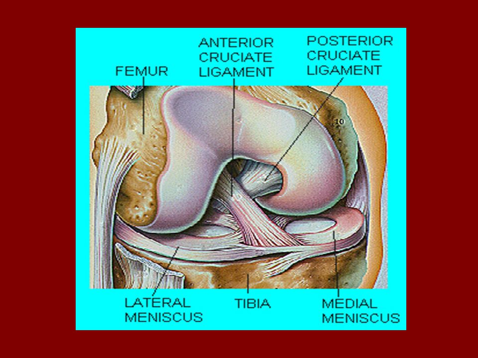
Inside Ligaments of the Knee
Anterior Cruciate Ligament ( ACL )- attaches on the front ( anterior ) aspect of the tibia, runs up and back to the posterior aspect of the femur. Controls anterior movement of the tibia under the femur. Posterior Cruciate Ligament ( PCL )- attaches to the front ( anterior) aspect of the femur, runs down and back to the posterior aspect of the tibia. Controls posterior movement of the tibia under the femur.
- attaches on the front ( anterior ) aspect of the tibia, runs up and back to the posterior aspect of the femur. Controls anterior movement of the tibia under the femur. Posterior Cruciate Ligament ( PCL )- attaches to the front ( anterior) aspect of the femur, runs down and back to the posterior aspect of the tibia. Controls posterior movement of the tibia under the femur.")
16
Cartilage of the Knee a.k.a. menisci
Medial Meniscus- forms a ½ moon shaped cushion base for the medial femoral condyle. The head of the femur. Lateral Meniscus- forms almost oval shaped cushion base for the lateral femoral condyle.

19
R. O. M. of the Knee Flexion- bending the knee
Extension- straightening the leg

Similar presentations


 articulating with 2 concave surfaces (tibia) Poor bony stability Stability increased.>")

 Kelly Heikkila (0305975) Allison Pruys (0310660)>")

 Tibia ◦ Main weight lower leg bone Medial malleolus comes off of.>")


>")


>")