Download presentation
Presentation is loading. Please wait.

1
Neuroscience and Behavior AP Psychology

2
The Brain Made up of neurons and glial cells. Glial cells support neural cells. Some scientists divide the brain up into three parts. Hindbrain Midbrain Forebrain

3
Techniques for Studying the Brain Lesion tissue destruction a brain lesion can be caused naturally or experimentally Brain tumors also lesion brain tissue

4
Techniques for Studying the Brain: Less Invasive Ways Electroencephalo gram (EEG) an amplified recording of the waves of electrical activity that sweep across the brain’s surface these waves are measured by electrodes placed on the scalp
 an amplified recording of the waves of electrical activity that sweep across the brain’s surface these waves are measured by electrodes placed on the scalp")
5
Less Invasive Ways to Study the Brain CT (computed tomography) Scan a series of x-ray photographs taken from different angles and combined by computer into a composite representation of a slice through the body; also called CAT scan PET (positron emission tomography) Scan a visual display of brain activity that detects where a radioactive form of glucose goes while the brain performs a given task MRI (magnetic resonance imaging) a technique that uses magnetic fields and radio waves to produce computer-generated images that distinguish among different types of soft tissue; allows us to see structures within the brain
 Scan a series of x-ray photographs taken from different angles and combined by computer into a composite representation of a slice through the body; also called CAT scan PET (positron emission tomography) Scan a visual display of brain activity that detects where a radioactive form of glucose goes while the brain performs a given task MRI (magnetic resonance imaging) a technique that uses magnetic fields and radio waves to produce computer-generated images that distinguish among different types of soft tissue; allows us to see structures within the brain")
6
Images Pet Scan of Brain MRI of brain (midsagittal) CT scan- brain tumor
 CT scan- brain tumor")
7
Brain Structures Brainstem the oldest part and central core of the brain, beginning where the spinal cord swells as it enters the skull responsible for automatic survival functions Medulla [muh-DUL- uh] base of the brainstem controls heartbeat and breathing
![Brain Structures Brainstem the oldest part and central core of the brain, beginning where the spinal cord swells as it enters the skull responsible for automatic survival functions Medulla [muh-DUL- uh] base of the brainstem controls heartbeat and breathing](http://images.slideplayer.com/26/8708562/slides/slide_7.jpg "Brain Structures Brainstem the oldest part and central core of the brain, beginning where the spinal cord swells as it enters the skull responsible for automatic survival functions Medulla [muh-DUL- uh] base of the brainstem controls heartbeat and breathing")
8
The Brainstem Reticular Formation a nerve network in the brainstem that plays an important role in controlling arousal and the ability to focus attention Thalamus the brain’s sensory switchboard, located on top of the brainstem Receives sensory information in the cortex and transmits replies to the cerebellum and medulla

9
Parts of the Brain Cerebellum the “little brain” attached to the rear of the brainstem it helps coordinate voluntary movement and balance

10
Limbic System a doughnut-shaped system of neural structures at the border of the brainstem and cerebral hemispheres associated with emotions such as fear and aggression and drives such as those for food and sex includes the hippocampus, amygdala, and hypothalamus. Amygdala two almond-shaped neural clusters that are components of the limbic system Emotional control center Memory Hippocampus-involved in memory processing

11
Brain Structures Hypothalamus neural structure lying below (hypo) the thalamus; directs several maintenance activities Hunger (eating) Thirst (drinking) body temperature Sexual arousal (libido) helps govern the endocrine system via the pituitary gland is linked to emotion
 the thalamus; directs several maintenance activities Hunger (eating) Thirst (drinking) body temperature Sexual arousal (libido) helps govern the endocrine system via the pituitary gland is linked to emotion")
12
Pituitary gland Hypothalamus

13
Brain Structures: The Cerebral Cortex Top layer of our brain. Contains wrinkles called fissures. The fissures increase surface area of our brain. the body’s ultimate control and information processing center

14
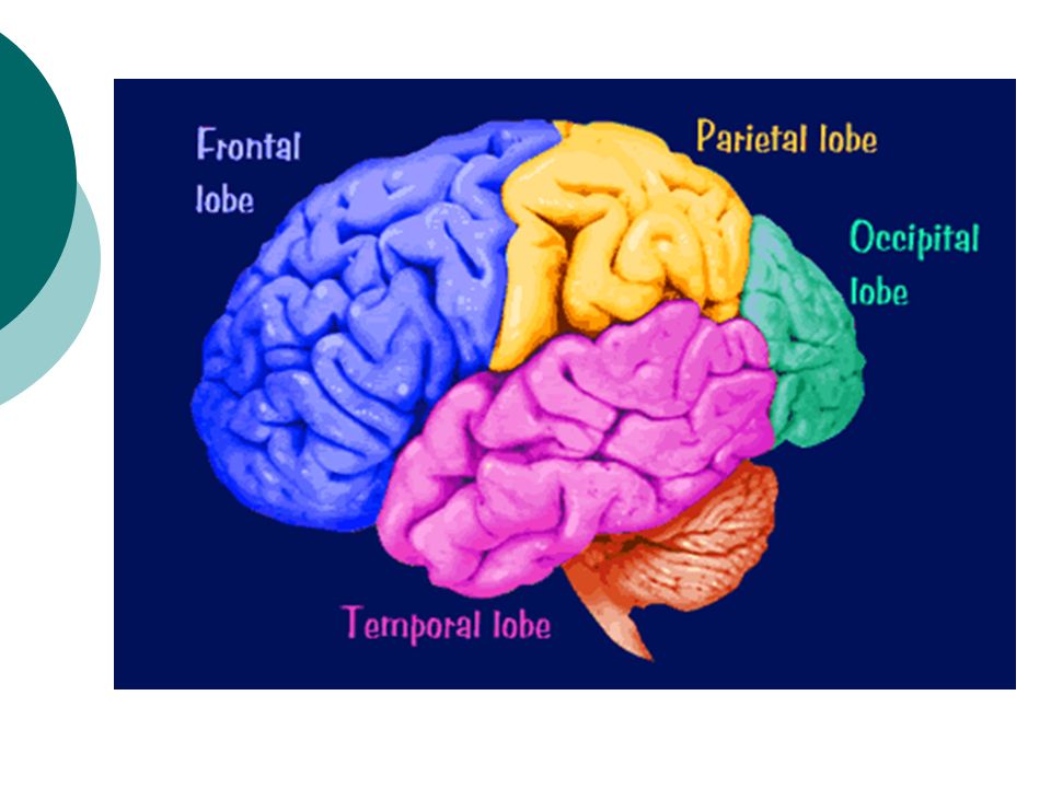
The Cerebral Cortex Divided into eight lobes, four in each hemisphere (frontal, parietal, occipital and temporal). Any area not dealing with our senses or muscle movements are called association areas.

15
The Cerebral Cortex Frontal Lobes involved in speaking and muscle movements and in making plans and judgments Contains Broca’s area (speech production) Parietal Lobes include the sensory cortex Occipital Lobes include the visual areas, which receive visual information from the opposite visual field Right half of each retina goes to left occipital lobe and vice versa Temporal Lobes include the auditory areas (process sound) Contains Wernicke’s area (comprehension of speech/language development)
 Parietal Lobes include the sensory cortex Occipital Lobes include the visual areas, which receive visual information from the opposite visual field Right half of each retina goes to left occipital lobe and vice versa Temporal Lobes include the auditory areas (process sound) Contains Wernicke’s area (comprehension of speech/language development)")
16
The Cerebral Cortex

18
Motor Cortex area at the rear of the frontal lobes that controls voluntary movements Sensory Cortex area at the front of the parietal lobes that registers and processes body sensations Aphasia (Disorders) impairment of language, usually caused by left hemisphere damage either to Broca’s area (impairing speaking) or to Wernicke’s area (impairing understanding)
 impairment of language, usually caused by left hemisphere damage either to Broca’s area (impairing speaking) or to Wernicke’s area (impairing understanding)")
19
The Cerebral Cortex

20
Association Areas More intelligent animals have increased “uncommitted” or association areas of the cortex

21
Specialization and Integration

22
Brain Plasticity The ability for our brains to form new connections after the neurons are damaged. the brain’s capacity for modification, as evident in brain reorganization following damage The younger you are, the more plastic your brain is.

23
Announcement! Your test will be Friday & Monday Multiple Choice –Friday Essay-Monday I will check reading notes Thursday Brain Models/Mobiles will be due Thursday!

24
Our Divided Brain Corpus Callosum large band of neural fibers connects the two brain hemispheres carries messages between the hemispheres Corpus callosum

25
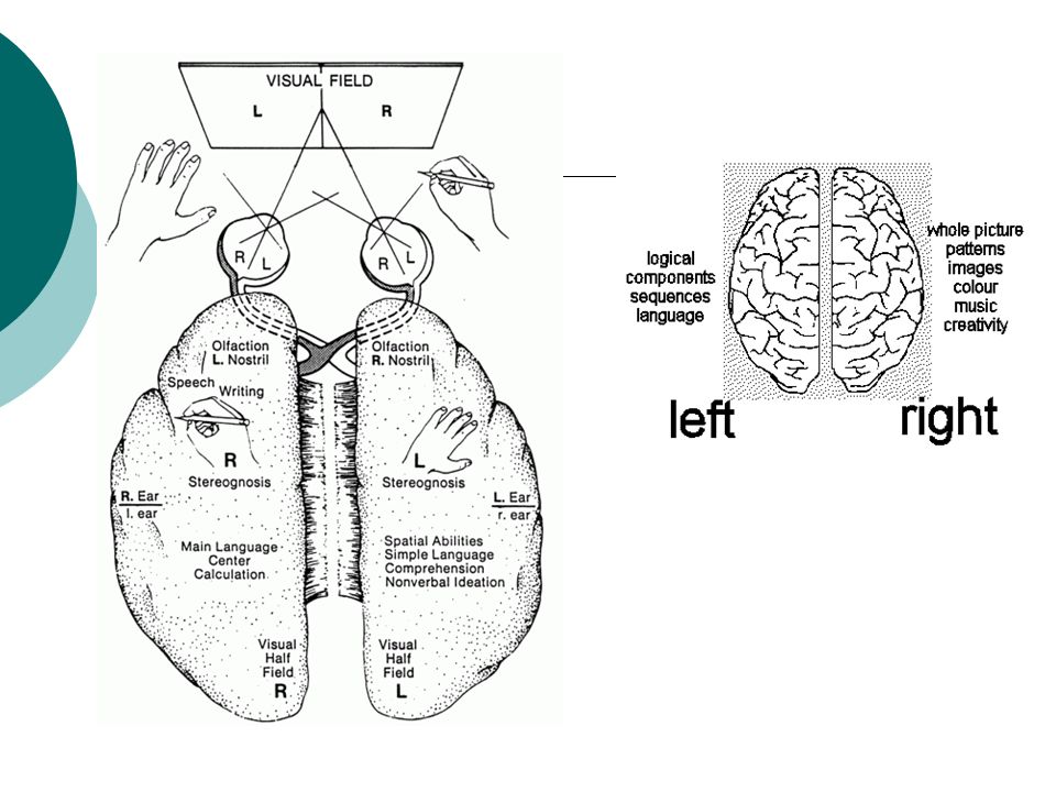
Our Divided Brain Images from the left visual field go to the brain’s right hemisphere Images from the right visual field go to the brain’s left hemisphere. Left hemisphere controls the right hand & vice versa The left hemisphere is dominant for language Broca’s, Wernicke’s, angular gyrus

27
Our Divided Brain Left Hemisphere Controls speech muscles Comprehension/ understanding of speech & writing Regulation of positive emotions Memory of words and numbers Spontaneous speaking and writing Logic & analytic thought Science and math Right Hemisphere Regulation of negative emotions Responses to simple commands Memory for shapes and music & creativity Interpreting spatial relationships & visual images Recognition of faces

28
Split Brain a condition in which the two hemispheres of the brain are isolated by cutting the connecting fibers (mainly those of the corpus callosum) between them Procedure done to treat severe epileptic seizures
 between them Procedure done to treat severe epileptic seizures")
29
So what happens to a “split brain”? When a split brain patient is asked what he sees, the left hemisphere sees the ring on the right side of the screen and can verbally say “ring”. The right hemisphere sees the left side of the screen, but cannot verbalize (say) what is seen (key). However, the patient can pick up the correct object using the left hand.
 what is seen (key). However, the patient can pick up the correct object using the left hand..")
30
Genetics Every human cell contains 46 chromosomes (23 pairs). Made up of genes. Made up of deoxyribonucleic acid- DNA. Made up of nucleotides. Your DNA (chromosomes) is found in the nucleus of every cell in your body. Your genotype is the genetic pattern that makes you different from anyone else Ex. Brown eyes- Bb or BB; blue eyes-bb Your phenotype is your observable characteristics Ex. You have brown eyes, blue eyes, tall, short Sex chromosomes (23 rd pair) XX-female; XY male Father determines sex of child because he can donate an X or Y.
 is found in the nucleus of every cell in your body. Your genotype is the genetic pattern that makes you different from anyone else Ex. Brown eyes- Bb or BB; blue eyes-bb Your phenotype is your observable characteristics Ex. You have brown eyes, blue eyes, tall, short Sex chromosomes (23 rd pair) XX-female; XY male Father determines sex of child because he can donate an X or Y..")
31
Twins Monozygotic twins- identical twins Results from one zygote (fertilized egg) dividing into two Each new zygote has the same chromosomes & same genes on each chromosome Dizygotic twins- fraternal twins Results from two separately fertilized eggs developing at the same time in utero Resulting twins are no more similar genetically than any other pair of siblings
 dividing into two Each new zygote has the same chromosomes & same genes on each chromosome Dizygotic twins- fraternal twins Results from two separately fertilized eggs developing at the same time in utero Resulting twins are no more similar genetically than any other pair of siblings")
32
Twins and Psychology Best way to really study genetics because they come from the same zygote. Bouchard Study .69 Correlational coefficient for IQ tests of identical twins raised apart. .88 raised together.

Similar presentations


. The more complex.>")


