Download presentation
Presentation is loading. Please wait.

1
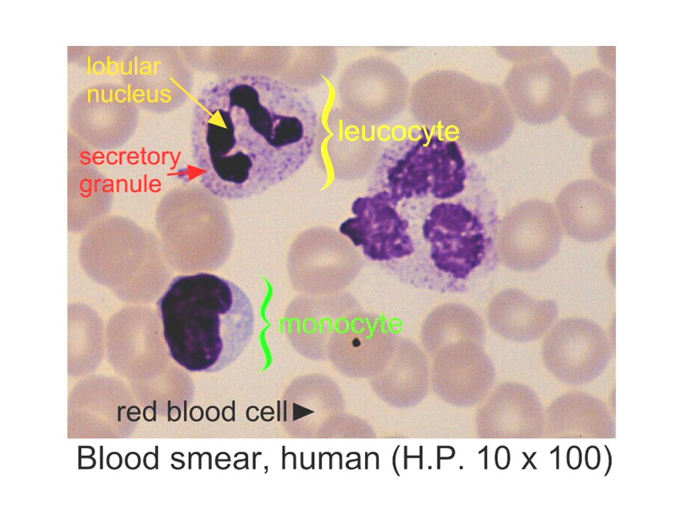
Blood Smear

6
This peripheral blood smear from a patient with essential thrombocythemia shows increased numbers of platelets, including some large forms. (H and E, 400x)
.")
9
Malaria

11
Atypical, or reactive, lymphocytes

12
Acanthocytes (red arrow) and schizocytes (black arrow) in a canine blood smear
 and schizocytes (black arrow) in a canine blood smear")
13
Keratocytes in a canine blood smear

14
Band neutrophil

15
Vacuolated monocyte

16
Lymphocyte with azurophilic granules

17
Hypersegmentation

18
Toxic granulation

19
Döhle body

20
Platelets - finger-stick

21
Giant platelet

22
Nucleated Red Blood Cells

23
Macrocytic Red Blood Cells Compared to Normal Red Blood Cells

24
Promyelocyte

25
Segmented Neutrophil, Metamyelocyte, Band

26
Sickle Cell Anemia

27
Pernicious Anemia

28
Plasma cell

29
Hairy cell

30
Larger than average RBCs are macrocytic (left), while those smaller than average are microcytic (right).
, while those smaller than average are microcytic (right).")
31
Pale cells (central pallor >1/3 dia) are referred to as hypochromic (right), while cells without central pallor are called hyperchromic (left).
 are referred to as hypochromic (right), while cells without central pallor are called hyperchromic (left).")
32
SCHISTOCYTES

33
TARGETS

34
OVALOCYTES

35
STOMATOCYTES

36
SPHEROCYTES

37
ACANTHOCYTES

38
BURR CELLS

39
Elliptocytosis

40
Tear Drop Cells

41
Rouleaux Formation

42
Basophilic stippling

43
Howell-Jolly Bodies

44
Platelet Clumping

45
Auer Rods

46
With iron deficiency anemia, the MCV of the red blood cells is decreased, the zone of central pallor is increased, and the overall sizes and shapes of the RBC's are less uniform (increased anisocytosis and poikilocytosis ).
.")
47
Heinz bodies (red arrows) and nucleated red blood cells (purple arrows) in scattered erythrocytes of a dog with zinc toxicosis (Dog, blood smear, Wright-Leishman stain).
 and nucleated red blood cells (purple arrows) in scattered erythrocytes of a dog with zinc toxicosis (Dog, blood smear, Wright-Leishman stain).")
48
Spherocytes (red arrows) and nucleated red blood cells (purple arrows) in the blood smear of a dog with zinc toxicosis (Dog, blood smear, Wright-Leishman stain).
 and nucleated red blood cells (purple arrows) in the blood smear of a dog with zinc toxicosis (Dog, blood smear, Wright-Leishman stain).")
53
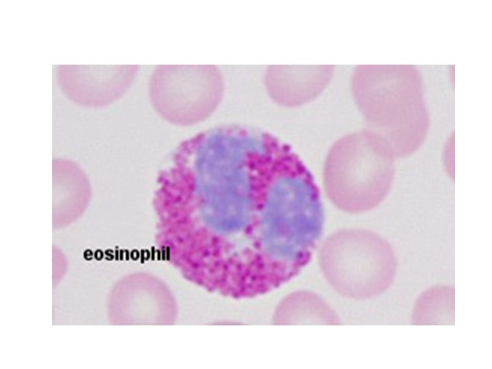
Can you identify the segmented neutrophil, band neutrophil, lymphocyte, monocyte, eosinophil, basophil, and platelet in this image?

56
This is a picture of a blood smear as seen by a microscope
This is a picture of a blood smear as seen by a microscope. The red cells are the smaller, more numerous round objects. There are two white cells in the middle (they are blue because they have been stained).
.")
66
Picture of bone marrow smear (control); Normal granulocytes and erythroblasts are evident.
; Normal granulocytes and erythroblasts are evident.")
67
Acute lymphoid leukemia (ALL); There is a marked proliferation of small lymphoblasts.
; There is a marked proliferation of small lymphoblasts.")
68
Acute myeloid leukemia (AML); There is a marked proliferation of large myeloblasts
; There is a marked proliferation of large myeloblasts")
69
Chronic myeloid leukemia (CML); There is a marked proliferation of granulocytes at various stages of maturation.
; There is a marked proliferation of granulocytes at various stages of maturation.")
70
AML (M0)
")
71
AML (M1)
")
72
AML (M2)
")
73
AML (M3)
")
74
AML (M4)
")
75
AML (M5)
")
76
AML (M5) -alpha-naphthyl butyrate esterase and chloroacetate esterase stains
 -alpha-naphthyl butyrate esterase and chloroacetate esterase stains")
77
AML (M6) -PAS stain
 -PAS stain")
78
AML (M6)
")
79
AML (M7)
")
80
Chronic myelocytic leukemia

81
Chronic lymphocytic leukemia (CLL)
")
82
Acute Lymphoblastic Leukemia (ALL)
")
83
Acute lymphocytic leukemia (ALL)
")
84
ALL-L1

85
ALL-L2

86
ALL-L3

87
thanks

Similar presentations