Download presentation
Presentation is loading. Please wait.

1
Jo Bartlett SSN, Clinical Educator Paediatric Critical Care, ORH
Introduction of Clare and me Introduction of PICU, our work environment About PICU PICU is staffed by a multidisciplinary team of anaesthetists, paediatricians, nurses, physiotherapists and dieticians, and is dedicated to the care of critically ill children from Oxfordshire, Berkshire, Buckinghamshire and Northamptonshire. It has six beds. The unit is capable of delivering the full range of intensive care therapies, including advanced techniques like High Frequency Oscillatory Ventilation, allowing us to provide specialist care to our patients. Children requiring intensive care support and with ages ranging from birth until their sixteenth birthday can be cared for in the PICU. The reasons for admission are varied. The main groups are: Medical : Respiratory, Neurology, Cardiology, Infection/Sepsis, Oncology, Endocrinology Surgical : Cardiothoracic surgery , Nurosurgery, Trauma, Neonatal Surgery, Paediatric Surgery, Craniofacial, ear, nose and throat The Paediatric Retrieval Team provides a safe environment to transport critically ill patient to the John Radcliffe Hospital. The service includes expert telephone advice to colleagues stabilising critically ill children in District General Hospitals. Our service includes High Dependency Care beds for those children that are not critically ill but require close monitoring. Admissions in the Paediatric Intensive Care Unit are always a very stressful time for parents and close relatives: we are keen to support the family as a whole. Colleagues in Social Services and Clinical Psychology provide us with valuable support. explanation that we are more focused at the acute setting, not on long term management, self study into long term management of complications of meningitis suggested but not our expertise Meningococcal Disease Jo Bartlett SSN, Clinical Educator Paediatric Critical Care, ORH

2
Objectives of lesson Learning outcomes of the module examine issues related to the care of children with complex health care needs in an acute setting. Knowledge to underpin professional skills development will focus particularly on: Caring for the highly dependent child Caring for children and families under stress Promoting optimal health and social care outcomes for the child and family Developing strategies for assessment, problem solving and care management Developing further expertise in accessing, selecting and applying appropriate evidence to support practice How we are going to run the session Understand causes, symptoms and clinical management of meningitis, in particular meningococcal meningitis / septicaemia Introduction to pathology and management of shock Focus on caring for the highly dependant child, caring for children and families under stress

3
Content of lesson Definitions of Meningitis / Meningoccocal disease and Septicaemia / Shock Causes Incidence Symptoms Treatment Nursing care of a child with meningitis, Septicaemia, and shock Case studies and discussion How we will work together with you to achieve these learning outcomes in this session Interactive session lots of chat and experience from you please

4
Definition Causes and Routes of infection: In newborns bacterial meningits is usually aquired by contact and aspiration of maternal intestinal and genital tract secretions during delivery (which bacterias are this?) Salmonella from pet reptiles in older infants and children most cases are caused by Strept pneumoniae, N. Meningitidis, Haemophilus influenza B, Routes of transmission: organisms penetrate through vulnerable sites of the blood-brain-barrier , infection spreads quickly around the coverings of the brain infection can also occur through direct invasion of CNS by bacteria ( skull fracture, penetrating injury, congenital malformations, complication of neurosurgery, lumbar puncture, placement of VP shunt much less cases of Haemophilus influenza since introduction of vaccin, before the 1990s 70% of cases of bacterial meningitis, decline by 95% now leading causes N meningitidis, and s. pneumoniae (vaccin available?) Meningococcal meningits is a notifiable disease which means Meningitis: Inflammation of the meninges (membranes which cover the brain and spinal cord) Can lead to raised ICP causing herniation of the brain stem and death (approx 20%)
 Salmonella from pet reptiles. in older infants and children most cases are caused by Strept pneumoniae, N. Meningitidis, Haemophilus influenza B, Routes of transmission: organisms penetrate through vulnerable sites of the blood-brain-barrier , infection spreads quickly around the coverings of the brain. infection can also occur through direct invasion of CNS by bacteria ( skull fracture, penetrating injury, congenital malformations, complication of neurosurgery, lumbar puncture, placement of VP shunt. much less cases of Haemophilus influenza since introduction of vaccin, before the 1990s 70% of cases of bacterial meningitis, decline by 95% now leading causes N meningitidis, and s. pneumoniae (vaccin available ) Meningococcal meningits is a notifiable disease which means. Meningitis: Inflammation of the meninges (membranes which cover the brain and spinal cord) Can lead to raised ICP causing herniation of the brain stem and death (approx 20%)")
5
Meningitis causative agents
Viral: Enterovirus, Herpes virus, Mumps virus Fungal: Candida Albians (preterm neonates) Crptocoocus neoforms and Histoplasma (immunocompromised patients) Bacterial: Haemophilus influenza B, Streptococcus pneumoniae, Strep B, Neisseria meningitidis, Meningococcus, TB, Salmonella and Listeria very rare Staphylococcal infection following surgery or skull fractures where the dura is torn Salmonella from pet reptiles Staphylococcal can happen years later
 Crptocoocus neoforms and Histoplasma (immunocompromised patients) Bacterial: Haemophilus influenza B, Streptococcus pneumoniae, Strep B, Neisseria meningitidis, Meningococcus, TB, Salmonella and Listeria very rare. Staphylococcal infection following surgery or skull fractures where the dura is torn. Salmonella from pet reptiles. Staphylococcal can happen years later.")
6
YUK Salmonella Candida Nisseria Meningitidis Haemophilus influenza
What are these piccies of. Match them to the names

7
Raised ICP: Signs Reduced or fluctuating level of consciousness (Glasgow Coma Scale score less than 9 or a GCS drop of 3 or more) Relative bradycardia and hypertension Focal neurological signs Abnormal posture or posturing Unequal, dilated or poorly responsive pupils Papilloedema Abnormal ‘doll’s eye’ movements Name them for me
 Relative bradycardia and hypertension. Focal neurological signs. Abnormal posture or posturing. Unequal, dilated or poorly responsive pupils. Papilloedema. Abnormal ‘doll’s eye’ movements. Name them for me.")
8
Not a lot of space for things to swell.
The pressure-volume relationship between ICP, volume of CSF, blood, and brain tissue, and cerebral perfusion pressure (CPP) is known as the Monro-Kellie doctrine or the Monro-Kellie hypothesis.3
 is known as the Monro-Kellie doctrine or the Monro-Kellie hypothesis.3.")
9
Meningococcal Meningitis
Meningococcal infection remains a major health problem in children, with a significant mortality and morbidity. Prompt recognition and early treatment are the only effective measure against invasive disease. This requires immediate administration of antibiotic therapy, and the recognition and treatment of patients who have complications such as septicaemia,shock, raised ICP . Risk factors for disease include: Younger age Winter or dry season Close contact with carrier or case Moving into new communities Exposure to resp infection Aktive or passive smoking Factors increasing susceptibilty and severity of disease include: -immunodeficiency Of children who progress to invasive meningococcal disease 30 – 50% have meningitis alone (mortality 5%) 7-10% have features of septicaemia (mortality 5 – 40%) 40% present a mixed picture of meningitis with septicaemia Meningococcal Meningitis Vaccines for Meningococcal B, Meningococcal C Pnemoccocus and Haemophilus influenza B 40% of healthy individuals are asymptomatic carriers of Neisseria meningitidis in their upper resp tract, Infection occurs most often in children <5 years, peak 6 – 12 months, another peak occurs in adolescence Transmission via droplets / resp secretions Persons in direct contact with patient should receive antibiotic prophylaxis (same household)
 7-10% have features of septicaemia (mortality 5 – 40%) 40% present a mixed picture of meningitis with septicaemia. Meningococcal Meningitis. Vaccines for Meningococcal B, Meningococcal C Pnemoccocus and Haemophilus influenza B. 40% of healthy individuals are asymptomatic carriers of Neisseria meningitidis in their upper resp tract, Infection occurs most often in children <5 years, peak 6 – 12 months, another peak occurs in adolescence. Transmission via droplets / resp secretions. Persons in direct contact with patient should receive antibiotic prophylaxis (same household)")
10
Signs of Meningitis 1 Vary considerably depending on the child´s age
Kernigs sign is pain with extension of the childs legs gradual onset of disease (more common) - preceded by several days of fever accompanied by upper resp tract or gastrointestinal symptoms followed by nonspecific signs of CNS infection such as increasing lethargy and irritability - recent ear or upper resp infection - history of fever or may not be present in neonates - apnoea, resp distress in neonates - vomiting, headache, poor feeding, photophobia - altered mental status: excessive lethargy or irritability in infants (does not quiet when comforted by caregiver) high fever, headache and stiff neck are common in children>2 years, symptoms may develop fast (1-2 hours) or over 1-2 days newborns and young babies may have subtle signs such as poor feeding, decreased activity, irritability, bulging fontanelle, high pitched cry Kernig´s and Brudzinki´s sign and stiff neck are signs of meningeal irritation Kernig´s: flex leg 90 degrees at hip, cannot be extended again without pain Brudzinki´s: involuntary flexion of legs when neck is flexed focal or generalised seizures occur in 20 – 30% of patients of all age rash may or may not be present but most proven cases develop a rash at some stage in their illness Signs of Meningitis 1 Vary considerably depending on the child´s age Fever Headache , photophobia (rare in young children), Altered mental status older child (lethargy, sleepy, irritability, combative, confused ‘drunk’) Stiff neck, Kernig´s sign, Brudzinki´s sign (rare in babies) Unsteady gait, Jitteriness Seizures Photophobia
 - preceded by several days of fever accompanied by upper resp tract or gastrointestinal symptoms followed by nonspecific signs of CNS infection such as increasing lethargy and irritability. - recent ear or upper resp infection. - history of fever or may not be present in neonates. - apnoea, resp distress in neonates. - vomiting, headache, poor feeding, photophobia. - altered mental status: excessive lethargy or irritability in infants (does not quiet when comforted by caregiver) high fever, headache and stiff neck are common in children>2 years, symptoms may develop fast (1-2 hours) or over 1-2 days. newborns and young babies may have subtle signs such as poor feeding, decreased activity, irritability, bulging fontanelle, high pitched cry. Kernig´s and Brudzinki´s sign and stiff neck are signs of meningeal irritation. Kernig´s: flex leg 90 degrees at hip, cannot be extended again without pain. Brudzinki´s: involuntary flexion of legs when neck is flexed. focal or generalised seizures occur in 20 – 30% of patients of all age. rash may or may not be present but most proven cases develop a rash at some stage in their illness. Signs of Meningitis 1. Vary considerably depending on the child´s age. Fever. Headache , photophobia (rare in young children), Altered mental status older child (lethargy, sleepy, irritability, combative, confused ‘drunk’) Stiff neck, Kernig´s sign, Brudzinki´s sign (rare in babies) Unsteady gait, Jitteriness. Seizures. Photophobia.")
11
Signs of Meningitis 2 Hypothermia (more common in babies)
Apnoea / cyanosis (common in babies) Vomiting, poor feeding Bulging fontanelle (in babies), high pitched cry, signs of a raised ICP Altered mental status (lethargy, irritability) Abnormal tone, floppy or stiff (in babies)
 Vomiting, poor feeding. Bulging fontanelle (in babies), high pitched cry, signs of a raised ICP. Altered mental status (lethargy, irritability) Abnormal tone, floppy or stiff (in babies)")
12
Kernigs sign

13
Brudzinki´s sign Raise the head and the legs will raise as well

14
Signs of Meningococcal Septicaemia
sudden onset ( less common presentation) - rapidly progressive manifestation of shock, purpura, disseminated intravascular coagulopathy, reduced levels of responsiveness, can result in death within 24 hours Many other pathologies (enterovirus, Schoenlein-Henoch-disease, bleeding disorders, or NAI) can present with a rash (and the meningococcal rash is not always non-blanching even) but fever and a noon-blanching rash should always prompt a serious consideration of the diagnosis of meningococcal disease and lead to antibiotics therapy unless another diagnose is apparent. Meningococcal disease maz progress rapidlz, even after appropriate antimicrobial treatment has commenced. All patients need to be closely monitored for deterioration Outcome may critically depend on the prompt recognition of 2 important complications: Shock Raised ICP Csonider level of parental concern, especially incomparison with previous illness, how quickly the illness is progressing, clinical judgement of the overall serverity of the illness Hyper or hypothermia Limb or joint pain Characteristic haemorrhagic rash (petechiae and / or purpura) Abnormal skin colour (pale or mottled), cold hands. Capillary refill >2sec Tachycardia, Hypotension (late sign) Tachypnoea, cyanosis (late sign) Rigors, fits, Decreasing level consciousness Decreased urine output, metabolic acidosis What are they As for any self limiting viral ilness. Difficult to spot, If a child is sent hmoe explain what they are looking out for and to come back if requuired even if this is soon But red Flag symptoms of LIMB PAIN, MOTTLED SKIN, COLD EXTREMITIES can appear 5 hours BEFORE classic symptoms
 - rapidly progressive manifestation of shock, purpura, disseminated intravascular coagulopathy, reduced levels of responsiveness, can result in death within 24 hours. Many other pathologies (enterovirus, Schoenlein-Henoch-disease, bleeding disorders, or NAI) can present with a rash (and the meningococcal rash is not always non-blanching even) but fever and a noon-blanching rash should always prompt a serious consideration of the diagnosis of meningococcal disease and lead to antibiotics therapy unless another diagnose is apparent. Meningococcal disease maz progress rapidlz, even after appropriate antimicrobial treatment has commenced. All patients need to be closely monitored for deterioration. Outcome may critically depend on the prompt recognition of 2 important complications: Shock. Raised ICP. Csonider level of parental concern, especially incomparison with previous illness, how quickly the illness is progressing, clinical judgement of the overall serverity of the illness. Hyper or hypothermia. Limb or joint pain. Characteristic haemorrhagic rash (petechiae and / or purpura) Abnormal skin colour (pale or mottled), cold hands. Capillary refill >2sec. Tachycardia, Hypotension (late sign) Tachypnoea, cyanosis (late sign) Rigors, fits, Decreasing level consciousness. Decreased urine output, metabolic acidosis. What are they. As for any self limiting viral ilness. Difficult to spot, If a child is sent hmoe explain what they are looking out for and to come back if requuired even if this is soon. But red Flag symptoms of LIMB PAIN, MOTTLED SKIN, COLD EXTREMITIES can appear 5 hours BEFORE classic symptoms.")
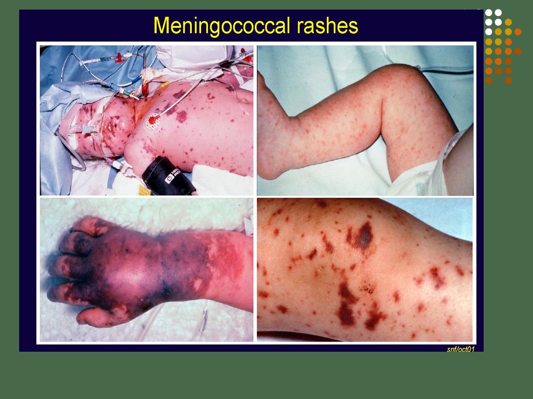
15
Meningococcal rash Note the arker centres

17
Diagnosis Clinical presentation LP with opening pressures recorded.
CSF analysis definitive diagnostic test, Bacterial meningitis will reveal? cloudy sample, glucose low, protein high, lots of neutrophils, culture and gram stains will be +VE Viral or fungal meningitis will reveal a normal glucose, slightly raised protein, leucocytes and lymphocytes will be present, Laboratory: Elevated WCC or Neutropenia, high CRP Blood culture, Throat secretions BEWARE OF RICP in lumber punctures CRP and WCC may take hours to respond following the onset of meningococcal infection and may be relatively normal early in the course of the disease

18
Treatment 1 Use personal protective equipment, initiate respiratory isolation, standard precautions gloves aprons, Assess accurately, Reassess, Reassess Record properly, Get appropriate people, senior Drs, ask for help. Consider masks, goggles Messy resuscitations

19
Treatment 2 A= Maintain airway, oxygen, B= Intubation and ventilation,
C= ECG monitor and pulse oximetry, Vascular access, if signs of dehydration or shock - fluid bolus, monitor fluid balance (urinary catheter) D= Pupils, AVPU, Temperature, Seizures? E= Serum glucose level, lab samples, lumbar puncture Medication as prescribed: antibiotics, antipyretics, inotropes, IVI. Be systematic. Reassess Resus guidelines
 D= Pupils, AVPU, Temperature, Seizures E= Serum glucose level, lab samples, lumbar puncture. Medication as prescribed: antibiotics, antipyretics, inotropes, IVI. Be systematic. Reassess. Resus guidelines.")
20
Definition Septicaemia: Presence of pathogens in the blood
Whole body inflammatory response or systematic inflammatory response syndrome Potentially deadly

21
Shock: Definition at risk of shock: - identifiable source of infection, duration and height of fever, associated features such as lethargy, vomiting, diarrhoea, decreased oral intake, decreased level of consciousness or awareness, Hypovolaemia Presence of shock in meningococcal disease is multifactorial septcaemia is the commonest cause of a child presenting in shock, unless an alternative diagnosis is very clear (trauma, anaphylaxis or poisoning), give antibiotic Maintenace of adequate tissue perfusion depends on a pump (the heart) delivering the correct type and volume of fluid (blood) through controlled vessels (arteries , veins and capillareis) without abnormal obstruction to flow. Is inadequate tissue perfusion. Resulting from the failure of the cardiovascular system to deliver sufficient oxygen and nutrients to sustain vital organ function. Underlying cause must be recognised and treated promptly, or cell and organ dysfunction and death may result
, give antibiotic. Maintenace of adequate tissue perfusion depends on a pump (the heart) delivering the correct type and volume of fluid (blood) through controlled vessels (arteries , veins and capillareis) without abnormal obstruction to flow. Is inadequate tissue perfusion. Resulting from the failure of the cardiovascular system to deliver sufficient oxygen and nutrients to sustain vital organ function. Underlying cause must be recognised and treated promptly, or cell and organ dysfunction and death may result.")
22
Shock: Types Hypovolaemic: Most common in Children. Inadequate circulating blood volume owing to blood or fluid loss (Septicaemia, Trauma, D+V, Burns) Cardiogenic: Cardiac compensatory mechanism fail, heart attacks, following surgery Distributive: In septic and anaphylactic shock, peripheral vasodilation, decreased venous return, hypotension (also Neurogenic, disrupted autonomic pathways from head injury, trauma to spinal cord)
 Cardiogenic: Cardiac compensatory mechanism fail, heart attacks, following surgery. Distributive: In septic and anaphylactic shock, peripheral vasodilation, decreased venous return, hypotension. (also Neurogenic, disrupted autonomic pathways from head injury, trauma to spinal cord)")
23
Signs of (Septic) Shock in Children
Shock is a clinical diagnosis, recogniton is problematic in children, Lactate is a good prognostic indicator. can maintain blood pressure until late stage of shock (hypotension is late sign of shock in children as opposed to adults) Heart rate: a raised heart rate is a common response to many types of stress (fever, anxiety, hypoxia, hypovolaemia). In shock, tachycardia is caused by catecolamine release and is an attempt to maintain cardiac output by increasing heart rate in the face of falling stroke volume. Bradycardia in a shocked child is caused by hypoxia and acidsis and is a preterminal sign.a early signs of brain hypoperfusin are agitation and confusion, often alternating with drowsiness Decreased urine output: kidneys receive second highest blood flow of any organ in the body,measurement or urine output can be useful indicator of adequate perfusion pressure progression of sepsis to multiorgan dysfunction, poor perfusion, hypoxia, hyperglycaemia and acidosis contribute to the process, risk of mortality increases with each additional organ system failure. Tachycardia (may be absent in hypothermic patients, No fever in neutropenic patients) Signs of decreased perfusion: Decreased peripheral pulses compared to central pulses Flash cap refill or cap refill >2 sec Mottled or cool extremities or vasodilation Tachypnoea Altered alertness, mental status Decreased urine output Metabolic acidosis, increased blood lactate
 Heart rate: a raised heart rate is a common response to many types of stress (fever, anxiety, hypoxia, hypovolaemia). In shock, tachycardia is caused by catecolamine release and is an attempt to maintain cardiac output by increasing heart rate in the face of falling stroke volume. Bradycardia in a shocked child is caused by hypoxia and acidsis and is a preterminal sign.a. early signs of brain hypoperfusin are agitation and confusion, often alternating with drowsiness. Decreased urine output: kidneys receive second highest blood flow of any organ in the body,measurement or urine output can be useful indicator of adequate perfusion pressure. progression of sepsis to multiorgan dysfunction, poor perfusion, hypoxia, hyperglycaemia and acidosis contribute to the process, risk of mortality increases with each additional organ system failure. Tachycardia (may be absent in hypothermic patients, No fever in neutropenic patients) Signs of decreased perfusion: Decreased peripheral pulses compared to central pulses. Flash cap refill or cap refill >2 sec. Mottled or cool extremities or vasodilation. Tachypnoea. Altered alertness, mental status. Decreased urine output. Metabolic acidosis, increased blood lactate.")
24
Management of Meningococcal Septacaemia
Monitor, ECG, Pulse oximetry, ABP, CVP, A+B= Reduce muscle oxygen demand and help restore ph balance by mechanical ventilation, Sedate- Morphine and Midazolam Paralyse-Vecuronium, Atracurium C= Support cardiovascular system: Inotropic drugs, Dopamine, Milrone, Noradrenaline, Adrenaline, Steroids (vasopressin) C=Restore intravascular volume with fluid resuscitation
 C=Restore intravascular volume with fluid resuscitation.")
25
Management of Meningococcal Septacaemia
C=Treat DIC,cristalloid/colloid/blood products: PRC, FFP,Platlets, Cryo,Vit K D= Antibiotics D= Neuro obs, ICP, Anticonvulsants for fits, Head circumference, Scan, PUPILS. D= Maintain normothermia: warm or cool E= Support other organs which fail (kidneys – haemofiltration) E= Fasciotomies for compartment syndrome release, measure tension of tissue F= Blood sugars, dextrose or insulin Support family how do you assess the paralysed patient for fits?
 E= Fasciotomies for compartment syndrome release, measure tension of tissue. F= Blood sugars, dextrose or insulin. Support family. how do you assess the paralysed patient for fits")
26
Aquarius CRRT Continouos renal replacement therapy.
CVVH choice on our unit, reduce imflammatory marhers and tumor necrosing factors

27
DIC Is a secondary process, which is poorly understood
Excess activation and subsequent depletion of clotting factors produces unrestrained clotting, then excessive bleeding (now disputed) Micro-thrombi are present causing ischemia then necrosis of extremities. Bigger clots cause pulmonary emboli, strokes and renal failure. Thrombocytopenia (low platelets), prolonged PT and APTT, decreased fibrinogen What is it. Have you seen it Possibly due to infections, trauma, crush injuries malignancies, vascular anomalies, transfusion reactions, snake bites,
 Micro-thrombi are present causing ischemia then necrosis of extremities. Bigger clots cause pulmonary emboli, strokes and renal failure. Thrombocytopenia (low platelets), prolonged PT and APTT, decreased fibrinogen. What is it. Have you seen it. Possibly due to infections, trauma, crush injuries malignancies, vascular anomalies, transfusion reactions, snake bites,")
28
Complications of Meningitis/ Meningococcal Septicaemia
Brain swelling, raised ICP, Death Seizures Subdural effusions, Brain abscess, Infarcts Hydrocephalus, Cranial nerve palsy’s Hearing and sight impairments Learning disability DIC causing tissue necrosis - Amputation of toes/fingers/limbs Mortality rate for bacterial meningitis after the neonatal period is <10% outcome depends on the patient´s age, duration of illness before initiating effective treatment, type of causative organism, intensity of the patients inflammatory response, number of bacteria or quantity of active bacterial products in CSF at time of diagnosis severe neurological sequelae occur in 10 – 20% of patients, most common are hearing loss, mental retardation, seizures, dealy in learning to speak, visual impairment and behavioural problems

29
Suggestions for further study
Treatment of shock Antibiotics used Age specific vital signs and laboratory variables Familiarize with crash trolley in placement area Consider long-term implications of complications of Meningitis for patient and family

30
References, Bibliography
Aehlert B (2007) Mosby´s Comprehensive Pediatric Emergency Care, revised edition, Elsevier Helfaer M and Nichols D (eds) (2009) Roger´s Handbook of Pediatric Intensive Care ( 4th edition) Lippincott Williams & Wilkins Hazinski M (1992) Nursing Care of the Critically Ill Child (2nd Edition) Mosby Barry P, Morris K and Ali T (eds) (2010) Paediatric Intensive Care, Oxford University Press NICE clinical guideline 102, Bacterial meningitis and meningococcal septicaemia, 2010 Meningococcal disease ppt, available from author (Dr. Shelley Segal, ORH) normal Heart Rates by age Infant (1 – 12 months) Toddler (1 – 3 years Preschooler (4 – 5 years) School age (6 – 12 years) Adolescent (13 – 18 years) normal resp rate for age normal BP for age Glasgow Coma Scale
 Mosby´s Comprehensive Pediatric Emergency Care, revised edition, Elsevier. Helfaer M and Nichols D (eds) (2009) Roger´s Handbook of Pediatric Intensive Care ( 4th edition) Lippincott Williams & Wilkins. Hazinski M (1992) Nursing Care of the Critically Ill Child (2nd Edition) Mosby. Barry P, Morris K and Ali T (eds) (2010) Paediatric Intensive Care, Oxford University Press. NICE clinical guideline 102, Bacterial meningitis and meningococcal septicaemia, Meningococcal disease ppt, available from author (Dr. Shelley Segal, ORH) normal Heart Rates by age. Infant (1 – 12 months) Toddler (1 – 3 years Preschooler (4 – 5 years) School age (6 – 12 years) Adolescent (13 – 18 years) normal resp rate for age. normal BP for age. Glasgow Coma Scale.")
31
Useful Websites www.meningitis-trust.org public support
produced leaflet (educational materials for health professionals) Nice guidelines
 Nice guidelines.")
Similar presentations


 leading to inadequate oxygen delivery to tissues.>")
