Download presentation
Presentation is loading. Please wait.

1
JOINTS Dr.LUBNA NAZLI ASST. PROF. ANATOMY RAK MHSU Dt. 1/10/2007
Mon am

2
Objectives: Definition. Classification of joints. Types of joints.
Movements of different joints. Structure of synovial joints. Nerve supply of joints. Blood supply of joints. Stability of joints. Applied anatomy.

3
Definition: The bones of the skeleton are joined to one another at different sites, such connections are termed Joints or Articulations.
4
Classification of Joints
The joints are divided into three types: 1. Fibrous or immovable joints.(Synarthroses) 2. Cartilaginous or slightly movable joints. (Amphiarthroses) 3. Synovial or freely movable joints. (Diarthroses)
 2. Cartilaginous or slightly movable joints. (Amphiarthroses) 3. Synovial or freely movable joints. (Diarthroses)")
5
Fibrous or immovable joints:
The joint surfaces of the bones are in direct contact, held together by connective tissue or hyaline cartilage. There is no movement. Eg: joints between the bones of the skull, excepting those of the mandible.

8
TYPES OF FIBROUS JOINTS
There are four types: Sutures Schindylesis Gomphosis Synchondrosis.

9
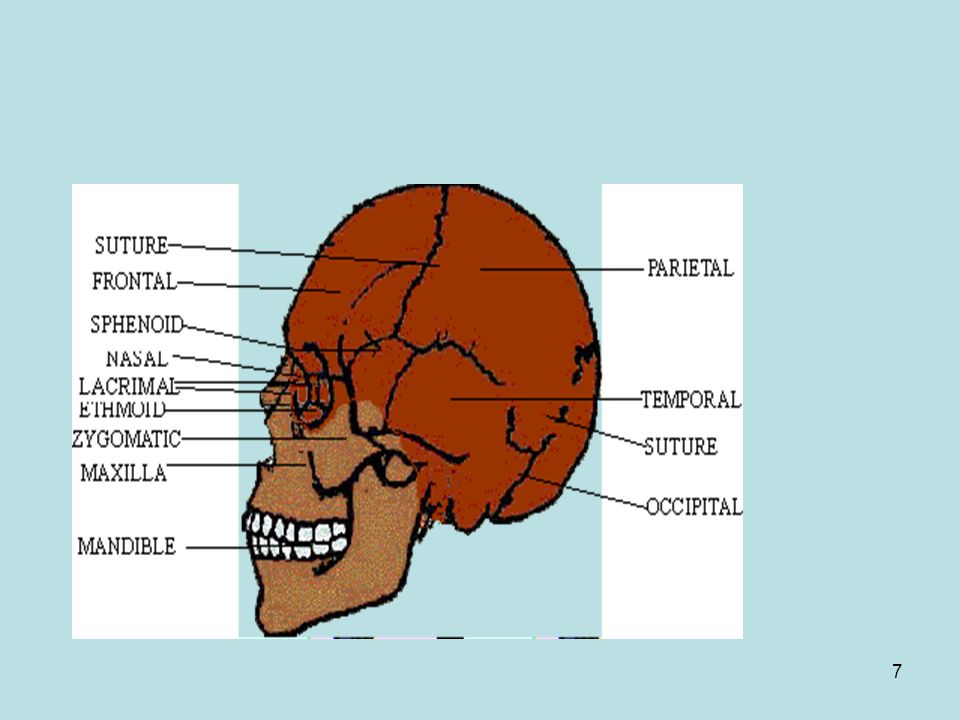
SUTURES Suture is a form of joint where the bones are united by a thin layer of fibrous tissue continuous externally with the pericranium and internally with the dura mater. Eg: skull joints.

10
JOINT BETWEEN SKULL BONES

11
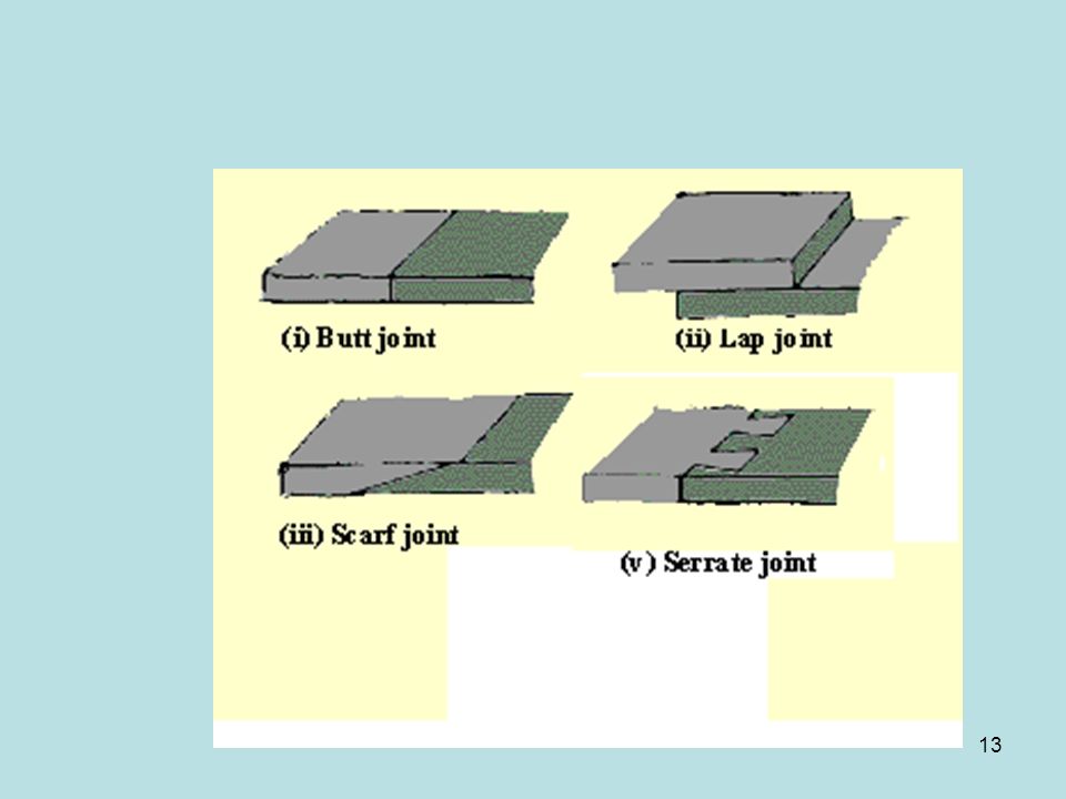
TYPES OF SUTURES There are three varieties: 1. Dentate suture.
2. Serrate suture. 3. Limbous suture.

12
The dentate suture has tooth-like form of the projecting processes,seen in the suture between the parietal bones. In the serrate suture the edges of the bones are serrated like the teeth of a fine saw, as between the two portions of the frontal bone. In the limbous suture, there is interlocking with a certain degree of bevelling of the articular surfaces, so that the bones overlap one another, as in the suture between the parietal and frontal bones.

14
Schindylesis Schindylesis is that form of articulation in which a thin plate of bone is received into a cleft or fissure formed by the separation of two laminae in another bone. Eg: articulation of the rostrum of the sphenoid and perpendicular plate of the ethmoid with the vomer.

15
Gomphosis Gomphosis is articulation by the insertion of a conical process into a socket. Eg: articulations of the roots of the teeth with the alveoli of the mandible and maxillae.

16
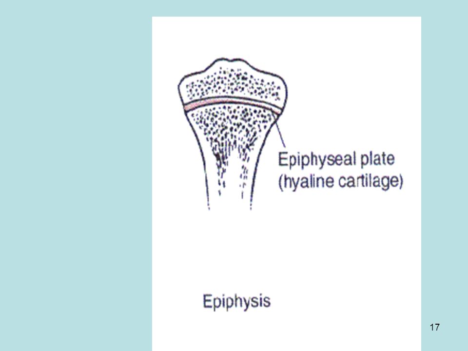
Synchondrosis Where the connecting medium is cartilage the joint is termed a synchondrosis. This is a temporary form of joint, for the cartilage is converted into bone before adult life. Eg: Between the epiphysis and bodies of long bones. Between the occipital and the sphenoid.

18
Cartilaginous or slightly movable joints
In these articulations the bony surfaces are connected by broad flattened discs of fibrocartilage. Eg: articulations between the bodies of the vertebrae. Eg: inferior tibiofibular articulation. United by an interosseous ligament. The first form is termed a symphysis. The second is syndesmosis.

19
Symphysis Fibrocartilage

20
Syndesmosis Interosseous membrane

21
Cartilaginous joint

22
SYNOVIAL JOINTS Structure of synovial joint :
The ends of the opposing bones are covered with hyaline cartilage, the articular cartilage. They are separated by a space called the joint cavity. The joints are enclosed in a dense fibrous joint capsule. The outer layer of the capsule consists of the ligaments that hold the bones together.

23
The inner layer is the synovial membrane that secretes synovial fluid into the joint cavity for lubrication. As these joints have a synovial membrane, they are called synovial joints. 7. The joint may be divided, completely or incompletely, by an articular disc or meniscus.

25
Synovial joint

26
Synovial joint with intra-articular disc

27
Types of synovial joints:
They are determined by the kind of movement permitted in each. Uniaxial: All movements take place around one axis. 1. Ginglymus joint. 2. Pivot joint. 3. Bicondylar joint.

28
Ginglymus or hinge joint
The axis is transverse. It permits movement only in one plane, forward and backward. Examples of ginglymus: 1. interphalangeal joints. 2. Elbow joint.

29
Ginglymus or Hinge joint

30
Trochoid or pivot-joint.
It is longitudinal. Movement is limited to rotation, the joint is formed by a pivot-like process turning within a ring. Eg: Proximal radioulnar Articulation of the odontoid process of the axis with the atlas.

31
Pivot joint

32
Condylar (Bicondylar)
This joint allows primary movement in one plane flexion, extension with small amounts of movement in another plane (rotation). Eg: knee joint temporomandibular joint.
. Eg: knee joint. temporomandibular joint.")
33
Bicondylar joint

34
Biaxial: Movement is around two horizontal axes at right angles to each other. 1. Condyloid joint. 2. Saddle-joint.

35
Condyloid / Ellipsoid joint — an ovoid articular surface or condyle, is received into an elliptical cavity to permit flexion, extension, adduction, abduction, and circumduction, but no axial rotation. Eg: wrist-joint. metacarpophalangeal joint.

36
Ellipsoid joint

37
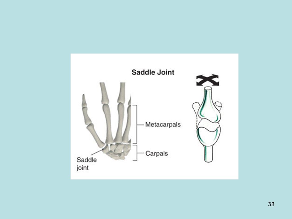
Saddle-joint : The opposing surfaces are reciprocally concavo-convex
Saddle-joint : The opposing surfaces are reciprocally concavo-convex. The movements are flexion, extension, Adduction, abduction, and circumduction are allowed; but no axial rotation. Eg: carpometacarpal joint of the thumb.

39
Polyaxial: movement is in 3 or more directions
Polyaxial: movement is in 3 or more directions. Flexion/Extension, adduction/abduction, medial and lateral rotation. Ball-and-socket joint is formed by the reception of a globular head into a cup-like cavity. Eg: hip joint. shoulder joint.

40
Ball and Socket joint

41
Gliding or plane joint. It is formed by the apposition of plane surfaces, one slightly concave, the other slightly convex, the movement between them is limited by the ligaments or bony processes surrounding the joint. Eg: between the articular processes of the vertebræ. The carpal joints. The tarsal joints.

42
Plane or Gliding joint

43
Nerve supply of joints A sensory nerve supplying the joint also supplies the muscles moving the joint and the skin overlying the joint. This is called HILTON’S LAW. Joints transmit a sensation called proprioception, which provides an awareness of movement and position of the parts of the body.

44
BLOOD SUPPLY OF JOINTS Joints receive blood supply from the periarticular arterial plexus whose branches pierce fibrous capsule and form subsynovial vascular plexus.

45
STABILITY OF JOINTS It depends on 3 factors.
Articular surfaces: shape, size and arrangement of bones. Ligaments: fibrous and elastic ligaments prevent excessive movement. 3. Muscles: tone of muscles acting on the joint.

46
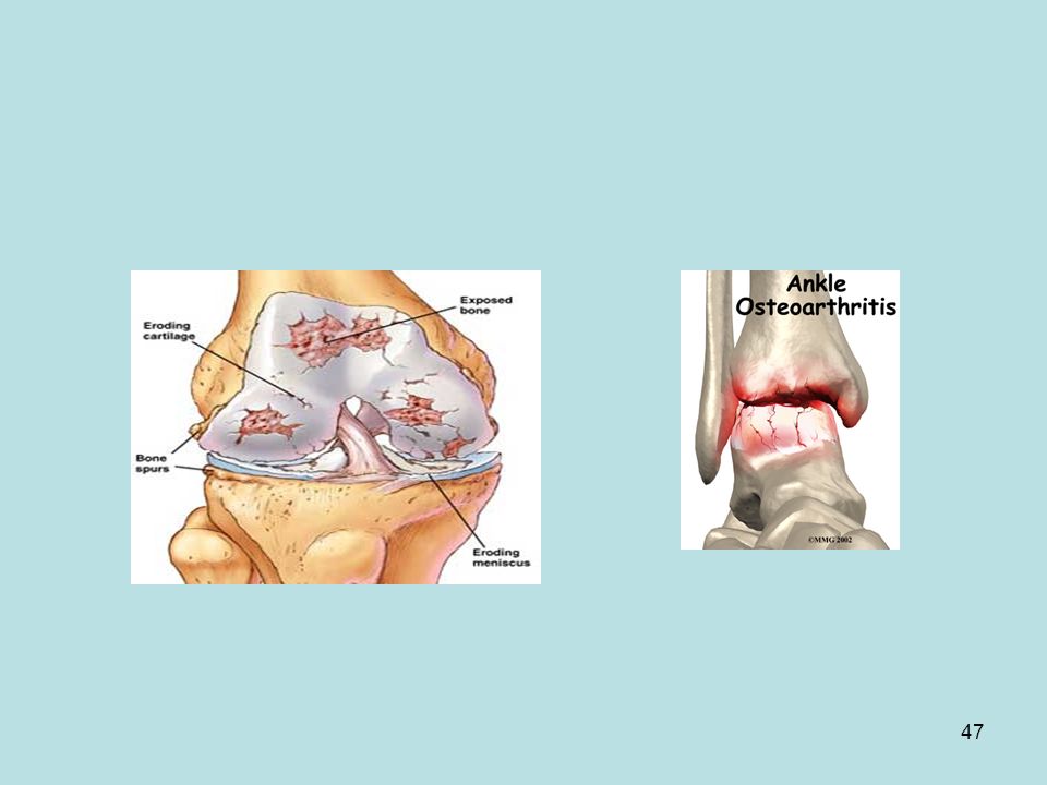
APPLIED ANATOMY JOINT DISLOCATIONS & SUBLUXATION
It is caused by trauma and characterized by pain, deformity and loss of function. SPRAIN is caused by ligamentous tear. ARTHRITIS is inflammation of joints. Osteoarthritis is due to degenerative changes in articular cartilage.

50
STIFFNESS OF JOINTS: It is related to weather
STIFFNESS OF JOINTS: It is related to weather. The viscosity of synovial fluid increases with fall in temperature leading to stiffness of joints. NEUROPATHIC JOINT: It is due to complete denervation of joint caused by leprosy and tabes dorsalis. The reflexes are eliminated and joint is unprotected.

Similar presentations


![Articulations. Articulations- points where two or more bones come together to form a joint [ maybe rigid or movable] Classified by Structure or Function.](/13/3878517/big_thumb.jpg "Articulations. Articulations- points where two or more bones come together to form a joint [ maybe rigid or movable] Classified by Structure or Function.>")


: a point of contact between bones. Some allow movement, others are immovable (sutures). Most joints.>")


 and type of substance.>")


