Download presentation
Presentation is loading. Please wait.

1
Hamburg High School EMT Program
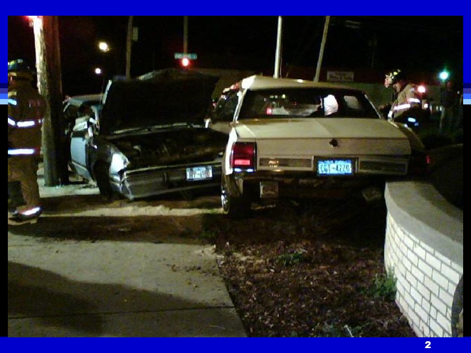
4/22/2017 Shock and Bleeding Hamburg High School EMT Program Temple College EMSP

5
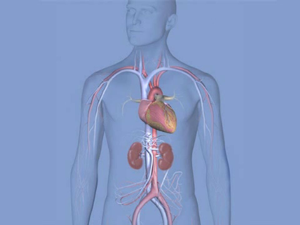
Circulatory System Anatomy Heart Arteries Capillary Veins
Muscular organ, size of your fist, located in the center of the thoracic cavity Arteries Carry blood rich in oxygen – all arteries carry blood away from the heart Capillary Place where exchange of oxygen and carbon dioxide take place Veins Carry blood high in carbon dioxide – all veins carry blood back to the heart

6
Function of the Blood Transportation of Gases Nutrition Excretion
Carries oxygen from lungs to body cells, carries carbon dioxide back to lungs to be exhaled Nutrition Circulates nutrients from tissue to other body cells Excretion Carries waste products from cells to organs, such as the kidneys

7
Function of the Blood Protection Regulation
Carries antibodies and WBC’s, which help fight disease and infection Regulation Blood carries substances that control the functions of the body, such as hormones, water, salt, enzymes, and chemicals Blood also helps regulate body temperature by carrying body heat to the lungs and skin

8
The average amount of blood found in a:
Male: 5 – 6 Liters Female: 4 – 5 Liters

9
Composition of Blood Plasma Red Blood Cells
Water, salty fluid that makes up over half the volume of the blood. (56% of the bloods volume) Red & white blood cells and platelets are carried in plasma Red Blood Cells RBC’s, erythrocytes, or red corpuscles. Carry oxygen to tissues and carbon dioxide away. Gives blood its red color
 Red & white blood cells and platelets are carried in plasma. Red Blood Cells. RBC’s, erythrocytes, or red corpuscles. Carry oxygen to tissues and carbon dioxide away. Gives blood its red color.")
10
Composition of Blood White Blood Cells Platelets WBC’s or leukocytes
Involved in destroying micro-organisms (germs) and produce antibodies that help the body resist infection Platelets Helps the blood to clot; when fragments are activated they release chemical factors Solid part of blood
 and produce antibodies that help the body resist infection. Platelets. Helps the blood to clot; when fragments are activated they release chemical factors. Solid part of blood.")
11
Perfusion Circulation of blood through an organ
Perfusion supplies oxygen and other nutrients to the cells of all organ systems and the removal of waste products

12
Hypoperfusion (SHOCK)
Inadequate circulation of blood through an organ

13
Types of Bleeding Arterial Bleed Venous Bleed Capillary Bleed
Bright red Spurting Venous Bleed Dark red/maroon Steady flow Easier to control Capillary Bleed Dark red Oozing Often clots spontaneously

14
Severity of Bleeding Considered severe when loss equals:
One liter (1000 cc) in an adult ½ liter (500 cc) in a child cc in an infant Uncontrolled bleeding or significant blood loss leads to shock (hypoperfusion) and possibly death
 in an adult. ½ liter (500 cc) in a child cc in an infant. Uncontrolled bleeding or significant blood loss leads to shock (hypoperfusion) and possibly death.")
15
Severity of Blood Loss Determined by:
General impression of the amount of blood loss Signs or symptoms of hypoperfusion If the patient exhibits signs and symptoms of shock (hypoperfusion), the bleeding is to be considered serious
, the bleeding is to be considered serious.")
16
Control of External Bleeding
PPE for BSI Maintain Airway Administer Oxygen 1. Direct Pressure gloved hand dressing/bandage 2. Elevation 3. Tourniquet CONSIDER: Arterial pressure points Upper extremity: Brachial Lower extremity: Femoral

17
Other Methods to Control Bleeding
Splints Air splints

18
Tourniquet Application can cause permanent damage to nerves, muscle, and blood vessels resulting in loss of the extremity Used for amputations 3-4” wide & 6-8 layers deep Wrap around extremity twice at a point above bleeding as close to the wound as possible Tie one knot in the bandage and place a stick or rod on top of the knot and tie it Twist the stick until the bleeding stops Once the bleeding has stopped secure in place

19
Tourniquet Blood pressure cuff may be used as a tourniquet until bleeding stops Write “TK” and time of application on forehead of patient Notify other personnel Do not loosen or remove until definitive care is available Do not use wire, rope, or any material that may cut into the skin Do not cover with sheets, blankets, etc. Do not apply over a joint, but as close to the injury as possible

20
Bleeding from the Nose, Ears, and Mouth
Possible causes Skull fracture Simple nose bleed – epistaxis Facial trauma

21
Skull Fracture May cause loss of blood or clear fluid (cerebrospinal fluid) from the nose and ears. Check for Halo Effect Do not stop the flow of fluid – collect in a loose dressing

22
Epistaxis Causes Fractured skull Facial injuries Sinusitis, other URIs
High BP Clotting disorders Digital trauma (nose picking)
")
23
Epistaxis Nosebleed Management Sit up, lean forward
Pinch the fleshy portion of the nostrils together Keep in sitting position Keep patient calm and quiet Apply ice over nose 15 min adequate

24
Epistaxis can result in life-threatening blood loss

25
Internal Bleeding 1. Can result in severe blood loss which can lead to shock and subsequent death 2. Injured or damaged internal organs are a common cause of extensive bleeding that is concealed 3. Painful, swollen, deformed extremities (fractures) may lead to serious internal blood loss 4. Suspicion and severity of internal bleeding should be based on mechanism of injury and clinical signs and symptoms
 may lead to serious internal blood loss. 4. Suspicion and severity of internal bleeding should be based on mechanism of injury and clinical signs and symptoms.")
26
Internal Bleeding Mechanism of Injury Blunt trauma Falls
4/22/2017 Internal Bleeding Mechanism of Injury Blunt trauma Falls Motorcycle Crashes Pedestrian Impacts Automobile Collision Blast Injuries Look for evidence of contusions, abrasions, deformities, impact marks and swelling Temple College EMSP

27
Internal Bleeding Mechanism of Injury Penetrating Trauma
Pain, tenderness, swelling or discoloration at site of injury Bleeding from mouth, rectum, vagina or other orifice Vomiting of bright red blood or dark coffee-ground-colored emesis Dark, tarry stools or stools with bright red blood Tender, rigid abdomen S/S of shock

28
Internal Bleeding Management BSI Open airway High concentration oxygen
4/22/2017 Internal Bleeding Management BSI Open airway High concentration oxygen Assist ventilations Control external bleeding with direct pressure Stabilize fractures Transport rapidly to appropriate facility Temple College EMSP

29
Abdominal Evisceration
Penetrating Trauma

30
SHOCK Inadequate perfusion (blood flow) of cells leading to inadequate oxygen delivery to tissues
 of cells leading to inadequate oxygen delivery to tissues.")
31
Cardiovascular System
Transports oxygen, fuel to cells Removes carbon dioxide, waste products for elimination from body Cardiovascular system must be able to maintain sufficient flow through capillary beds to meet cell’s oxygen and fuel needs

32
How can perfusion fail, thus causing shock?
Heart fails as a pump Blood vessels dilate Loss of Volume

33
3 Types of Shock and Their Causes
Hypovolemic Shock Cardiogenic Shock Distributive Shock

35
1. Hypovolemic Shock A type of shock that is the result of low blood volume

36
A. Hemorrhagic Shock Shock most often seen by EMT’s
The natural response to bleeding is blood vessel constriction and clotting. Uncontrolled or significant bleeding can lead to shock and possibly death Loss of volume

37
A. Hemorrhagic Shock Signs and Symptoms AMS, restlessness, anxiety
Increased respiratory rate Weak, rapid pulse Pale, moist/clammy, cool skin Nausea, vomiting Thirst Falling blood pressure (LATE SIGN) Infants and children maintain their blood pressure until their blood volume is more than half gone, so by the time their blood pressure drops they are close to death
 Infants and children maintain their blood pressure until their blood volume is more than half gone, so by the time their blood pressure drops they are close to death.")
38
B. Metabolic Shock Form of Hypovolemic shock that is the result of electrolyte or plasma loss and does not involve the loss of whole blood. It can occur due to dehydration or from burns

39
B. Metabolic Shock Signs and Symptoms Sunken eyes
Poor skin turgor or tenting of the skin Recent episodes of vomiting and diarrhea Presence of burns

40
2. Cardiogenic Shock Result of a massive myocardial infarction
Heart is unable to pump effectively and as a result blood begins to back up in the system Heart’s output depends on How often it beats (heart rate) How hard it beats (contractibility)
 How hard it beats (contractibility)")
41
2. Cardiogenic Shock Signs and Symptoms Emergency care
Those symptoms commonly seen during a heart attack Emergency care CPR with AED

42
3. Distributive Shock Blood vessels suddenly dilate causing the blood pressure to fall

43
A. Anaphylactic Shock Form of shock caused by a violent systemic reaction to an allergen/toxin Signs and Symptoms Those seen with a severe allergic reaction

44
B. Neurogenic Shock Spinal cord severed
Uncontrolled dilation of the blood vessels and drop in blood pressure Compensatory mechanisms are not activated because nerve impulses cannot be conducted through the spinal cord

45
C. Septic Shock Caused by a severe bacterial infection
Toxins (poisons) released into the bloodstream Vessels dilate and there is a drop in blood pressure Increase in fluid seeping into the tissues from the capillaries Urinary tract infection is a common cause Seldom seen in the field
 released into the bloodstream. Vessels dilate and there is a drop in blood pressure. Increase in fluid seeping into the tissues from the capillaries. Urinary tract infection is a common cause. Seldom seen in the field.")
46
Psychogenic Shock Simple fainting (syncope)
Caused by stress, pain, fright Heart rate slows, vessels dilate Brain becomes hypoperfused Loss of consciousness occurs

47
Shock: Signs and Symptoms
Anxiety, restlessness, combativeness, or altered mental status Weakness, faintness or dizziness Thirst Shallow rapid breathing Rapid, weak pulse Pale, cool, clammy skin

48
Shock: Signs and Symptoms
Capillary refill >2 seconds (infants and children ONLY) Dropping blood pressure (late sign) Dilated pupils that are sluggish to respond Nausea & vomiting
 Dropping blood pressure (late sign) Dilated pupils that are sluggish to respond. Nausea & vomiting.")
49
Shock: Signs and Symptoms
Shock is NOT the same thing as a low blood pressure! A falling blood pressure is a LATE sign of shock!

50
Severity of Shock Compensated shock (Early) Decompensated shock (Late)
Body senses a decrease in perfusion and attempts to compensate Increased heart rate & respirations to increase flow of blood and oxygen Decompensated shock (Late) Body can no longer compensate for low blood volume (late sign is falling B/P)
 Body can no longer compensate for low blood volume (late sign is falling B/P)")
51
Severity of Shock Irreversible shock
Body has lost the battle to maintain perfusion Cell damage occurs in liver & kidneys. Even if vital signs can be restored patient may die

52
Emergency Care for Shock
Golden Hour From the time the injury occurs, until the patient is in the operating room Platinum Ten Minutes Goal for on-scene time when caring for a trauma patient

53
Treatment Secure, maintain airway Apply high concentration oxygen
Control bleeding (MAST if needed) Splint fractures Prevent loss of body heat
 Splint fractures. Prevent loss of body heat.")
54
Treatment Elevate lower extremities 8 to 12 inches in hypovolemic shock if no fractures Do NOT elevate the lower extremities in cardiogenic shock Administer nothing by mouth, even if the patient complains of thirst

55
Anti-Shock Garment MAST – military anti-shock trousers
PASG – pneumatic anti-shock garment Purpose: to create artificial vasoconstriction in the lower extremities and the lower abdomen

56
Indications Systolic B/P of 50 mmhg with signs of shock
Systolic B/P of 90 mmhg with a pelvic injury

57
S/S of Shock Restlessness Cool clammy skin Tachycardia
Delayed capillary refill (children & infants)
")
58
Absolute Contra-Indications
Pulmonary Edema Penetrating Chest Wound

59
Partial Contra-Indications
Pregnancy (legs only) Abdominal injury (evisceration) or impaled object (legs only) Impaled object in leg (abdominal and one leg only)
 Abdominal injury (evisceration) or impaled object (legs only) Impaled object in leg (abdominal and one leg only)")
60
MAST Use You must remove all undergarments before putting MAST in place Inflate all 3 compartments at one time to 100 to 106 mmhg MAST can only be deflated under Medical Control IVs established Surgical team ready Abdomen then one leg at a time SLOWLY

61
Never deflate entire suit at once

62
Adequate Flow = Adequate Perfusion
Flow = Perfusion Adequate Flow = Adequate Perfusion Inadequate Flow = Inadequate Perfusion (Hypoperfusion) Hypoperfusion = Shock
 Hypoperfusion = Shock.")
Similar presentations


 leading to inadequate oxygen delivery to tissues.>")


