Download presentation
Presentation is loading. Please wait.

1
The Cornea Light enters the eye through the cornea
The cornea is transparent so that light can travel through it. Seventy-five percent of the eye’s power rests in the cornea, although it is very small. It is only about half a millimeter thick. The cornea is connected to the sclera, the tough white part of the eye. The cornea acts as a lens, bending rays of light as they enter the eye Each time you blink, your eyelids cleanse and moisten the cornea

2
The Iris The iris is a ring of muscle that contracts and expands to change the amount of light that enters the eye The iris contracts when light coming in is bright: The iris expands when the light coming in is dim: Iris gives your eye color

3
The pupil The pupil is the part of the eye that looks black
It is actually a hole covered by the clear cornea The pupil looks black because it is an opening into the dark inside of the eye The iris determines how large the pupil is depending on the amount of light entering the eye.

4
The Lens Directly behind the pupil is the lens
The lens of your eye is convex, however, muscles attached to the lens will change shape to help focus the light entering the eye (this process begins with the cornea). The lens refracts light, forming an image on the lining of your eyeball When you focus on a distant object, the ciliary muscles holding the lens contract, making the lens longer and thinner When you focus on a nearby object, the muscles relax and the lens becomes shorter and fatter Behind the lens is the vitreous humor. This is a thick, white fluid that gives the eyes their firm, rubbery texture. Most of your eye is filled with vitreous humor fluid.
. The lens refracts light, forming an image on the lining of your eyeball. When you focus on a distant object, the ciliary muscles holding the lens contract, making the lens longer and thinner. When you focus on a nearby object, the muscles relax and the lens becomes shorter and fatter. Behind the lens is the vitreous humor. This is a thick, white fluid that gives the eyes their firm, rubbery texture. Most of your eye is filled with vitreous humor fluid.")
5
The Retina The layer of the cells lining the inside of the eyeball is the retina As the cornea and lens refract light, an upside-down image is formed on the retina The retina is made up of light sensitive cells called rods and cones Rods contain pigment that reacts to small amounts of light and distinguish between black, white, and shades of gray. Rods are responsible for night vision Cones respond to color. Three types of cones: red, green, and blue Cone cells only function in bright light (this is why it is difficult to see colored in dim lighting)
")
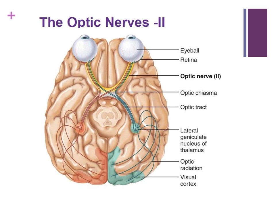
7
Optic Nerve and the brain
Signals generated by the rods and cones travel to your brain along a short, thick nerve call the optic nerve. When the signals reach your brain, it automatically turns the image right-side-up Your brain combines both images (one from each eye) into a single three-dimensional image Your optic nerve creates a blind spot- one spot of the retina where there are no cones or rods and where the optic nerve begins
 into a single three-dimensional image. Your optic nerve creates a blind spot- one spot of the retina where there are no cones or rods and where the optic nerve begins.")
9
Diagram

10
Correcting Vision Some lenses in eyeglasses are convex and some are concave . The type of lens used depends on whether the eyeball is too long or too short If the eyeball is too long it results in nearsightedness If the eyeball is too short, it results in farsightedness

11
Nearsightedness A nearsighted person can see nearby things clearly, but objects at a distance appear blurry Lens focuses the image in front of the retina Concave lenses can help correct nearsightedness because the lens spreads out the rays a little before they enter the lens of the eye

12
Farsightedness A farsighted person can see distant objects, but nearby objects appear blurry The lens focuses the rays of light so that they would meet behind the retina, the image that falls on the retina is out of focus Convex lenses are used to help correct farsightedness because they make the rays bend toward each other a little before they enter the eye

Similar presentations