Download presentation
Presentation is loading. Please wait.

1
The Central Nervous System

2
The Cerebrum I. The Brain Function Thinking and consciousness
Willed movements Memory Vision Hearing Sensory perception Emotions Speech

3
A. The Cerebrum 2. General Comments 83% of total weight of brain
Paired cerebral hemispheres

4
A. The Cerebrum 3. Anatomy Lobes Named for bones over them Frontal
Temporal Parietal Occipital Insula (limbic) –inside lateral sulcus
 –inside lateral sulcus.")
5
Parietal Lobe - Responsible for the sensation of pain, touch, taste, temperature, pressure. It is also related with mathematics and logics. Limbic Lobe - involved in the emotional and sexual aspects of behavior and in the processing of memory. Temporal Lobe - involved with hearing, also a role in memory and emotion processing. Frontal Lobe - Responsible for thinking, planning, programming individual needs and emotion. Occipital Lobe - Responsible for vision. Damage to this area results in partial or complete blindness.

6
A. The Cerebrum 3. Anatomy Fissures c.Sulci (sulcus singular)
Deep grooves Longitudinal (median, between cerebral hemispheres) Transverse (between cerebrum and cerebellum) c.Sulci (sulcus singular) Shallow grooves Central: between frontal/parietal Parieto-occipital Lateral: between temporal/frontal/ parietal
 Transverse (between cerebrum and cerebellum) c.Sulci (sulcus singular) Shallow grooves. Central: between frontal/parietal. Parieto-occipital. Lateral: between temporal/frontal/ parietal.")
9
A. The Cerebrum 3. Anatomy Gyri (gyrus singular) c.Basic Regions
Elevated ridges of tissue Precentral (anterior to central sulcus) Postcentral (posterior to central sulcus) c.Basic Regions Cortex Outer layer Gray Matter White Matter – internal Basal Nuclei Islands of gray matter inside white matter
 Postcentral (posterior to central sulcus) c.Basic Regions. Cortex. Outer layer. Gray Matter. White Matter – internal. Basal Nuclei. Islands of gray matter inside white matter.")
10
A. The Cerebrum 4. Cortex Gyri (gyrus singular) c.Basic Regions
Elevated ridges of tissue Precentral (anterior to central sulcus) Postcentral (posterior to central sulcus) c.Basic Regions Cortex Outer layer Gray Matter White Matter – internal Basal Nuclei Islands of gray matter inside white matter
 Postcentral (posterior to central sulcus) c.Basic Regions. Cortex. Outer layer. Gray Matter. White Matter – internal. Basal Nuclei. Islands of gray matter inside white matter.")
11
The Diencephalon VI. The Brain Consists of Thalamus Hypothalamus
Epithalamus

12
The Diencephalon VI. The Brain Thalamus
“Gateway of the Cerebral Cortex” for afferent (sensory) fibers Relay station for sensory and motor impulses
 fibers. Relay station for sensory and motor impulses.")
13
The Diencephalon VI. The Brain Hypothalamus Autonomic control center
Main visceral control center of the body Involved in regulation of Body temperature Water balance and thirst Food intake and metabolism Sleep and wake cycles

14
The Diencephalon VI. The Brain Hypothalamus Involved in regulation of
Endocrine functions Regulates pituitary Produces releasing and inhibiting hormones Produces hormones Antidiuretic hormone (ADH) Oxytocin
 Oxytocin.")
15
The Diencephalon Hypothalamus Involved in regulation of
Endocrine functions Center for emotional response and behavior Thirst center Appetite center Sex (sexual arousal) center Pain center Pleasure center Fear Anger
 center. Pain center. Pleasure center. Fear. Anger.")
16
The Diencephalon Epithalamus Forms roof of 3rd ventricle
Pineal Gland or Body Secretes melatonin Regulates sleep-wake cycles and moods Choroid Plexus Forms cerebral spinal fluid

17
VI. The Brain The Brain Stem Midbrain Pons Medulla Oblongata

18
The Brain Stem VI. The Brain Midbrain
Conduction pathway between higher and lower brain centers Cranial Nerves III & IV

19
The Brain Stem VI. The Brain Pons
Conduction pathway between higher and lower brain centers Regulates breathing Cranial Nerves V - VII

20
The Brain Stem Medulla Autonomic reflex center for body homeostasis
Centers Cardiac Vasomotor Respiratory Vomiting Hiccupping Swallowing Coughing Sneezing Cranial Nerves VIII - XII

21
VI. The Brain Cerebellum Assists in maintaining Balance Posture
Equilibrium Coordinates skeletal muscle

22
VI. The Brain Functional Brain Systems Limbic System
Our emotional or affective (feelings) brain Includes hypothalamus and parts of the cerebrum
 brain. Includes hypothalamus and parts of the cerebrum.")
23
VI. The Brain Functional Brain Systems The Reticular Formation
Involved in arousal of the brain Filters the flood of sensory input (99% filtered out) Filters out repetitive, familiar or weak signals Passes on unussual, strong, or significant signals
 Filters out repetitive, familiar or weak signals. Passes on unussual, strong, or significant signals.")
24
Functional Brain Systems
VI. The Brain Functional Brain Systems The Ventricles Cavities within the brain through which cerebral-spinal fluid flows . Ventricles Lateral (1st & 2nd) (1) Cerebrum b. Interventricular foramen (foramen of Monro) c. Third (1) Starts in Cerebrum (2) Diencephalon (Thalamus, etc.) d. Cerebral aqueduct (1) Brain stem: midbrain e. Fourth (1) Aperatures (a) Lateral (2) (b) Median (1) (2) Brain stem: pons (3) Cerebellum (4) Brain stem: medulla oblongata f. Central Canal (1) Spinal cord
 (1) Cerebrum b. Interventricular foramen (foramen of Monro) c. Third (1) Starts in Cerebrum (2) Diencephalon (Thalamus, etc.) d. Cerebral aqueduct (1) Brain stem: midbrain e. Fourth (1) Aperatures (a) Lateral (2) (b) Median (1) (2) Brain stem: pons (3) Cerebellum (4) Brain stem: medulla oblongata f. Central Canal (1) Spinal cord.")
25
Function – involved with
VII. The Spinal Cord Function – involved with Sensory pathways to brain Motor pathways to body Spinal cord reflexes . Ventricles Lateral (1st & 2nd) (1) Cerebrum b. Interventricular foramen (foramen of Monro) c. Third (1) Starts in Cerebrum (2) Diencephalon (Thalamus, etc.) d. Cerebral aqueduct (1) Brain stem: midbrain e. Fourth (1) Aperatures (a) Lateral (2) (b) Median (1) (2) Brain stem: pons (3) Cerebellum (4) Brain stem: medulla oblongata f. Central Canal (1) Spinal cord
 (1) Cerebrum b. Interventricular foramen (foramen of Monro) c. Third (1) Starts in Cerebrum (2) Diencephalon (Thalamus, etc.) d. Cerebral aqueduct (1) Brain stem: midbrain e. Fourth (1) Aperatures (a) Lateral (2) (b) Median (1) (2) Brain stem: pons (3) Cerebellum (4) Brain stem: medulla oblongata f. Central Canal (1) Spinal cord.")
26
Gray Matter at center of cord
VII. The Spinal Cord Gray Matter at center of cord Dorsal (posterior) horns = cell bodies of the sensory neurons Ventral (anterior) horns = cell bodies of the motor neurons . Ventricles Lateral (1st & 2nd) (1) Cerebrum b. Interventricular foramen (foramen of Monro) c. Third (1) Starts in Cerebrum (2) Diencephalon (Thalamus, etc.) d. Cerebral aqueduct (1) Brain stem: midbrain e. Fourth (1) Aperatures (a) Lateral (2) (b) Median (1) (2) Brain stem: pons (3) Cerebellum (4) Brain stem: medulla oblongata f. Central Canal (1) Spinal cord
 horns = cell bodies of the sensory neurons. Ventral (anterior) horns = cell bodies of the motor neurons. . Ventricles. Lateral (1st & 2nd) (1) Cerebrum b. Interventricular foramen (foramen of Monro) c. Third (1) Starts in Cerebrum (2) Diencephalon (Thalamus, etc.) d. Cerebral aqueduct (1) Brain stem: midbrain e. Fourth (1) Aperatures (a) Lateral (2) (b) Median (1) (2) Brain stem: pons (3) Cerebellum (4) Brain stem: medulla oblongata f. Central Canal (1) Spinal cord.")
27
Gray Matter at center of cord
VII. The Spinal Cord Gray Matter at center of cord Lateral horns = cell bodies of preganglionic neurons of the autonomic nervous system . Ventricles Lateral (1st & 2nd) (1) Cerebrum b. Interventricular foramen (foramen of Monro) c. Third (1) Starts in Cerebrum (2) Diencephalon (Thalamus, etc.) d. Cerebral aqueduct (1) Brain stem: midbrain e. Fourth (1) Aperatures (a) Lateral (2) (b) Median (1) (2) Brain stem: pons (3) Cerebellum (4) Brain stem: medulla oblongata f. Central Canal (1) Spinal cord
 (1) Cerebrum b. Interventricular foramen (foramen of Monro) c. Third (1) Starts in Cerebrum (2) Diencephalon (Thalamus, etc.) d. Cerebral aqueduct (1) Brain stem: midbrain e. Fourth (1) Aperatures (a) Lateral (2) (b) Median (1) (2) Brain stem: pons (3) Cerebellum (4) Brain stem: medulla oblongata f. Central Canal (1) Spinal cord.")
28
B. Gray Matter at center of cord
VII. The Spinal Cord B. Gray Matter at center of cord Gray commisure = connects the lateral halves of gray matter . Ventricles Lateral (1st & 2nd) (1) Cerebrum b. Interventricular foramen (foramen of Monro) c. Third (1) Starts in Cerebrum (2) Diencephalon (Thalamus, etc.) d. Cerebral aqueduct (1) Brain stem: midbrain e. Fourth (1) Aperatures (a) Lateral (2) (b) Median (1) (2) Brain stem: pons (3) Cerebellum (4) Brain stem: medulla oblongata f. Central Canal (1) Spinal cord
 (1) Cerebrum b. Interventricular foramen (foramen of Monro) c. Third (1) Starts in Cerebrum (2) Diencephalon (Thalamus, etc.) d. Cerebral aqueduct (1) Brain stem: midbrain e. Fourth (1) Aperatures (a) Lateral (2) (b) Median (1) (2) Brain stem: pons (3) Cerebellum (4) Brain stem: medulla oblongata f. Central Canal (1) Spinal cord.")
29
VII. The Spinal Cord C. Spinal Cord Injuries Flaccid Paralysis
occurs when there is damage to lower motor neurons (i.e. anterior horns of gray matter) results in a total loss of muscle tone and atrophy of the muscle tissue . Ventricles Lateral (1st & 2nd) (1) Cerebrum b. Interventricular foramen (foramen of Monro) c. Third (1) Starts in Cerebrum (2) Diencephalon (Thalamus, etc.) d. Cerebral aqueduct (1) Brain stem: midbrain e. Fourth (1) Aperatures (a) Lateral (2) (b) Median (1) (2) Brain stem: pons (3) Cerebellum (4) Brain stem: medulla oblongata f. Central Canal (1) Spinal cord
 results in a total loss of muscle tone and atrophy of the muscle tissue. . Ventricles. Lateral (1st & 2nd) (1) Cerebrum b. Interventricular foramen (foramen of Monro) c. Third (1) Starts in Cerebrum (2) Diencephalon (Thalamus, etc.) d. Cerebral aqueduct (1) Brain stem: midbrain e. Fourth (1) Aperatures (a) Lateral (2) (b) Median (1) (2) Brain stem: pons (3) Cerebellum (4) Brain stem: medulla oblongata f. Central Canal (1) Spinal cord.")
30
VII. The Spinal Cord C. Spinal Cord Injuries Spastic Paralysis
occurs when there is damage to upper motor neurons results in increased muscle tone, due to reduced inhibition of, but no voluntary control over, skeletal muscle . Ventricles Lateral (1st & 2nd) (1) Cerebrum b. Interventricular foramen (foramen of Monro) c. Third (1) Starts in Cerebrum (2) Diencephalon (Thalamus, etc.) d. Cerebral aqueduct (1) Brain stem: midbrain e. Fourth (1) Aperatures (a) Lateral (2) (b) Median (1) (2) Brain stem: pons (3) Cerebellum (4) Brain stem: medulla oblongata f. Central Canal (1) Spinal cord
 (1) Cerebrum b. Interventricular foramen (foramen of Monro) c. Third (1) Starts in Cerebrum (2) Diencephalon (Thalamus, etc.) d. Cerebral aqueduct (1) Brain stem: midbrain e. Fourth (1) Aperatures (a) Lateral (2) (b) Median (1) (2) Brain stem: pons (3) Cerebellum (4) Brain stem: medulla oblongata f. Central Canal (1) Spinal cord.")
31
VIII. CNS Protective Structures
Skull and Vertebral Column Cranial Bones (8) Vertebral Column (33) .
 Vertebral Column (33) .")
32
VIII. CNS Protective Structures
Meninges three, thin membranes that completely cover the brain and the spinal cord. Spinal fluid flows in the space between two of the membranes. Include the dura, arachnoid, pia .

33
VIII. CNS Protective Structures
Meninges Dura Matter Outer layer In the skull, a double layered outer layer = periosteal layer attached to periosteium of skull inner layer = meningeal layer is outermost brain covering . Ventricles Lateral (1st & 2nd) (1) Cerebrum b. Interventricular foramen (foramen of Monro) c. Third (1) Starts in Cerebrum (2) Diencephalon (Thalamus, etc.) d. Cerebral aqueduct (1) Brain stem: midbrain e. Fourth (1) Aperatures (a) Lateral (2) (b) Median (1) (2) Brain stem: pons (3) Cerebellum (4) Brain stem: medulla oblongata f. Central Canal (1) Spinal cord
 (1) Cerebrum b. Interventricular foramen (foramen of Monro) c. Third (1) Starts in Cerebrum (2) Diencephalon (Thalamus, etc.) d. Cerebral aqueduct (1) Brain stem: midbrain e. Fourth (1) Aperatures (a) Lateral (2) (b) Median (1) (2) Brain stem: pons (3) Cerebellum (4) Brain stem: medulla oblongata f. Central Canal (1) Spinal cord.")
34
VIII. CNS Protective Structures
Skull and Vertebral Column Meninges Dura Mater In vertebral column no outer layer Arachnoid mater- middle layer Pia mater - inner layer . Ventricles Lateral (1st & 2nd) (1) Cerebrum b. Interventricular foramen (foramen of Monro) c. Third (1) Starts in Cerebrum (2) Diencephalon (Thalamus, etc.) d. Cerebral aqueduct (1) Brain stem: midbrain e. Fourth (1) Aperatures (a) Lateral (2) (b) Median (1) (2) Brain stem: pons (3) Cerebellum (4) Brain stem: medulla oblongata f. Central Canal (1) Spinal cord
 (1) Cerebrum b. Interventricular foramen (foramen of Monro) c. Third (1) Starts in Cerebrum (2) Diencephalon (Thalamus, etc.) d. Cerebral aqueduct (1) Brain stem: midbrain e. Fourth (1) Aperatures (a) Lateral (2) (b) Median (1) (2) Brain stem: pons (3) Cerebellum (4) Brain stem: medulla oblongata f. Central Canal (1) Spinal cord.")
35
VIII. CNS Protective Structures
Skull and Vertebral Column Meninges C.Spaces between the meninges 1.Epidural Above the dura Only in spinal column 2.Subdural Between dura and arachanoid 3.Subarachnoid Between dura and pia .

37
VIII. CNS Protective Structures
D. Ventricles 1.fluid filled cavities in the brain 2.Include 2 Lateral Ventricles: in the cerebral hemispheres Third ventricle: in diencephalon 4th ventricle: between the Pons & Cerebellum . Ventricles Lateral (1st & 2nd) (1) Cerebrum b. Interventricular foramen (foramen of Monro) c. Third (1) Starts in Cerebrum (2) Diencephalon (Thalamus, etc.) d. Cerebral aqueduct (1) Brain stem: midbrain e. Fourth (1) Aperatures (a) Lateral (2) (b) Median (1) (2) Brain stem: pons (3) Cerebellum (4) Brain stem: medulla oblongata f. Central Canal (1) Spinal cord
 (1) Cerebrum b. Interventricular foramen (foramen of Monro) c. Third (1) Starts in Cerebrum (2) Diencephalon (Thalamus, etc.) d. Cerebral aqueduct (1) Brain stem: midbrain e. Fourth (1) Aperatures (a) Lateral (2) (b) Median (1) (2) Brain stem: pons (3) Cerebellum (4) Brain stem: medulla oblongata f. Central Canal (1) Spinal cord.")
39
VIII. CNS Protective Structures
D. Ventricles 3. Contain tufts of vascular tissue called the choroid plexus Produce cerebral spinal fluid b.CSF flows flows from ventricles to surface of brain and then is returned to the blood. .

41
VIII. CNS Protective Structures
Cerebral Spinal Fluid Nourishes brain and spinal cord Gives buoyancy to brain Prevents brain from being crushed by its own weight Produced in choroid plexuses volume = 150 mL (1/2 cup) Replaced every 3-4 hrs arachanoid . Ventricles Lateral (1st & 2nd) (1) Cerebrum b. Interventricular foramen (foramen of Monro) c. Third (1) Starts in Cerebrum (2) Diencephalon (Thalamus, etc.) d. Cerebral aqueduct (1) Brain stem: midbrain e. Fourth (1) Aperatures (a) Lateral (2) (b) Median (1) (2) Brain stem: pons (3) Cerebellum (4) Brain stem: medulla oblongata f. Central Canal (1) Spinal cord
 Replaced every 3-4 hrs. arachanoid . Ventricles. Lateral (1st & 2nd) (1) Cerebrum b. Interventricular foramen (foramen of Monro) c. Third (1) Starts in Cerebrum (2) Diencephalon (Thalamus, etc.) d. Cerebral aqueduct (1) Brain stem: midbrain e. Fourth (1) Aperatures (a) Lateral (2) (b) Median (1) (2) Brain stem: pons (3) Cerebellum (4) Brain stem: medulla oblongata f. Central Canal (1) Spinal cord.")
42
Blood Brain Barrier Is the relative impermeability of brain capillaries Due to tight junctions, and endothelial lining of blood vessels in brain .

43
Blood Brain Barrier Prevents passage of proteins, blood borne metabolic wastes (urea, creatine), some toxins, most drugs . Allows passage of nutrients: glucose, essential amino acids, some electrolytes

44
VIII. CNS Protective Structures
Blood Brain Barrier Protects brain against fluctuations in Hormones Ions Nutrients Toxic substances . Ventricles Lateral (1st & 2nd) (1) Cerebrum b. Interventricular foramen (foramen of Monro) c. Third (1) Starts in Cerebrum (2) Diencephalon (Thalamus, etc.) d. Cerebral aqueduct (1) Brain stem: midbrain e. Fourth (1) Aperatures (a) Lateral (2) (b) Median (1) (2) Brain stem: pons (3) Cerebellum (4) Brain stem: medulla oblongata f. Central Canal (1) Spinal cord
 (1) Cerebrum b. Interventricular foramen (foramen of Monro) c. Third (1) Starts in Cerebrum (2) Diencephalon (Thalamus, etc.) d. Cerebral aqueduct (1) Brain stem: midbrain e. Fourth (1) Aperatures (a) Lateral (2) (b) Median (1) (2) Brain stem: pons (3) Cerebellum (4) Brain stem: medulla oblongata f. Central Canal (1) Spinal cord.")
45
HIV

46
VIII. CNS Disorders Meningitis 1.Infection of the CSF
Viral: less dangerous Bacterial: can lead to brain damage, hearing loss, learning disabilities, death . Ventricles Lateral (1st & 2nd) (1) Cerebrum b. Interventricular foramen (foramen of Monro) c. Third (1) Starts in Cerebrum (2) Diencephalon (Thalamus, etc.) d. Cerebral aqueduct (1) Brain stem: midbrain e. Fourth (1) Aperatures (a) Lateral (2) (b) Median (1) (2) Brain stem: pons (3) Cerebellum (4) Brain stem: medulla oblongata f. Central Canal (1) Spinal cord 2.Infection of the CSF Causes inflamation of the meninges
 (1) Cerebrum b. Interventricular foramen (foramen of Monro) c. Third (1) Starts in Cerebrum (2) Diencephalon (Thalamus, etc.) d. Cerebral aqueduct (1) Brain stem: midbrain e. Fourth (1) Aperatures (a) Lateral (2) (b) Median (1) (2) Brain stem: pons (3) Cerebellum (4) Brain stem: medulla oblongata f. Central Canal (1) Spinal cord. 2.Infection of the CSF. Causes inflamation of the meninges.")
47
VIII. CNS Protective Structures
Meningitis 3.Symptoms High fever Stiff neck Intolerance for light . Ventricles Lateral (1st & 2nd) (1) Cerebrum b. Interventricular foramen (foramen of Monro) c. Third (1) Starts in Cerebrum (2) Diencephalon (Thalamus, etc.) d. Cerebral aqueduct (1) Brain stem: midbrain e. Fourth (1) Aperatures (a) Lateral (2) (b) Median (1) (2) Brain stem: pons (3) Cerebellum (4) Brain stem: medulla oblongata f. Central Canal (1) Spinal cord
 (1) Cerebrum b. Interventricular foramen (foramen of Monro) c. Third (1) Starts in Cerebrum (2) Diencephalon (Thalamus, etc.) d. Cerebral aqueduct (1) Brain stem: midbrain e. Fourth (1) Aperatures (a) Lateral (2) (b) Median (1) (2) Brain stem: pons (3) Cerebellum (4) Brain stem: medulla oblongata f. Central Canal (1) Spinal cord.")
48
VIII. CNS Protective Structures
Encephalitis Inflamation of brain tissue and surrounding meninges Cause – viral infections Outcome Destruction of gray matter Can be fatal Alzheimer Disease Accumulation of plaque and tangles in brain Cause – unknown . Ventricles Lateral (1st & 2nd) (1) Cerebrum b. Interventricular foramen (foramen of Monro) c. Third (1) Starts in Cerebrum (2) Diencephalon (Thalamus, etc.) d. Cerebral aqueduct (1) Brain stem: midbrain e. Fourth (1) Aperatures (a) Lateral (2) (b) Median (1) (2) Brain stem: pons (3) Cerebellum (4) Brain stem: medulla oblongata f. Central Canal (1) Spinal cord
 (1) Cerebrum b. Interventricular foramen (foramen of Monro) c. Third (1) Starts in Cerebrum (2) Diencephalon (Thalamus, etc.) d. Cerebral aqueduct (1) Brain stem: midbrain e. Fourth (1) Aperatures (a) Lateral (2) (b) Median (1) (2) Brain stem: pons (3) Cerebellum (4) Brain stem: medulla oblongata f. Central Canal (1) Spinal cord.")
49
VIII. CNS Protective Structures
Parkinson characterized by a decrease in spontaneous movements, gait difficulty, postural instability, rigidity and tremor Cause the degeneration of the neurons producing dopamine . Ventricles Lateral (1st & 2nd) (1) Cerebrum b. Interventricular foramen (foramen of Monro) c. Third (1) Starts in Cerebrum (2) Diencephalon (Thalamus, etc.) d. Cerebral aqueduct (1) Brain stem: midbrain e. Fourth (1) Aperatures (a) Lateral (2) (b) Median (1) (2) Brain stem: pons (3) Cerebellum (4) Brain stem: medulla oblongata f. Central Canal (1) Spinal cord
 (1) Cerebrum b. Interventricular foramen (foramen of Monro) c. Third (1) Starts in Cerebrum (2) Diencephalon (Thalamus, etc.) d. Cerebral aqueduct (1) Brain stem: midbrain e. Fourth (1) Aperatures (a) Lateral (2) (b) Median (1) (2) Brain stem: pons (3) Cerebellum (4) Brain stem: medulla oblongata f. Central Canal (1) Spinal cord.")
50
IX. CRANIAL AND SPINAL NERVES
Cranial Nerves Visible on ventral surface 12 pair . Ventricles Lateral (1st & 2nd) (1) Cerebrum b. Interventricular foramen (foramen of Monro) c. Third (1) Starts in Cerebrum (2) Diencephalon (Thalamus, etc.) d. Cerebral aqueduct (1) Brain stem: midbrain e. Fourth (1) Aperatures (a) Lateral (2) (b) Median (1) (2) Brain stem: pons (3) Cerebellum (4) Brain stem: medulla oblongata f. Central Canal (1) Spinal cord
 (1) Cerebrum b. Interventricular foramen (foramen of Monro) c. Third (1) Starts in Cerebrum (2) Diencephalon (Thalamus, etc.) d. Cerebral aqueduct (1) Brain stem: midbrain e. Fourth (1) Aperatures (a) Lateral (2) (b) Median (1) (2) Brain stem: pons (3) Cerebellum (4) Brain stem: medulla oblongata f. Central Canal (1) Spinal cord.")
51
IX. CRANIAL AND SPINAL NERVES
Cranial Nerves 12 Pair Olfactory: smell Optic: vision Oculomotor: eye movement pupil dilation Trochlear: eye movement Trigeminal: chewing; somatosensory (pain touch) for head . Ventricles Lateral (1st & 2nd) (1) Cerebrum b. Interventricular foramen (foramen of Monro) c. Third (1) Starts in Cerebrum (2) Diencephalon (Thalamus, etc.) d. Cerebral aqueduct (1) Brain stem: midbrain e. Fourth (1) Aperatures (a) Lateral (2) (b) Median (1) (2) Brain stem: pons (3) Cerebellum (4) Brain stem: medulla oblongata f. Central Canal (1) Spinal cord
 for head. . Ventricles. Lateral (1st & 2nd) (1) Cerebrum b. Interventricular foramen (foramen of Monro) c. Third (1) Starts in Cerebrum (2) Diencephalon (Thalamus, etc.) d. Cerebral aqueduct (1) Brain stem: midbrain e. Fourth (1) Aperatures (a) Lateral (2) (b) Median (1) (2) Brain stem: pons (3) Cerebellum (4) Brain stem: medulla oblongata f. Central Canal (1) Spinal cord.")
52
A. Cranial Nerves 2. 12 pair Abducens: eye movement
Facial Nerve: taste, facial expressions, somatosensory from ear Vestibulocochlear: hearing and balance Glossopharyngeal: taste, swallowing, sematosensory from tongue . Ventricles Lateral (1st & 2nd) (1) Cerebrum b. Interventricular foramen (foramen of Monro) c. Third (1) Starts in Cerebrum (2) Diencephalon (Thalamus, etc.) d. Cerebral aqueduct (1) Brain stem: midbrain e. Fourth (1) Aperatures (a) Lateral (2) (b) Median (1) (2) Brain stem: pons (3) Cerebellum (4) Brain stem: medulla oblongata f. Central Canal (1) Spinal cord
 (1) Cerebrum b. Interventricular foramen (foramen of Monro) c. Third (1) Starts in Cerebrum (2) Diencephalon (Thalamus, etc.) d. Cerebral aqueduct (1) Brain stem: midbrain e. Fourth (1) Aperatures (a) Lateral (2) (b) Median (1) (2) Brain stem: pons (3) Cerebellum (4) Brain stem: medulla oblongata f. Central Canal (1) Spinal cord.")
53
A. Cranial Nerves pair Vegas: sensory, motor, autonomic functions of viscera Spinal Accessory: head movement Hypoglossal: tongue muscles . Here is a mnemonic: On Old Olympus Towering Top A Famous Vocal German Viewed Some Hops.

54
IX. CRANIAL AND SPINAL NERVES
B. Spinal Nerves – 31 pair (1) Cervical Thoracic Lumbar Sacral Coccygeal Sciatic Lumbar plexus Intercostal Brachial plexus Cervical plexus . Ventricles Lateral (1st & 2nd) (1) Cerebrum b. Interventricular foramen (foramen of Monro) c. Third (1) Starts in Cerebrum (2) Diencephalon (Thalamus, etc.) d. Cerebral aqueduct (1) Brain stem: midbrain e. Fourth (1) Aperatures (a) Lateral (2) (b) Median (1) (2) Brain stem: pons (3) Cerebellum (4) Brain stem: medulla oblongata f. Central Canal (1) Spinal cord
 Cervical. Thoracic. Lumbar. Sacral. Coccygeal. Sciatic. Lumbar plexus. Intercostal. Brachial plexus. Cervical plexus. . Ventricles. Lateral (1st & 2nd) (1) Cerebrum b. Interventricular foramen (foramen of Monro) c. Third (1) Starts in Cerebrum (2) Diencephalon (Thalamus, etc.) d. Cerebral aqueduct (1) Brain stem: midbrain e. Fourth (1) Aperatures (a) Lateral (2) (b) Median (1) (2) Brain stem: pons (3) Cerebellum (4) Brain stem: medulla oblongata f. Central Canal (1) Spinal cord.")
55
IX. CRANIAL AND SPINAL NERVES
B. Spinal Nerves – 31 pair A nerve plexus is a network of interwoven nerves. . Ventricles Lateral (1st & 2nd) (1) Cerebrum b. Interventricular foramen (foramen of Monro) c. Third (1) Starts in Cerebrum (2) Diencephalon (Thalamus, etc.) d. Cerebral aqueduct (1) Brain stem: midbrain e. Fourth (1) Aperatures (a) Lateral (2) (b) Median (1) (2) Brain stem: pons (3) Cerebellum (4) Brain stem: medulla oblongata f. Central Canal (1) Spinal cord
 (1) Cerebrum b. Interventricular foramen (foramen of Monro) c. Third (1) Starts in Cerebrum (2) Diencephalon (Thalamus, etc.) d. Cerebral aqueduct (1) Brain stem: midbrain e. Fourth (1) Aperatures (a) Lateral (2) (b) Median (1) (2) Brain stem: pons (3) Cerebellum (4) Brain stem: medulla oblongata f. Central Canal (1) Spinal cord.")
56
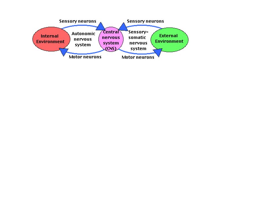
Recall Divisions of the N.S. Central N.S. Peripheral N.S. Efferent
Afferent Spinal Cord Brain CNS: brain and spinal cord PNS: all nerves coming from/to the CNS Afferent: carries impulses towards CNS Efferent: carries impulses away from CNS Somatic: goes to skeletal muscles (voluntary; responds to external stimuli; one ganglia) Autonomic: goes to viscera and glands (involuntary; responds to internal stimuli; two ganglia) Parasympathetic: rest and digest Sympathetic: fight or flight Autonomic Somatic Parasympathetic Sympathetic
 Autonomic: goes to viscera and glands (involuntary; responds to internal stimuli; two ganglia) Parasympathetic: rest and digest. Sympathetic: fight or flight. Autonomic. Somatic. Parasympathetic. Sympathetic.")
57
X. THE PERIPHERAL NS CNS PNS

58
X. THE PERIPHERAL NS A. Afferent vs Efferent

59
X. THE PERIPHERAL NS A. Afferent vs Efferent Afferent NS
Sensory neurons. Proprioceptors: sensitive to position and movement Pick up stimulus and carry it toward the CNS . Ventricles Lateral (1st & 2nd) (1) Cerebrum b. Interventricular foramen (foramen of Monro) c. Third (1) Starts in Cerebrum (2) Diencephalon (Thalamus, etc.) d. Cerebral aqueduct (1) Brain stem: midbrain e. Fourth (1) Aperatures (a) Lateral (2) (b) Median (1) (2) Brain stem: pons (3) Cerebellum (4) Brain stem: medulla oblongata f. Central Canal (1) Spinal cord
 (1) Cerebrum b. Interventricular foramen (foramen of Monro) c. Third (1) Starts in Cerebrum (2) Diencephalon (Thalamus, etc.) d. Cerebral aqueduct (1) Brain stem: midbrain e. Fourth (1) Aperatures (a) Lateral (2) (b) Median (1) (2) Brain stem: pons (3) Cerebellum (4) Brain stem: medulla oblongata f. Central Canal (1) Spinal cord.")
60
X. THE PERIPHERAL NS A. Afferent vs Efferent 2.Efferent NS
Motor neurons Carry response impulses from the CNS to the effector . Ventricles Lateral (1st & 2nd) (1) Cerebrum b. Interventricular foramen (foramen of Monro) c. Third (1) Starts in Cerebrum (2) Diencephalon (Thalamus, etc.) d. Cerebral aqueduct (1) Brain stem: midbrain e. Fourth (1) Aperatures (a) Lateral (2) (b) Median (1) (2) Brain stem: pons (3) Cerebellum (4) Brain stem: medulla oblongata f. Central Canal (1) Spinal cord
 (1) Cerebrum b. Interventricular foramen (foramen of Monro) c. Third (1) Starts in Cerebrum (2) Diencephalon (Thalamus, etc.) d. Cerebral aqueduct (1) Brain stem: midbrain e. Fourth (1) Aperatures (a) Lateral (2) (b) Median (1) (2) Brain stem: pons (3) Cerebellum (4) Brain stem: medulla oblongata f. Central Canal (1) Spinal cord.")
61
A. Afferent vs Efferent X. THE PERIPHERAL NS
3. Disorders of Afferent & Efferent systems Myasthenia Gravis: immune system attacks Ach; leads to muscle weakness and fatigue Multiple sclerosis: loss of myelin sheath of motor and sensory neurons; leads to limb weakness, tremors, vision disorders, vertigo . Ventricles Lateral (1st & 2nd) (1) Cerebrum b. Interventricular foramen (foramen of Monro) c. Third (1) Starts in Cerebrum (2) Diencephalon (Thalamus, etc.) d. Cerebral aqueduct (1) Brain stem: midbrain e. Fourth (1) Aperatures (a) Lateral (2) (b) Median (1) (2) Brain stem: pons (3) Cerebellum (4) Brain stem: medulla oblongata f. Central Canal (1) Spinal cord
 (1) Cerebrum b. Interventricular foramen (foramen of Monro) c. Third (1) Starts in Cerebrum (2) Diencephalon (Thalamus, etc.) d. Cerebral aqueduct (1) Brain stem: midbrain e. Fourth (1) Aperatures (a) Lateral (2) (b) Median (1) (2) Brain stem: pons (3) Cerebellum (4) Brain stem: medulla oblongata f. Central Canal (1) Spinal cord.")
62
B. Somatic vs Autonomic NS
X. THE PERIPHERAL NS B. Somatic vs Autonomic NS Somatic Controls skeletal muscles. Can be conscious or subconscious Has a single neuron between CNS and effector . Ventricles Lateral (1st & 2nd) (1) Cerebrum b. Interventricular foramen (foramen of Monro) c. Third (1) Starts in Cerebrum (2) Diencephalon (Thalamus, etc.) d. Cerebral aqueduct (1) Brain stem: midbrain e. Fourth (1) Aperatures (a) Lateral (2) (b) Median (1) (2) Brain stem: pons (3) Cerebellum (4) Brain stem: medulla oblongata f. Central Canal (1) Spinal cord
 (1) Cerebrum b. Interventricular foramen (foramen of Monro) c. Third (1) Starts in Cerebrum (2) Diencephalon (Thalamus, etc.) d. Cerebral aqueduct (1) Brain stem: midbrain e. Fourth (1) Aperatures (a) Lateral (2) (b) Median (1) (2) Brain stem: pons (3) Cerebellum (4) Brain stem: medulla oblongata f. Central Canal (1) Spinal cord.")
63
B. Divisions of the Efferent NS
X. THE PERIPHERAL NS B. Divisions of the Efferent NS 2. Autonomic Controls smooth muscles of viscera and glands Is subconscious Has two neurons between CNS and effector . Ventricles Lateral (1st & 2nd) (1) Cerebrum b. Interventricular foramen (foramen of Monro) c. Third (1) Starts in Cerebrum (2) Diencephalon (Thalamus, etc.) d. Cerebral aqueduct (1) Brain stem: midbrain e. Fourth (1) Aperatures (a) Lateral (2) (b) Median (1) (2) Brain stem: pons (3) Cerebellum (4) Brain stem: medulla oblongata f. Central Canal (1) Spinal cord
 (1) Cerebrum b. Interventricular foramen (foramen of Monro) c. Third (1) Starts in Cerebrum (2) Diencephalon (Thalamus, etc.) d. Cerebral aqueduct (1) Brain stem: midbrain e. Fourth (1) Aperatures (a) Lateral (2) (b) Median (1) (2) Brain stem: pons (3) Cerebellum (4) Brain stem: medulla oblongata f. Central Canal (1) Spinal cord.")
64
B. Divisions of the Efferent NS
X. THE PERIPHERAL NS B. Divisions of the Efferent NS 2. Autonomic c.Has two neurons between CNS and effector Preganglionic neurons Originate in spinal cord Ganglion neurons Nerve cell bodies Neurons in effector . Ventricles Lateral (1st & 2nd) (1) Cerebrum b. Interventricular foramen (foramen of Monro) c. Third (1) Starts in Cerebrum (2) Diencephalon (Thalamus, etc.) d. Cerebral aqueduct (1) Brain stem: midbrain e. Fourth (1) Aperatures (a) Lateral (2) (b) Median (1) (2) Brain stem: pons (3) Cerebellum (4) Brain stem: medulla oblongata f. Central Canal (1) Spinal cord
 (1) Cerebrum b. Interventricular foramen (foramen of Monro) c. Third (1) Starts in Cerebrum (2) Diencephalon (Thalamus, etc.) d. Cerebral aqueduct (1) Brain stem: midbrain e. Fourth (1) Aperatures (a) Lateral (2) (b) Median (1) (2) Brain stem: pons (3) Cerebellum (4) Brain stem: medulla oblongata f. Central Canal (1) Spinal cord.")
65
C. Sympathetic and Parasympathetic
X. THE PERIPHERAL NS C. Sympathetic and Parasympathetic 1. Sympathetic “Fight or Flight” Consists of Preganglionic neurons 2+ Ganglionic neurons Specialized neurons in adrenal gland (secretes hormone controlling production of Ach) . Ventricles Lateral (1st & 2nd) (1) Cerebrum b. Interventricular foramen (foramen of Monro) c. Third (1) Starts in Cerebrum (2) Diencephalon (Thalamus, etc.) d. Cerebral aqueduct (1) Brain stem: midbrain e. Fourth (1) Aperatures (a) Lateral (2) (b) Median (1) (2) Brain stem: pons (3) Cerebellum (4) Brain stem: medulla oblongata f. Central Canal (1) Spinal cord
 . Ventricles. Lateral (1st & 2nd) (1) Cerebrum b. Interventricular foramen (foramen of Monro) c. Third (1) Starts in Cerebrum (2) Diencephalon (Thalamus, etc.) d. Cerebral aqueduct (1) Brain stem: midbrain e. Fourth (1) Aperatures (a) Lateral (2) (b) Median (1) (2) Brain stem: pons (3) Cerebellum (4) Brain stem: medulla oblongata f. Central Canal (1) Spinal cord.")
67
A. Gate Control Theory XI. Pain Perception
Without any stimulation, both large and small nerve fibers are quiet and the inhibitory interneuron (I) blocks the signal in the projection neuron (P) that connects to the brain. The "gate is closed" and therefore NO PAIN . Ventricles Lateral (1st & 2nd) (1) Cerebrum b. Interventricular foramen (foramen of Monro) c. Third (1) Starts in Cerebrum (2) Diencephalon (Thalamus, etc.) d. Cerebral aqueduct (1) Brain stem: midbrain e. Fourth (1) Aperatures (a) Lateral (2) (b) Median (1) (2) Brain stem: pons (3) Cerebellum (4) Brain stem: medulla oblongata f. Central Canal (1) Spinal cord
 blocks the signal in the projection neuron (P) that connects to the brain. The gate is closed and therefore NO PAIN. . Ventricles. Lateral (1st & 2nd) (1) Cerebrum b. Interventricular foramen (foramen of Monro) c. Third (1) Starts in Cerebrum (2) Diencephalon (Thalamus, etc.) d. Cerebral aqueduct (1) Brain stem: midbrain e. Fourth (1) Aperatures (a) Lateral (2) (b) Median (1) (2) Brain stem: pons (3) Cerebellum (4) Brain stem: medulla oblongata f. Central Canal (1) Spinal cord.")
68
A. Gate Control Theory XI. Pain Perception
2.With non-painful stimulation, large nerve fibers are activated primarily. This activates the projection neuron (P), BUT it ALSO activates the inhibitory interneuron (I) which then BLOCKS the signal in the projection neuron (P) that connects to the brain. The "gate is closed" and therefore NO PAIN. . Ventricles Lateral (1st & 2nd) (1) Cerebrum b. Interventricular foramen (foramen of Monro) c. Third (1) Starts in Cerebrum (2) Diencephalon (Thalamus, etc.) d. Cerebral aqueduct (1) Brain stem: midbrain e. Fourth (1) Aperatures (a) Lateral (2) (b) Median (1) (2) Brain stem: pons (3) Cerebellum (4) Brain stem: medulla oblongata f. Central Canal (1) Spinal cord
, BUT it ALSO activates the inhibitory interneuron (I) which then BLOCKS the signal in the projection neuron (P) that connects to the brain. The gate is closed and therefore NO PAIN. . Ventricles. Lateral (1st & 2nd) (1) Cerebrum b. Interventricular foramen (foramen of Monro) c. Third (1) Starts in Cerebrum (2) Diencephalon (Thalamus, etc.) d. Cerebral aqueduct (1) Brain stem: midbrain e. Fourth (1) Aperatures (a) Lateral (2) (b) Median (1) (2) Brain stem: pons (3) Cerebellum (4) Brain stem: medulla oblongata f. Central Canal (1) Spinal cord.")
69
A. Gate Control Theory XI. Pain Perception
3.With pain stimulation, small nerve fibers become active. They activate the projection neurons (P) and BLOCK the inhibitory interneuron (I). Because activity of the inhibitory interneuron is blocked, it CANNOT block the output of the projection neuron that connects with the brain. The "gate is open", therefore, PAIN!! . Ventricles Lateral (1st & 2nd) (1) Cerebrum b. Interventricular foramen (foramen of Monro) c. Third (1) Starts in Cerebrum (2) Diencephalon (Thalamus, etc.) d. Cerebral aqueduct (1) Brain stem: midbrain e. Fourth (1) Aperatures (a) Lateral (2) (b) Median (1) (2) Brain stem: pons (3) Cerebellum (4) Brain stem: medulla oblongata f. Central Canal (1) Spinal cord
 and BLOCK the inhibitory interneuron (I). Because activity of the inhibitory interneuron is blocked, it CANNOT block the output of the projection neuron that connects with the brain. The gate is open , therefore, PAIN!! . Ventricles. Lateral (1st & 2nd) (1) Cerebrum b. Interventricular foramen (foramen of Monro) c. Third (1) Starts in Cerebrum (2) Diencephalon (Thalamus, etc.) d. Cerebral aqueduct (1) Brain stem: midbrain e. Fourth (1) Aperatures (a) Lateral (2) (b) Median (1) (2) Brain stem: pons (3) Cerebellum (4) Brain stem: medulla oblongata f. Central Canal (1) Spinal cord.")
70
I = "Inhibitory Interneuron"; P = "Projection Neuron" - = inhibition (blocking); + = excitation (activation)
; + = excitation (activation)")
71
XI. Pain Perception B. Controlling Pain
1.Aspirin: acts mostly in PNS; reduces inflammation 2.Morphine: acts in CNS to block pain messages 3.Acupuncture: stimulates large diameter fibers that inhibit pain (closes the gate) . Other pain drugs act on a variety of neurotransmitter systems
 . Other pain drugs act on a variety of neurotransmitter systems.")
Similar presentations


>")


 Pia mater -inner membrane, contains.>")


 Bone – Cranium, Vertebrae 2) Meninges – Three connective tissue membranes covering the brain and spinal cord a) Dura Mater – outermost,>")
 Brain and spinal cord Peripheral Nervous System (PNS) ◦ nerves.>")


