Download presentation
Presentation is loading. Please wait.

1
ESOPHAGEAL CA EZE BLESSING 1466

3
introduction Esophageal cancer (or oesophageal cancer) is cancer arising from the foodpipe known as the esophagus that runs between the throat and the stomach. Pnemonia can be a complication.
 is cancer arising from the foodpipe known as the esophagus that runs between the throat and the stomach. Pnemonia can be a complication.")
4
BENIGN Squamous Papilloma Esophageal leiomyoma
Papillary fronds lined by several layers of hyperplastic squamous epithelium around a fibrovascular core May be associated with HPV Demarcated intramural nodule, composed of composed of irregularly oriented bundles of well-differentiated smooth-muscle cells Normal Squamous epithelium is seen on top

5
TYPES The two main sub-types of the disease are esophageal squamous-cell carcinoma (often abbreviated to ESCC), which is more common in the developing world, and esophageal adenocarcinoma (EAC), which is more common in the developed world. A general rule of thumb is that a cancer in the upper two-thirds and middle one 3rd is likely to be ESCC and one in the lower one-third EAC.
, which is more common in the developing world, and esophageal adenocarcinoma (EAC), which is more common in the developed world. A general rule of thumb is that a cancer in the upper two-thirds and middle one 3rd is likely to be ESCC and one in the lower one-third EAC.")
7
SQUAMOUS CELL CANCER Arises from epithelial cells of the esophagus
RISK FACTORS:The most common causes of the squamous-cell type are: tobacco, alcohol, achalasia,very hot drinks, and a poor diet. High levels of dietary exposure to nitrosamines.oral hygiene Chewing betel nut (areca) is an important risk factor in Asia Genetic factors: p16/INK4 tumor suppressor gene and EGFR, p53 in 50% of esophageal cancers
 is an important risk factor in Asia. Genetic factors: p16/INK4 tumor suppressor gene and EGFR, p53 in 50% of esophageal cancers.")
8
Squamous cell carcinoma – morphology
Mucosal epithelial dysplasia -> carcinoma in situ -> invasive cancer polypoid exophytic masses necrotizing ulcerations diffuse infiltrative neoplasms

9
ESCC STAGING Stage 1A:Cancer has grown through the inner layer and invaded the wall of the esophagus. The grade is 1. Stage IB Cancer has invaded the wall of the esophagus and is grade 2 or 3. Or, cancer is found in the lower part of the esophagus, it has invaded the muscle layer or outer layer of the esophagus, and the grade is 1. Stage IIA Cancer is found in the upper or middle part of the esophagus, it has invaded the muscle layer or outer layer of the esophagus, and the grade is 1. Or, cancer is found in the lower part of the esophagus, it has invaded the muscle layer or outer layer of the esophagus, and the grade is 2 or 3. Stage IIB Cancer is found in the upper or middle part of the esophagus, it has invaded the muscle layer or outer layer of the esophagus, and the grade is 2 or 3. Or, cancer has not invaded the outer layer, and cancer cells are found in one or two nearby lymph nodes.

10
ADENO CA The most common causes of the adenocarcinoma type are GERD smoking tobacco, obesity, and acid reflux RISK FACTORS: male gender, acid reflux, obesity. Adenocarcinoma is the more common type of esophageal cancer. Having Barrett esophagus increases the risk of this type of cancer. Acid reflux disease (gastroesophageal reflux disease, or GERD) can develop into Barett esophagus. Other risk factors include smoking, being male, or being obese. PROTECTIVE EFFECT: Female hormone, h pylori, prolonged period of breastfeeding declines risk
 can develop into Barett esophagus. Other risk factors include smoking, being male, or being obese. PROTECTIVE EFFECT: Female hormone, h pylori, prolonged period of breastfeeding declines risk.")
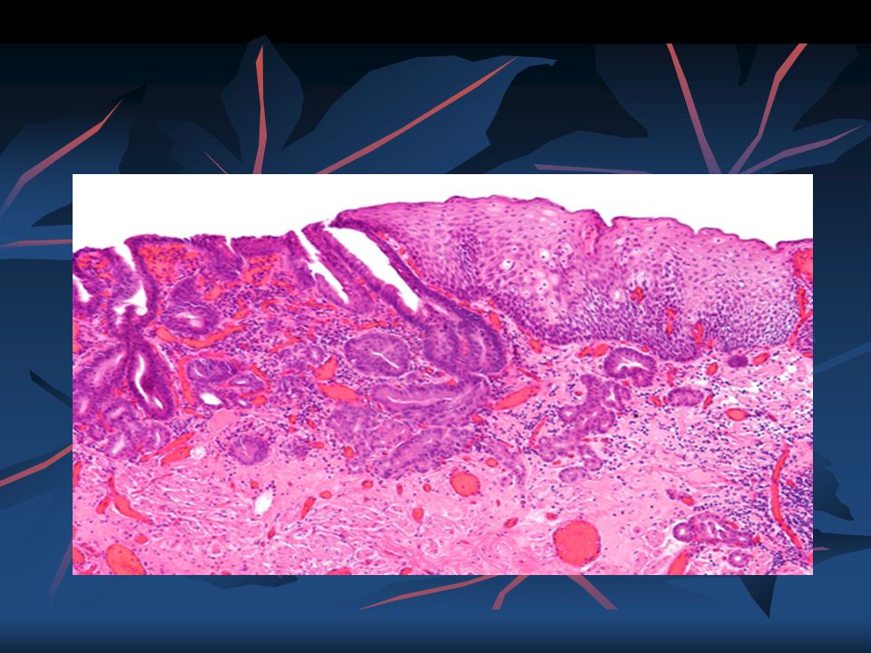
11
Barrett esophagus - sequelae
Ulceration, Bleeding, Stricture Adenocarcinoma

13
Usually distal esophageal tumor with invasion of gastric cardia; appears as flat patches to nodular masses; may have adjacent Barrett mucosa

15
EAC STAGING Stage IA Cancer has grown through the inner layer and invades the wall of the esophagus. The grade is 1 or 2. Stage IB Cancer has invaded the wall of the esophagus and is grade 3. Or, cancer has invaded more deeply into the muscle layer of the esophagus, and the grade is 1 or 2. Stage IIA Cancer has invaded the muscle layer of the esophagus, and the grade is 3. Stage IIB Cancer has invaded the outer layer of the esophagus. Or, cancer has not invaded the outer layer, but cancer cells are also found in one or two nearby

16
GERD cont. The long-term erosive effects of acid reflux (an extremely common condition, also known as gastroesophageal reflux disease or GERD) have been strongly linked to this type of cancer. Longstanding GERD can induce a change of cell type in the lower portion of the esophagus in response to erosion of its squamous lining.This phenomenon, known as Barrett's esophagus, seems to appear about 20 years later in women than in men, maybe due to hormonal factors. Having symptomatic GERD or bile reflux makes Barrett's esophagus more likely, which in turn raises the risk of further changes that can ultimately lead to adenocarcinoma. The risk of developing adenocarcinoma in the presence of Barrett's esophagus is unclear, and may in the past have been overestimated.
 have been strongly linked to this type of cancer. Longstanding GERD can induce a change of cell type in the lower portion of the esophagus in response to erosion of its squamous lining.This phenomenon, known as Barrett s esophagus, seems to appear about 20 years later in women than in men, maybe due to hormonal factors. Having symptomatic GERD or bile reflux makes Barrett s esophagus more likely, which in turn raises the risk of further changes that can ultimately lead to adenocarcinoma. The risk of developing adenocarcinoma in the presence of Barrett s esophagus is unclear, and may in the past have been overestimated.")
17
sypmtoms Painful or difficult swallowing Weight loss
A hoarse voice or cough that doesn't go away enlarged lymph nodes (glands) around the collarbone, a dry cough, and possibly coughing up or vomiting blood Backwards movement of food through the esophagus and possibly mouth (regurgitation) Chest pain and heart burns Vomiting blood
 around the collarbone, a dry cough, and possibly coughing up or vomiting blood. Backwards movement of food through the esophagus and possibly mouth (regurgitation) Chest pain and heart burns. Vomiting blood.")
18
EPIDERMIOLOGY Esophageal cancer is the eighth most frequently diagnosed cancer worldwide, and because of its poor prognosis it is the sixth most common cause of cancer-related death ESCC comprises 60–70% of all cases of esophageal cancer worldwide, while EAC accounts for a further 20–30% (melanomas, leiomyosarcomas, carcinoids and lymphomas are less common types). In general, ESCC is more common in the developing world, and EAC is more common in the developed world In Western countries, EAC has become the dominant form of the disease, following an increase in incidence over recent decades (in contrast to the incidence of ESCC, which has remained largely stable). In 2012, the global incidence rate for EAC was 0.7 per 100,000 with a strong male predominance (1.1 per 100,000 in men vs. 0.3 in women) Areas with particularly high incidence rates include northern and western Europe, north America and Oceania. The countries with highest recorded rates were the UK, Netherlands, Ireland, Iceland and New Zealand
. In general, ESCC is more common in the developing world, and EAC is more common in the developed world. In Western countries, EAC has become the dominant form of the disease, following an increase in incidence over recent decades (in contrast to the incidence of ESCC, which has remained largely stable). In 2012, the global incidence rate for EAC was 0.7 per 100,000 with a strong male predominance (1.1 per 100,000 in men vs. 0.3 in women) Areas with particularly high incidence rates include northern and western Europe, north America and Oceania. The countries with highest recorded rates were the UK, Netherlands, Ireland, Iceland and New Zealand.")
19
Doctor uses imaging tests and a biopsy to diagnose esophageal cancer
The disease is diagnosed by biopsy done by an endoscope (a fiberoptic camera) generally tend to be fairly poor, as diagnosis is often late. Five-year survival rates are around 13% to 18% Staging. CT scan Esophagogastroduodenoscopy (EGD) and biopsy PET scan (sometimes useful for determining the stage of disease, and whether surgery is possible DIAGNOSES
 generally tend to be fairly poor, as diagnosis is often late. Five-year survival rates are around 13% to 18% Staging. CT scan. Esophagogastroduodenoscopy (EGD) and biopsy. PET scan (sometimes useful for determining the stage of disease, and whether surgery is possible. DIAGNOSES.")
20
PREVENTION Do not smoke Limit or do not drink alcoholic beverages
Get checked by your doctor if you have severe GERD Get regular checkups if you have Barrett esophagus

21
TREATMENT Treatment is based on the cancer's stage and location, together with the person's general condition and individual preferences. Chemotherapy surgery

22
A 58-year-old man had dysphagia
Irregular reddish, ulcerated exophytic mid-esophageal mass seen on mucosal surface Diagnosis? Risk factors?

23
A 57 years old man present with cough and weight loss
A 57 years old man present with cough and weight loss. She has noticed a gradual onset difficulty with swallowing both solid and luquid food. She has been soking for the past 20 yrs and also ingest hard liqueur. He has lost 20lbs in last few months. Esophageal biopsy reveals presence of squamous cells with keratin. Which is correct A. Barret esophagus is the major risk factor B. GERD is a common risk factor. C. The tumour is most likely n the upper one third of the esophagus D. Primary ciliary dyskinesia

24
A 37 years old male with frequent episodes of gastro-esophageal reflux disease noticed a dramatic improvement in the frequency and severity of her heart burn. She consulted Dr.Blessing who ordered endoscopy. Esophageal biopsy taken reveals the presence of columner type epithelium with increased Goblet cells in gastro eso junction. Which is correct A. risk of squamous cell cancer is increased B. it is an example of a dysplasia C. It represents malignant transformtion D.Risk of adeno-carcinoma is incresed

25
REFERENCES Das A. Tumors of the esophagus. In: Feldman M, Friedman LS, Brandt LJ, eds. Sleisenger and Fordtran's Gastrointestinal and Liver Disease. 9th ed. Philadelphia, PA: Elsevier Saunders; 2010:chap 46. PATOMA Robins patholgy Dr Jeevan’s slides

Similar presentations


 has Barrett’s esophagus diagnosed on upper endoscopy. Which of.>")


 primary gastric lymphoma gastric polyps.>")


 Cancer>")

