Download presentation
Presentation is loading. Please wait.

1
ENDOCRINE PART 2 Chapter 51

2
The Pituitary http://www. youtube. com/watch
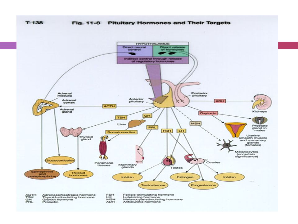
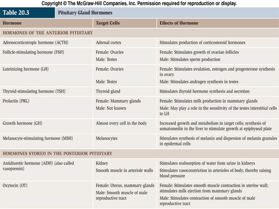
The pituitary is located at the base of the brain, in a small depression of the sphenoid bone (sella turcica). Purpose: control the activity of many other endocrine glands. “ Master gland” Has two lobes, the anterior & posterior lobes.
. Purpose: control the activity of many other endocrine glands. Master gland Has two lobes, the anterior & posterior lobes.")
3
Anatomy Anterior lobe: glandular tissue, accounts for 75% of total weight. Hormones in this lobe are controlled by regulating hormones from the hypothalmus (stimulate or inhibit). Posterior: contains axons that originate in the hypothalmus. Therefore this lobe does not produce hormones but stores those produced by the neurosecretory cells in the hypothalmus. Release of hormones is triggered by receptors in the hypothalmus. Pituitary = hypophysis Anterior lobe called the adenohypophysis Posterior lobe called the neurohypophysis
. Posterior: contains axons that originate in the hypothalmus. Therefore this lobe does not produce hormones but stores those produced by the neurosecretory cells in the hypothalmus. Release of hormones is triggered by receptors in the hypothalmus. Pituitary = hypophysis. Anterior lobe called the adenohypophysis. Posterior lobe called the neurohypophysis.")
5
Terms Trophic hormones: hormones that control the secretion of hormones by other glands. Example: TSH stimulates the thyroid to secrete hormones. Effector hormones: produce an effect directly when secreted. Example ADH stimulates kidneys

6
Review – Hormones Posterior Anterior

8
Anterior Pituitary Secretes:
GH: stimulates growth of bone and muscle , promotes protein synthesis and fat metabolism. ACTH (Adrenocorticotropin ): stimulates adrenal cortex secretion of mineralcorticoids (aldosterone) & glucocorticoids (cortisol), & androgens. TSH: stimulates thyroid to increase secretion of thyroxine, its control is from regulating hormones in the hypothalmus. GH: stimulates growth; promotes active transport of amino acids into cell & influences the rate at which CHO and fats are catabolized. Stimulates growth in childhood, is important for maintaining a healthy body composition and well-being in adults. In adults it is important for maintaining muscle mass as well as bone mass. It also plays a role in maintenance of constant blood sugar levels (through conversion of conversion of glycogen to glucose) ACTH (Adrenocorticotropin ): stimulates adrenal gland cortex secretion of mineralcorticoids (aldosterone) & glucocorticoids (cortisol). Cortisol, a so-called "stress hormone" is vital to survival. It helps maintain blood pressure and blood glucose levels. Aldosterone maintain electrolyte/water balance. TSH: stimulates thyroid to increase secretion of thyroxine, regulates the body's metabolism, energy, growth and development, and nervous system activity- vital to survival
: stimulates adrenal cortex secretion of mineralcorticoids (aldosterone) & glucocorticoids (cortisol), & androgens. TSH: stimulates thyroid to increase secretion of thyroxine, its control is from regulating hormones in the hypothalmus. GH: stimulates growth; promotes active transport of amino acids into cell & influences the rate at which CHO and fats are catabolized. Stimulates growth in childhood, is important for maintaining a healthy body composition and well-being in adults. In adults it is important for maintaining muscle mass as well as bone mass. It also plays a role in maintenance of constant blood sugar levels (through conversion of conversion of glycogen to glucose) ACTH (Adrenocorticotropin ): stimulates adrenal gland cortex secretion of mineralcorticoids (aldosterone) & glucocorticoids (cortisol). Cortisol, a so-called stress hormone is vital to survival. It helps maintain blood pressure and blood glucose levels. Aldosterone maintain electrolyte/water balance. TSH: stimulates thyroid to increase secretion of thyroxine, regulates the body s metabolism, energy, growth and development, and nervous system activity- vital to survival.")
9
Anterior Pituitary Cont’d
Prolactin: stimulates milk production from the breasts after childbirth to enable nursing. Oxytoxin from posterior lobe controls milk ejection. FSH: promotes sperm production in men and stimulates the ovaries to enable ovulation in women. LH and FSH work together to cause normal function of the ovaries and testes. LH: regulates testosterone in men and estrogen, progesterone in women. Prolactin: stimulates milk production from the breasts after childbirth to enable nursing; controlled by regulating hormones from hypothalmus.can FSH: promotes sperm production in men and stimulates the ovaries to enable ovulation in women. LH and FSH work together to cause normal function of the ovaries and testes. LH: regulates testosterone in men and estrogen in women

10
Posterior Pituitary Antidiuretic hormone or ADH - also called vasopressin, constricts arterioles to increase arterial pressure; increases water reabsorption in distal tubules. Oxytocin: stimulates uterus to contract at childbirth; stimulates mammary ducts to contract (milk ejection in lactation). ADH & oxytocin are called EFFECTOR hormones b/c they produce an effect when secreted. Whereas: Hypothalmic hormones stimulate the posterior pituitary to release TROPHIC (gland-stimulating) hormones.
. ADH & oxytocin are called EFFECTOR hormones b/c they produce an effect when secreted. Whereas: Hypothalmic hormones stimulate the posterior pituitary to release TROPHIC (gland-stimulating) hormones.")
11
Anterior Pituitary Disorders
Hormone Increased level Decreased level GH Gigantism (child) Acromegaly (adult) Dwarfism (child) Lethargy, premature aging ACTH Cushing’s Disease Addison’s Disease TSH Goiter, increased BMR, HR, BP Graves disease Decreased BMR, HR, CO, BP Cretinism (children) Prolactin Amenorrhea,glactorhea Too little milk FSH Late puberty, infertility LH Menstrual cycle disturbance Amenorrhea, impotence Trophic hormones: stimulate another gland. i.e. ACTH, TSH, FSH, LH. Major causes of pituitary disease include tumors, pituitary infarction, genetic disorders and trauma. Pituitary tumors produce local (increased pressure in the cranium – visual field abnormalities, headaches and somnolence) and systemic effects from over or under production of hormones.. Treatment involves removal of pituitary tumors.
 Acromegaly (adult) Dwarfism (child) Lethargy, premature aging. ACTH. Cushing’s Disease. Addison’s Disease. TSH. Goiter, increased BMR, HR, BP. Graves disease. Decreased BMR, HR, CO, BP. Cretinism (children) Prolactin. Amenorrhea,glactorhea. Too little milk. FSH. Late puberty, infertility. LH. Menstrual cycle disturbance. Amenorrhea, impotence. Trophic hormones: stimulate another gland. i.e. ACTH, TSH, FSH, LH. Major causes of pituitary disease include tumors, pituitary infarction, genetic disorders and trauma. Pituitary tumors produce local (increased pressure in the cranium – visual field abnormalities, headaches and somnolence) and systemic effects from over or under production of hormones.. Treatment involves removal of pituitary tumors.")
12
Posterior Pituitary Disorders
Hormone Increased Decreased Oxytocin Precipitates childbirth, excess milk Prolonged childbirth, diminished milk ADH (vasopressin) Increased BP, decreased urinary output, edema. SIADH Diabetes insipidus, dilute urine & increased urine output
 Increased BP, decreased urinary output, edema. SIADH. Diabetes insipidus, dilute urine & increased urine output.")
13
Pituitary Disorders Disorders occur most often in the anterior pituitary The anterior pituitary hormones regulates growth, metabolic activity and sexual development. Major causes include: tumors, pituitary infarction, genetic disorders. Pathologic consequences of pituitary disorders are 1) hyperpituitarism, 2) hypopituitarism, 3) local compression of brain tissue by expanding tumor
 hyperpituitarism, 2) hypopituitarism, 3) local compression of brain tissue by expanding tumor.")
14
Hyperpituitarism

15
Hyperfunction Results in excess production/secretion of one or more hormones: GH, PRL, ACTH. Most common cause is a benign adenoma. Three main types of pituitary tumors represent overgrowth of 1) eosinophilic cells (gigantism, acromegaly), 2) basophilic cells (Cushing’s disease) or 3) chromophobic cells (cause destruction of pituitary gland).
 eosinophilic cells (gigantism, acromegaly), 2) basophilic cells (Cushing’s disease) or 3) chromophobic cells (cause destruction of pituitary gland).")
16
Pituitary Adenoma Anterior pituitary adenoma, a benign tumor which is classified according to size, degree of invasiveness and the hormone secreted. Prolactin and GH are the hormones most commonly over-produced by adenomas.

17
Adenoma’s Cont’d Changes in neurological function may occur as adenomas compress surrounding tissue. Manifestations include headaches, visual defects and increased ICP.

18
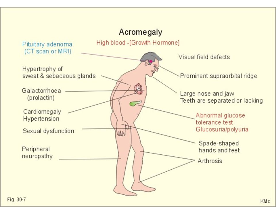
Increased GH Gigantism & Acromegaly
Gigantism is the result of GH hypersecretion before the closure of the epiphyseal plates (childhood). Abnormally tall but body proportions are normal GH is a hormone necessary for growth, regulates cell division, synthesis of protein. It exerts metabolic effects on organs, skin, muscle and connective tissue. GH is a peptide anterior pituitary hormone essential for growth. GH-releasing hormone stimulates release of GH. GH-inhibiting hormone suppresses the release of GH. The hypothalamus maintains homeostatic levels of GH. Cells under the action of GH increase in size (hypertrophy) and number (hyperplasia). GH also causes increase in bone length and thickness by deposition of cartilage at the ends of bones. During adolescence, sex hormones cause replacement of cartilage by bone, halting further bone growth even though GH is still present. Too little or two much GH can cause dwarfism or gigantism, respectively.
. Abnormally tall but body proportions are normal. GH is a hormone necessary for growth, regulates cell division, synthesis of protein. It exerts metabolic effects on organs, skin, muscle and connective tissue. GH is a peptide anterior pituitary hormone essential for growth. GH-releasing hormone stimulates release of GH. GH-inhibiting hormone suppresses the release of GH. The hypothalamus maintains homeostatic levels of GH. Cells under the action of GH increase in size (hypertrophy) and number (hyperplasia). GH also causes increase in bone length and thickness by deposition of cartilage at the ends of bones. During adolescence, sex hormones cause replacement of cartilage by bone, halting further bone growth even though GH is still present. Too little or two much GH can cause dwarfism or gigantism, respectively.")
19
Acromegaly is over secretion of GH in adulthood
Continued growth of boney, connective tissue leads to disproportionate enlargement of tissue..

20
Acromegaly Acromegaly is a very rare condition and usually develops between the ages of 30 and 50, with excess production of GH after the person has stopped growing (which usually occurs between the ages of 15 to 17 years of age).
.")
21
Acromegaly Rare condition – develops between ages 30-50
Manifestations: Coarsening of facial features Enlarged hands & feet Carpel tunnel syndrome Excessive sweating & oily skin Headaches Vision disturbance Sleep apnea General tiredness Oligomenorrhea or amenorrhea Impotence (adult males) Decreased libido
 Decreased libido.")
23
Diagnosis History & physical exam Investigation includes:
GH analysis (glucose tolerance) Normally GH concentarion falls with oral glucose; in acromegaly it does not. Prolactin levels as well as other pituitary function tests MRI or CT & visual field tests to determine size and position of the adenoma. Bone scan History: note change in hat, glove, shoe or ring size. Report fatigue and lethargy Backache & arthralgia Visual defects and headaches Laboratory Analysis In hyperpituitarism, usually only one hormone is produced in excess, most common being PRL, ACTH, GH Physical Exam Changes in facial features; in head, hand & foot size Arthritic type changes Vision changes from compression on optic nerves HTN Organomegaly (cardiac or hepatic) Deepened voice Dysphagia from enlarged toungue
 Normally GH concentarion falls with oral glucose; in acromegaly it does not. Prolactin levels as well as other pituitary function tests. MRI or CT & visual field tests to determine size and position of the adenoma. Bone scan. History: note change in hat, glove, shoe or ring size. Report fatigue and lethargy. Backache & arthralgia. Visual defects and headaches. Laboratory Analysis. In hyperpituitarism, usually only one hormone is produced in excess, most common being PRL, ACTH, GH. Physical Exam. Changes in facial features; in head, hand & foot size. Arthritic type changes. Vision changes from compression on optic nerves. HTN. Organomegaly (cardiac or hepatic) Deepened voice. Dysphagia from enlarged toungue.")
24
Treatment Surgery (primary choice) Stereotactic Radiation therapy
Drug treatment – when surgery is not feasible Combinations of above Acromegaly may be treated by operating on the pituitary gland, by radiotherapy, by drug treatment, or a combination of these. The aim of all treatments is to reduce growth hormone levels to normal as this is associated with the disappearance of symptoms and improvement of your general wellbeing. Transphenoidal pituitary surgery and removal of adenoma. Surgery is the gold standard of treatment in clients who are acceptable surgical risks. Radiation is indicated in clients where surgery failed to bring GH down to normal levels or in poor surgical candidates. There is a higher risk of panhypopituitarism with this. However neurologic complications are possible such as visual loss, weakness and memory impairment.

25
Transsphenoidal Surgery

26
Drug treatment of Acromegaly
Somatostatin analogs: stop GH production (octreocide acetate) GH receptor antagonists: (GHRAs) interfere with the action of GH & decreases the action of GH on target tissues. (pegvisomant) Dopamine agonists: Dopamine agonists work on dopamine receptors on the surface of the tumor to inhibit GH release from the tumour (cabergoline). Drug Treatment Octreocert acetate: Somavert Dopamine agonist: Parlodel Medical treatment: Octreotide causes tumor shrinkage; Dostinex & Parlodel (dopamine agonists) lower GH secretion; Somavert blocks the action of GH New developments Pegvisomant (Somavert) is a completely new way of treating acromegaly. All current forms of treatment attempt to lower the amount of GH released by the pituitary gland. Pegvisomant is a blocker of the action of GH. It does not try to inhibit the release of GH from the pituitary into the blood but instead stops the GH leaving the blood to stick to cells throughout the body. This should block all the unwanted effects of GH and studies in patients with acromegaly suggest it is very effective. It is hoped it will be available in the near future.
 GH receptor antagonists: (GHRAs) interfere with the action of GH & decreases the action of GH on target tissues. (pegvisomant) Dopamine agonists: Dopamine agonists work on dopamine receptors on the surface of the tumor to inhibit GH release from the tumour (cabergoline). Drug Treatment. Octreocert acetate: Somavert. Dopamine agonist: Parlodel. Medical treatment: Octreotide causes tumor shrinkage; Dostinex & Parlodel (dopamine agonists) lower GH secretion; Somavert blocks the action of GH. New developments. Pegvisomant (Somavert) is a completely new way of treating acromegaly. All current forms of treatment attempt to lower the amount of GH released by the pituitary gland. Pegvisomant is a blocker of the action of GH. It does not try to inhibit the release of GH from the pituitary into the blood but instead stops the GH leaving the blood to stick to cells throughout the body. This should block all the unwanted effects of GH and studies in patients with acromegaly suggest it is very effective. It is hoped it will be available in the near future.")
28
Hypopituitarism: Anterior Pituitary
Hormone deficiency caused by the inadequate secretion of one or more of the hormones normally secreted by the pituitary, is known as hypopituitarism. It may be caused by compression of the normal tissue by a developing tumour, surgery, or radiotherapy. If hypopituitarism is caused by tumour compression, function may be partially or fully recovered after surgery or medical therapy to reduce the size of the tumour. Surgically-induced hypopituitarism may occur in some cases, particularly if the tumour is large, but this can be transitory and some or all of the function may recover. Hypopituitarism may also develop after several years of treatment of a pituitary tumour by radiotherapy.

29
Decreased GH in child: Dwarfism
Condition of being undersized There are many forms of dwarfism, some are genetic. Dwarfism related to pituitary gland is the result of insufficient GH Pituitary dwarfism is successfully treated by administering human growth hormone Dwarfism, condition of being undersized, or less than 127 cm (50 in) in height. Some dwarfs have been less than 64 cm (24 in) tall when fully grown. The term midget is usually applied to physically well-proportioned dwarfs. The term pygmy is applied to people whose shortness of stature is a racial trait and not caused by disease. Cretinism, a result of a disease of the thyroid gland, is the cause of most dwarfism in Europe, Canada, and the United States . Other causes of dwarfism are Down syndrome, a congenital condition with symptoms similar to those of cretinism; achondroplasia, a disease characterized by short extremities resulting from absorption of cartilaginous tissue during the fetal stage; hypochondroplasia, a milder form of achondroplasia; spinal tuberculosis; and deficiency of the secretions of the pituitary gland or of the ovary. Geneticists have recently found the gene responsible for achondroplasia and hypochondroplasia, the most common forms of dwarfism. Treatment of cretinism with thyroxine or thyroid extract early in infancy results in normal growth and development. Pituitary dwarfism is successfully treated by administering human growth hormone.
 in height. Some dwarfs have been less than 64 cm (24 in) tall when fully grown. The term midget is usually applied to physically well-proportioned dwarfs. The term pygmy is applied to people whose shortness of stature is a racial trait and not caused by disease. Cretinism, a result of a disease of the thyroid gland, is the cause of most dwarfism in Europe, Canada, and the United States . Other causes of dwarfism are Down syndrome, a congenital condition with symptoms similar to those of cretinism; achondroplasia, a disease characterized by short extremities resulting from absorption of cartilaginous tissue during the fetal stage; hypochondroplasia, a milder form of achondroplasia; spinal tuberculosis; and deficiency of the secretions of the pituitary gland or of the ovary. Geneticists have recently found the gene responsible for achondroplasia and hypochondroplasia, the most common forms of dwarfism. Treatment of cretinism with thyroxine or thyroid extract early in infancy results in normal growth and development. Pituitary dwarfism is successfully treated by administering human growth hormone.")
30
Hypopituitarism (Adult)- GH
Lack of GH leads to: Increased CV disease Excessive tiredness Anxiety Depression Reduced quality of life Possible premature death Complete lack of GH is not fatal in adults as would be the case with some hormones, but it can have major detrimental effects that may cause disease some years earlier than might otherwise have been the case. Studies have shown that patients with underactive pituitary glands (hypopituitarism) replaced with all hormones except GH have an increased risk of dying prematurely, possible because of cardiovascular disease. Lack of GH causes changes in blood cholesterol concentrations (and also a number of other chemicals involved in blood clotting) which, in other studies, have been associated with an increased risk of heart disease. GH deficient adults have been shown to suffer from excessive tiredness, anxiety, depression and generally feeling unwell, as well as having feelings of social isolation and a tendency to be easily upset. This can be severe enough to lead to an inability to work or to maintain a basic acceptable lifestyle. These factors have led most studies to conclude that GH deficiency results in reduced 'quality of life'. Symptoms of Hormone Deficiency Symptoms Deficient Hormone Growth retardation in children, excessive tiredness, muscle weakness GH Hypogonadism - reduced body hair, low libido, impotence in men; menorrhoea, dyspareunia and hot flushes in women FSH/LH: Weight gain, decreased energy, sensitivity to cold, constipation, dry skinTSHPale appearance, weight loss, low blood pressure, dizziness, tiredness ACTH thirst and polyuriaAVP Acute hypopituitarism - sudden headache, collapse, hypothermia, hypoglycaemia and hypotension. This may be a life-threatening emergency.
 replaced with all hormones except GH have an increased risk of dying prematurely, possible because of cardiovascular disease. Lack of GH causes changes in blood cholesterol concentrations (and also a number of other chemicals involved in blood clotting) which, in other studies, have been associated with an increased risk of heart disease. GH deficient adults have been shown to suffer from excessive tiredness, anxiety, depression and generally feeling unwell, as well as having feelings of social isolation and a tendency to be easily upset. This can be severe enough to lead to an inability to work or to maintain a basic acceptable lifestyle. These factors have led most studies to conclude that GH deficiency results in reduced quality of life . Symptoms of Hormone Deficiency Symptoms Deficient Hormone Growth retardation in children, excessive tiredness, muscle weakness GH Hypogonadism - reduced body hair, low libido, impotence in men; menorrhoea, dyspareunia and hot flushes in women FSH/LH: Weight gain, decreased energy, sensitivity to cold, constipation, dry skinTSHPale appearance, weight loss, low blood pressure, dizziness, tiredness ACTH thirst and polyuriaAVP. Acute hypopituitarism - sudden headache, collapse, hypothermia, hypoglycaemia and hypotension. This may be a life-threatening emergency.")
31
Hyperprolactemia Prolactin levels are normally high during pregnancy and lactation. Symptoms of hyperprolactemia include; discharge from breasts (galactorrhoea) oligomenorrhoea or amenorrhoea in women reduced libido and potency in men pressure effects (e.g. headache and visual disturbance) - more commonly in men Increased levels caused by: prolactin-secreting pituitary tumor A non-secreting tumor that prevents dopamine (PRIH) from reaching normal prolactin-producing cells.
 oligomenorrhoea or amenorrhoea in women. reduced libido and potency in men. pressure effects (e.g. headache and visual disturbance) - more commonly in men. Increased levels caused by: prolactin-secreting pituitary tumor. A non-secreting tumor that prevents dopamine (PRIH) from reaching normal prolactin-producing cells.")
32
Treatment May be surgery, radiation, or medical therapy with drugs that will suppress the production of prolactin Urgent: deterioration in vision Important: successful RX. results in restoration of fertility Patients may be predisposed to problems related to osteoporosis Ask about erectile function & reassure client that it is part of the disease and can be treated. Transphenoidal hypophysectomy – anterior pituitary Transphenoidal neurophysectomy – posterior pituitary.

33
Increased ACTH: Cushing’s Disease
Cushing’s Disease is caused by pituitary hypersecretion of ACTH which causes the adrenal glands to produce too much cortisol (hypercotisolism). Cushing's Syndrome If the source of the increased cortisol is not with the pituitary gland, e.g. (adrenal tumors, long term steroid administration) then the correct name is Cushing's Syndrome. Cushing's disease. This is a problem arising in the pituitary gland caused by a tumour which overproduces a hormone called ACTH. This in turn stimulates the adrenal glands to overproduce the steroid hormone cortisol. Cushing's syndrome can also be caused by a small growth in one, or both, of the adrenal glands. Cushing's is rare and is more often found in women than in men. It can affect all age groups, but the peak incidence is in middle age. Typical symptoms behaviourial changes, depression and mood swings, occasionally psychological problems can be severe face tends to be rounder (moon face) and redder weight gain around the trunk (central obesity) muscle wasting and proximal myopathy (patients have difficulty standing from a seated position without use of arms) tendency to bruise easily appearance of red 'stretch marks' on the abdomen, similar to those which occur during pregnancy irregular periods (oligomenorrhoea) or loss of normal menstrual function (amenorrhoea) - females impotence - males reduced fertility decrease in sex drive increase in hair growth on the face and body (hirsutism) increase in blood pressure development of mild diabetes mellitus Because Cushing's progresses slowly and gradually in most cases, it can go unrecognised for some time.
. Cushing s Syndrome If the source of the increased cortisol is not with the pituitary gland, e.g. (adrenal tumors, long term steroid administration) then the correct name is Cushing s Syndrome. Cushing s disease. This is a problem arising in the pituitary gland caused by a tumour which overproduces a hormone called ACTH. This in turn stimulates the adrenal glands to overproduce the steroid hormone cortisol. Cushing s syndrome can also be caused by a small growth in one, or both, of the adrenal glands. Cushing s is rare and is more often found in women than in men. It can affect all age groups, but the peak incidence is in middle age. Typical symptoms. behaviourial changes, depression and mood swings, occasionally psychological problems can be severe. face tends to be rounder (moon face) and redder. weight gain around the trunk (central obesity) muscle wasting and proximal myopathy (patients have difficulty standing from a seated position without use of arms) tendency to bruise easily. appearance of red stretch marks on the abdomen, similar to those which occur during pregnancy. irregular periods (oligomenorrhoea) or loss of normal menstrual function (amenorrhoea) - females. impotence - males. reduced fertility. decrease in sex drive. increase in hair growth on the face and body (hirsutism) increase in blood pressure. development of mild diabetes mellitus. Because Cushing s progresses slowly and gradually in most cases, it can go unrecognised for some time.")
34
Posterior Pituitary Disorders

35
Deficiency or excess of ADH
Diabetes insipidus – losing fluid SIADH - retaining fluid

36
Normal urine production

37
Posterior Lobe Disorders
SIADH & diabetes insipidus are major disorders of the posterior pituitary, but even if posterior lobe becomes damaged, hormonal deficiencies may not develop because……?? the hypothalmus continues to produce the ADH & oxytocin.

38
Hyper – Posterior Pituitary

39
SIADH (Syndrome of Inappropriate Anti-Diuretic Hormone)
Too much ADH produced or secreted. SIADH commonly results from malignancies. Also from CHF & CVA causing damage to the hypothalamus or pituitary which results in failure of the feedback loop that regulates ADH. Client retains water causing dilutional hyponaetremia & decreased serum osmolality. Decreased serum osmolality causes water to move into cells. Excessive production of ADH secreted from posterior pituitary or an ectopic source. Often the result of a carcinoma (i.e. lung oat cell ca), also infections, trauma, drugs. Key features are water retention, hyponatremia and hypo-osmolality
, also infections, trauma, drugs. Key features are water retention, hyponatremia and hypo-osmolality.")
41
http://video. google. ca/videosearch
8&sa=N&tab=iv#

42
SIADH Signs and Symptoms
Water intoxication

43
Assessment Serum sodium low Serum osmolality low
Urine osmolality disproportionately elevated in relation to the serum osmolality Urine specific gravity elevated Plasma ADH elevated Serum sodium (hyponatremia) Serum osmolality (low) Urine specific gravity (increased)
 Serum osmolality (low) Urine specific gravity (increased)")
44
Water intoxication, cerebral edema, severe hyponatremia
cause altered neurological status, untreated may cause death!

45
Treatment of SIADH Treat underlying cause Restrict fluid intake
Hypertonic or isotonic IV solution Monitor for signs of fluid and electrolyte imbalance Monitor for neurological effects Monitor in and out Weigh Medic Alert Lithium (inhibits action of ADH and thus promotes water excretion). Medications: diuretics Water deprivation via fluid restriction decreases plasma volume and increases plasma osmolality
. Medications: diuretics. Water deprivation via fluid restriction decreases plasma volume and increases plasma osmolality.")
46
Hypofunction – Posterior pituitary

47
Diabetes Insipitus (DI)
DI is usually insidious but can occur with damage to the hypothalamus or the pituitary. (neurogenic DI) May be a result of defect in renal tubules, do not respond to ADH (nephrogenic DI) Decreased production or release of ADH results in massive water loss Leads to hypovolemia & dehydration. Diabetes Insipidus Diabetes insipidus (DI) literally means the passage of copious volumes of urine 'lacking taste', in contrast to the 'sweet tasting' urine typical of diabetes mellitus. DI is characterized by the production of large volumes of dilute urine (more than 3 litres per day) and constant thirst. It can have a number of causes but inadequate secretion of AVP (ADH), the pituitary hormone that controls kidney urine flow and concentration, is the most common. Water balance is normally achieved by three mechanisms: adequate vasopressin secretion thirst appreciation and drinking vasopressin-responsive kidneys The hypothalmus normally detects dehydration, sends a message to the pituitary, which in turn releases ADH, and sends it to the kidney. The kidney acts on the collecting and distal tubules to reabsorb to restore fluid balance. With excess fluid balance, the hypothalmus sends a message to the pituitary to inhibit secretion of ADH, promoting excretion of fluid. Trauma, infection or tumor growth in the region of the hypothalamus or pituitary may reduce the secretion of vasopressin. Pituitary surgery can result in transitory DI, but in some cases it may be permanent and may also be accompanied by the loss of other pituitary hormones. A deficiency of ADH results in inability to conserve water. Vasopression is given in acute phase.
 May be a result of defect in renal tubules, do not respond to ADH (nephrogenic DI) Decreased production or release of ADH results in massive water loss. Leads to hypovolemia & dehydration. Diabetes Insipidus. Diabetes insipidus (DI) literally means the passage of copious volumes of urine lacking taste , in contrast to the sweet tasting urine typical of diabetes mellitus. DI is characterized by the production of large volumes of dilute urine (more than 3 litres per day) and constant thirst. It can have a number of causes but inadequate secretion of AVP (ADH), the pituitary hormone that controls kidney urine flow and concentration, is the most common. Water balance is normally achieved by three mechanisms: adequate vasopressin secretion. thirst appreciation and drinking. vasopressin-responsive kidneys. The hypothalmus normally detects dehydration, sends a message to the pituitary, which in turn releases ADH, and sends it to the kidney. The kidney acts on the collecting and distal tubules to reabsorb to restore fluid balance. With excess fluid balance, the hypothalmus sends a message to the pituitary to inhibit secretion of ADH, promoting excretion of fluid. Trauma, infection or tumor growth in the region of the hypothalamus or pituitary may reduce the secretion of vasopressin. Pituitary surgery can result in transitory DI, but in some cases it may be permanent and may also be accompanied by the loss of other pituitary hormones. A deficiency of ADH results in inability to conserve water. Vasopression is given in acute phase.")
48
Clinical Manifestations
Polyuria of more than 3 litres per 24 hours in adults (may be up to 20!) Urine specific gravity low Polydipsia (excessive drinking) Weight loss Dry skin & mucous membranes Possible hypovolemia, hypotension, electrolyte imbalance
 Urine specific gravity low. Polydipsia (excessive drinking) Weight loss. Dry skin & mucous membranes. Possible hypovolemia, hypotension, electrolyte imbalance.")
49
Diagnostic Tests Serum sodium Urine specific gravity Serum osmolality
Urine osmolality Serum ADH levels Vasopressin test and water deprivation test: increased hyperosmolality is diagnostic for DI. Serum sodium (hypernatremia) Urine specific gravity (low) Serum osmolality (high) Urine osmolality (decreased) Serum ADH levels: decreased Vasopressin test and water deprivation test: increased hyperosmolality is diagnostic for DI. After administering vassopressin – normally people will show a concentration of urine but not as pronounced as those with DI.
 Urine specific gravity (low) Serum osmolality (high) Urine osmolality (decreased) Serum ADH levels: decreased. Vasopressin test and water deprivation test: increased hyperosmolality is diagnostic for DI. After administering vassopressin – normally people will show a concentration of urine but not as pronounced as those with DI.")
50
Management Medical management includes
Rehydration IV fluids (hypotonic) Symptom management ADH replacement (vasopressin) For nephrogenic DI: thiazide diuretics, mild salt depletion, prostaglandin inhibitors (i.e. ibuprophen)
 Symptom management. ADH replacement (vasopressin) For nephrogenic DI: thiazide diuretics, mild salt depletion, prostaglandin inhibitors (i.e. ibuprophen)")
51
Nursing Care Monitor for signs of fluid and electrolyte imbalance
Monitor in and out Daily weight Monitor for excessive thirst or output Assess serum and urine values (decreased SG, decreased urine osmolality, high serum osmolality) are early indicators
 are early indicators.")
52
POSSIBLE NURSING DIAGNOSIS
Fluid Volume Deficit Risk for Injury r/t altered LOC Risk for Altered Health Maintenance Sleep Pattern Disturbance r/t urinary frequency or anxiety Altered Urinary Elimination r/t excess urinary output Altered Body Image Altered sexuality Risk for injury – LOC related to degree of hyponatremia causing confusion, lethargy, irritability, seizures and coma. Also related to post-operative complications. Need to monitor for hormonal imbalance, ICP, meningitis, adrenal insufficiency, monitor for CSF drainage.How we test for test serous drainage for CSF? Others: PC electrolyte imbalance

53
Panhypopituitarism When both the anterior and posterior fail to secrete hormones, the condition is called panhypopituitarism. Causes include tumors, infection, injury, iatrogenic (radiation, surgery), infarction Manifestations don’t occur until 75% of pituitary has been obliterated. Treatment involves removal of cause and hormone replacement (adrenaocortical insufficiency, thyroid hormone, sex hormones).
, infarction. Manifestations don’t occur until 75% of pituitary has been obliterated. Treatment involves removal of cause and hormone replacement (adrenaocortical insufficiency, thyroid hormone, sex hormones).")
54
Know The what these conditions are & difference b/t
a) Cushings’ Disease & Cushings’ Syndrome b) Giantism & Acromegaly c) Dwarfism d) Diabetes Insipidus & Diabetes Mellitus Consider Nursing Diagnoses related to these conditions
 Cushings’ Disease & Cushings’ Syndrome. b) Giantism & Acromegaly. c) Dwarfism. d) Diabetes Insipidus & Diabetes Mellitus. Consider Nursing Diagnoses related to these conditions.")
55
What role does the pituitary gland play in fluid and electrolyte balance?

56
How BV is regulated: When the HYPOTHALMUS senses a decrease in serum sodium or increase in serum potassium, it sends a message to the PITUITARY to release adenocorticotropic hormone (ACTH). ACTH stimulates ADRENAL CORTEX to release ALDOSTERONE. It regulates water balance by increasing sodium reabsorption in renal tubules. As sodium is reabsorbed potassium is excreted by kidneys. As sodium is reabsorbed, the circulating blood volume increases through water reabsorbtion resulting in increased BV and BP.

57
Endocrine system and sodium balance?

58
Water Balance Maintained by ADH secreted from posterior pituitary

59
Sodium imbalance? Abdominal cramps Altered LOC
Muscle twitching, weakness Nausea Dry mucous membrane BP alterations depending on depletional or dilutional hyponatremia Poor skin turgor, weight changes r/t fluid Tachycardia

60
Potassium is responsible for:
a) Neuromuscular excitability and muscle contraction b) Important in glycogen formation and protein synthesis c) Correction of imbalances of acid-base metabolism
 Neuromuscular excitability and muscle contraction b) Important in glycogen formation and protein synthesis c) Correction of imbalances of acid-base metabolism")
61
Potassium imbalance? Has profound implications for neuromuscular and cardiac function. N/G suction, recent ileostomy, villous adenoma, inadequate intake, excess output, drugs e.g. diuretics, corticosteroids, insulin, some antibiotics, as well as diseases can lower K. Foods high in potassium: chocolate, dried fruit, nuts & seeds, oranges, bananas, apricots, cantaloupes, potatoes, mushrooms, tomatoes, carrots Because aldosterone and other steroids cause a retention of Na and an increased excretion of potassium, conditions in which hormones are decreased may cause an increased serum K level, For example- in Addison’s disease malfunctioning of the adrenal cortex reduces the corticosteroid level. Serum Na levels are decreased, and serum K levels may increase.

62
Potassium: Hypokalemia Watch for: (SUCTION)
Skeletal muscle weakness U wave- Electrocardiogram changes Constipation/ileus Toxic effects of digoxin (hypocalemia) Irregular weak pulse Orthostatic hypotension Numbness (paraesthesia)
 Irregular weak pulse. Orthostatic hypotension. Numbness (paraesthesia)")
63
Neuromuscular signs & symptoms of hypokalemia include:
a) Confusion & irritability b) Diminished deep tendon reflexes c) Parkinsonian type tremors
 Confusion & irritability. b) Diminished deep tendon reflexes. c) Parkinsonian type tremors.")
64
Questions to ask when assessing potassium imbalance in clients
Is client taking antacids? - may interfere Is clients renal status worsening? Is the client taking meds that could raise or lower potassium? Was the blood sample valid? (IV site) How is fluid intake/output
 How is fluid intake/output.")
65
If you were walking across the Sahara Desert with no water
If you were walking across the Sahara Desert with no water. The amount of ADH hormone secreted would be: a) Increased b) Decreased c) Stay the same
 Increased. b) Decreased. c) Stay the same.")
67
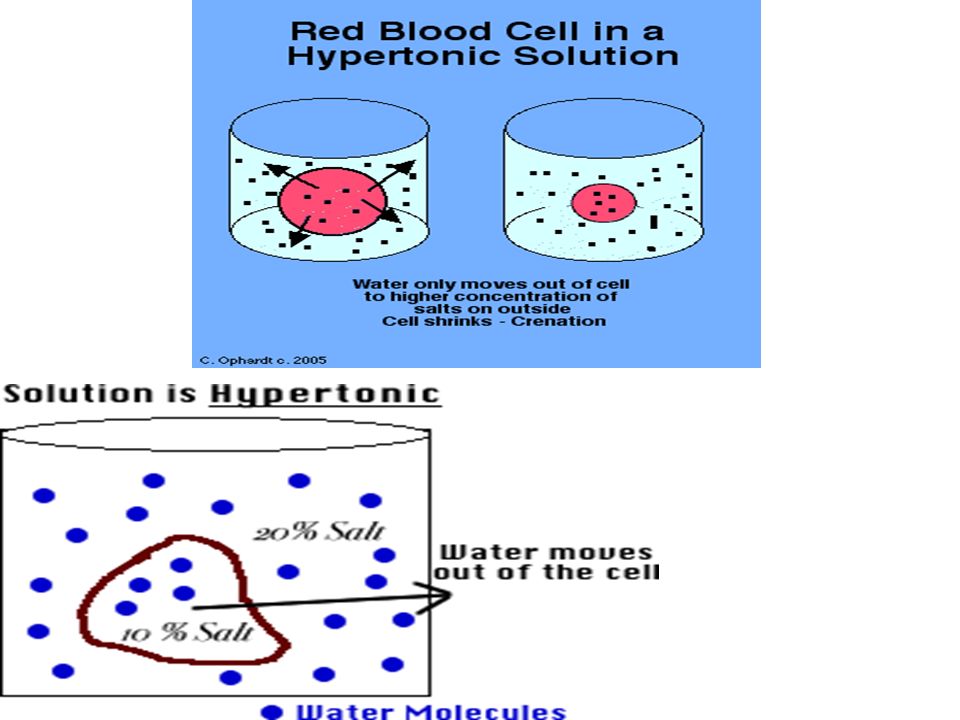
Giving a hypertonic IV solution to a client may cause fluid to be:
a) pulled from cells into the bloodstream b) pulled out of the bloodstream into the cells c) pushed out of the bloodstream into extravascular space
 pulled from cells into the bloodstream. b) pulled out of the bloodstream into the cells. c) pushed out of the bloodstream into extravascular space.")
70
Thirst Eating highly salty foods and/or losing fluids lead to an increase in extracellular fluid osmolality. This leads to drying of mucous membrane, which stimulates the thirst center in hypothalamus. This mechanism is less effective in elderly, thus they are more prone to dehydration. Also it takes a while for this response to occur. Anticipate!

Similar presentations


>")

