Download presentation
Presentation is loading. Please wait.

1
Fluid & Electrolyte Imbalance

2
Fluid Imbalance

3
Fluid Volume Deficit (Hypovolemia, Isotonic Dehydration)
Common Causes Hemorrhage Vomiting Diarrhea Burns Diuretic therapy Fever Impaired thirst

4
Clinical Manifestations
Signs/Symptoms Weight loss Thirst Orthostatic changes in pulse rate and bp Weak, rapid pulse Decreased urine output Dry mucous membranes Poor skin turgor

5
Treatment/Interventions (FVD)
Fluid Management Diet therapy – Mild to moderate dehydration. Correct with oral fluid replacement. Oral rehydration therapy – Solutions containing glucose and electrolytes. E.g., Pedialyte, Rehydralyte. IV therapy – Type of fluid ordered depends on the type of dehydration and the clients cardiovascular status.

6
Safety Alert

7
Nursing Implications Monitor postural heart rate and bp when getting patients out of bed

8
Fluid Volume Excess Common Causes: Congestive Heart Failure
Early renal failure IV therapy Excessive sodium ingestion SIADH Corticosteroid

9
Clinical Manifestations
Signs/Symptoms Increased BP Bounding pulse Venous distention Pulmonary edema Dyspnea Orthopnea (diff. breathing when supine) crackles
 crackles.")
10
Treatment/Interventions (FVE)
Drug therapy Diuretics may be ordered if renal failure is not the cause. Restriction of sodium and saline intake I/O Weight

11
More to consider? Age Prior medical history Infants Older adults
Acute illness Chronic illness Environmental factors Diet Lifestyle Medications

12
Physical Assessment Body systems I/O Weight Labs

13
Electrolyte Imbalance

14
Hypokalemia (<3.5mEq/L)
Pathophysiology – Decrease in K+ causes decreased excitability of cells, therefore cells are less responsive to normal stimuli

15
Hypokalemia (<3.5mEq/L)
Contributing factors: Diuretics Shift into cells Digitalis Water intoxication Corticosteroids Diarrhea Vomiting

17
Hypokalemia (<3.5mEq/L)
Interventions Assess and identify those at risk Encourage potassium-rich foods K+ replacement (IV or PO) Monitor lab values D/c potassium-wasting diuretics Treat underlying cause
 Monitor lab values. D/c potassium-wasting diuretics. Treat underlying cause.")
18
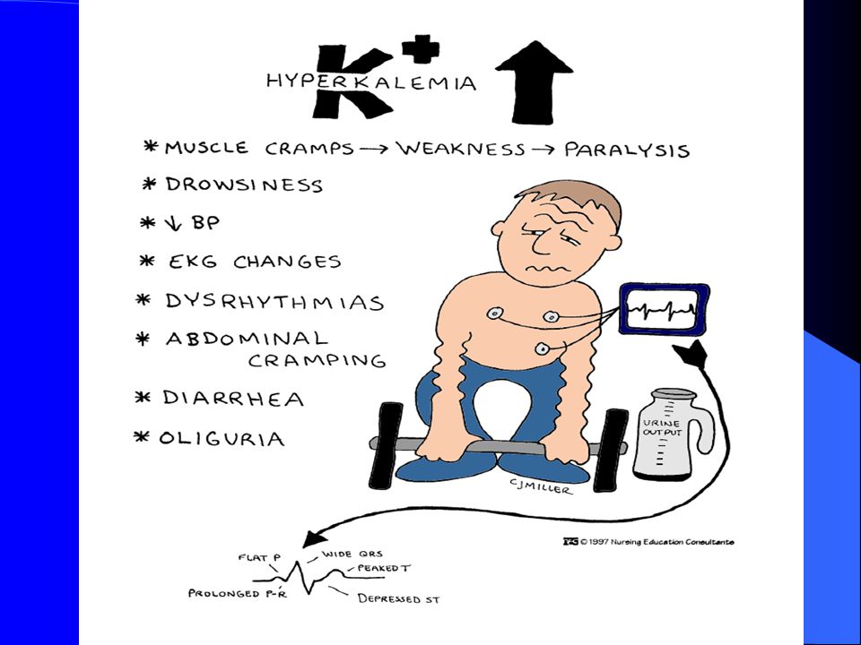
Hyperkalemia (>5.0mEq/L)
Pathophysiology – An inc. in K+ causes increased excitability of cells.

19
Hyperkalemia (>5.0mEq/L)
Contributing factors: Increase in K+ intake Renal failure K+ sparing diuretics Shift of K+ out of the cells

21
Hyperkalemia (>5.0mEq/L)
Interventions Need to restore normal K+ balance: Eliminate K+ administration Inc. K+ excretion Lasix Kayexalate (Polystyrene sulfonate) Infuse glucose and insulin Cardiac Monitoring
 Infuse glucose and insulin. Cardiac Monitoring.")
22
Hyponatremia (<135mEq/L)
Contributing Factors Excessive diaphoresis Wound Drainage NPO CHF Low salt diet Renal Disease Diuretics

23
Hyponatremia (<135mEq/L)
Assessment findings: Neuro - Generalized skeletal muscle weakness. Headache / personality changes. Resp.- Shallow respirations CV - Cardiac changes depend on fluid volume GI – Increased GI motility, Nausea, Diarrhea (explosive) GU - Increased urine output
 GU - Increased urine output.")
24
Hyponatremia (<135mEq/L)
Interventions/Treatment Restore Na levels to normal and prevent further decreases in Na. Drug Therapy – (FVD) - IV therapy to restore both fluid and Na. If severe may see 2-3% saline. (FVE) – Administer osmotic diuretic (Mannitol) to excrete the water rather than the sodium. Increase oral sodium intake and restrict oral fluid intake.
 - IV therapy to restore both fluid and Na. If severe may see 2-3% saline. (FVE) – Administer osmotic diuretic (Mannitol) to excrete the water rather than the sodium. Increase oral sodium intake and restrict oral fluid intake.")
25
Hypernatremia (>145mEq/L)
Contributing Factors Hyperaldosteronism Renal failure Corticosteroids Increase in oral Na intake Na containing IV fluids Decreased urine output with increased urine concentration

26
Hypernatremia (>145mEq/L)
Contributing factors (cont’d): Diarrhea Dehydration Fever Hyperventilation
: Diarrhea. Dehydration. Fever. Hyperventilation.")
27
Hypernatremia (>145mEq/L)
Assessment findings: Neuro - Spontaneous muscle twitches. Irregular contractions. Skeletal muscle wkness. Diminished deep tendon reflexes Resp. – Pulmonary edema CV – Diminished CO. HR and BP depend on vascular volume.

28
Hypernatremia (>145mEq/L)
GU – Dec. urine output. Inc. specific gravity Skin – Dry, flaky skin. Edema r/t fluid volume changes.

29
Hypernatremia (>145mEq/L)
Interventions/Treatment Drug therapy (FVD) .45% NSS. If caused by both Na and fluid loss, will administer NaCL. If inadequate renal excretion of sodium, will administer diuretics. Diet therapy Mild – Ensure water intake
 .45% NSS. If caused by both Na and fluid loss, will administer NaCL. If inadequate renal excretion of sodium, will administer diuretics. Diet therapy. Mild – Ensure water intake.")
30
Hypocalcemia (<9.0mg/dL)
Contributing factors: Dec. oral intake Lactose intolerance Dec. Vitamin D intake End stage renal disease Diarrhea

31
Hypocalcemia (<9.0mg/dL)
Contributing factors (cont’d): Acute pancreatitis Hyperphosphatemia Immobility Removal or destruction of parathyroid gland
: Acute pancreatitis. Hyperphosphatemia. Immobility. Removal or destruction of parathyroid gland.")
32
Hypocalcemia (<9.0mg/dL)
Assessment findings: Neuro –Irritable muscle twitches. Positive Trousseau’s sign. Positive Chvostek’s sign. Resp. – Resp. failure d/t muscle tetany. CV – Dec. HR., dec. BP, diminished peripheral pulses GI – Inc. motility. Inc. BS. Diarrhea

33
Positive Trousseau’s Sign

34
Positive Chvostek’s Sign

35
Hypocalcemia (<9.0mg/dL)
Interventions/Treatment Drug Therapy Calcium supplements Vitamin D Diet Therapy High calcium diet Prevention of Injury Seizure precautions

36
Hypercalcemia (>10.5mg/dL)
Contributing factors: Excessive calcium intake Excessive vitamin D intake Renal failure Hyperparathyroidism Malignancy Hyperthyroidism

37
Hypercalcemia (>10.5mg/dL)
Assessment findings: Neuro – Disorientation, lethargy, coma, profound muscle weakness Resp. – Ineffective resp. movement CV - Inc. HR, Inc. BP. , Bounding peripheral pulses, Positive Homan’s sign. Late Phase – Bradycardia, Cardiac arrest GI – Dec. motility. Dec. BS. Constipation GU – Inc. urine output. Formation of renal calculi

38
Hypercalcemia (>10.5mg/dL)
Interventions/Treatment Eliminate calcium administration Drug Therapy Isotonic NaCL (Inc. the excretion of Ca) Diuretics Calcium reabsorption inhibitors (Phosphorus) Cardiac Monitoring
 Diuretics. Calcium reabsorption inhibitors (Phosphorus) Cardiac Monitoring.")
39
Hypophosphatemia (<2.5mg/L)
Contributing Factors: Malnutrition Starvation Hypercalcemia Renal failure Uncontrolled DM

40
Hypophosphatemia (<2.5mg/L)
Assessment findings: (Chart 13-7) Neuro – Irritability, confusion CV – Dec. contractility Resp. – Shallow respirations Musculoskeletal - Rhabdomyolysis Hematologic – Inc. bleeding Dec. platelet aggregation
 Neuro – Irritability, confusion. CV – Dec. contractility. Resp. – Shallow respirations. Musculoskeletal - Rhabdomyolysis. Hematologic – Inc. bleeding. Dec. platelet aggregation.")
41
Hypophosphatemia (<2.5mg/L)
Interventions Treat underlying cause Oral replacement with vit. D IV phosphorus (Severe) Diet therapy Foods high in oral phosphate
 Diet therapy. Foods high in oral phosphate.")
42
Hyperphosphatemia (>4.5mg/L)
Causes few direct problems with body function. Care is directed to hypocalcemia. Rarely occurs

43
Hypomagnesemia (<1.4mEq/L)
Contributing factors: Malnutrition Starvation Diuretics Aminoglcoside antibiotics Hyperglycemia Insulin administration

44
Hypomagnesemia (<1.4mEq/L)
Assessment findings: *Neuro - Positive Trousseau’s sign. Positive Chvostek’s sign. Hyperreflexia. Seizures *CV – ECG changes. Dysrhythmias. HTN *Resp. – Shallow resp. *GI – Dec. motility. Anorexia. Nausea

45
Hypomagnesemia (<1.4mEq/L)
Interventions: Eliminate contributing drugs IV MgSO4 Assess DTR’s hourly with MgSO4 Diet Therapy

46
Hypermagnesemia (>2.0mEq/L)
Contributing factors: Increased Mag intake Decreased renal excretion

47
Hypermagnesemia (>2.0mEq/L)
Assessment findings: Neuro – Reduced or weak DTR’s. Weak voluntary muscle contractions. Drowsy to the point of lethargy CV – Bradycardia, peripheral vasodilatation, hypotension. ECG changes.

48
Hypermagnesemia (>2.0mg/dL)
Interventions Eliminate contributing drugs Administer diuretic Calcium gluconate reverses cardiac effects Diet restrictions

Similar presentations


 Integumentary System (chapters 44- 46)>")


