Download presentation
Presentation is loading. Please wait.

1
COLONY MORPHOLOGY ON AGAR PLATE CULTURES

2
Form of Colony

3
Elevation of Colony

4
Margin of Colony

5
Surface of Colony

6
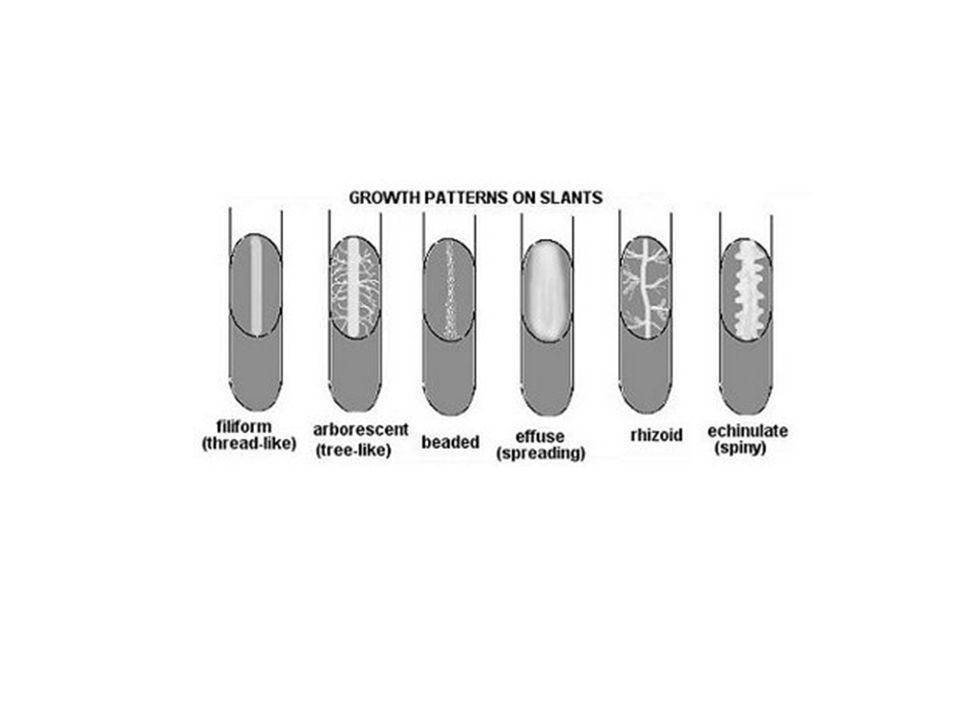
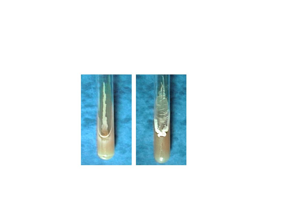
MORPHOLOGY ON slant medium

9
In a liquid medium, the region in which the organism grows depends on the oxygen requirement of that particular species.

10
Liquid medium. Turbid. Pellicle ((thick growth at the top of the tube
Liquid medium * Turbid * Pellicle ((thick growth at the top of the tube * Sediment

11
There are three common shapes of bacteria 1-Coccus
having one of the following arrangements Diplococcus: a pair of cocci Streptococcus: a chain of cocci Tetrad: a square of 4 cocci Sarcina: a cube of 8 cocci Staphylococcus: cocci in irregular, often grape-like clusters

12
Bacillus (rod) 2- Bacillus: a single bacillus Streptobacillus: bacilli in chains Coccobacillus: oval and similar to a coccus

13
Spiral 3- Vibrio: an incomplete spiral or comma-shaped
Spirillum: a thick, rigid spiral Spirochete: a thin, flexible spiral

14
Simple Stai Simple Staining and Bacterial Cell Morphology

15
Preparing a smear for staining
Preparing a smear for staining. (The following procedure is used for all of our staining) 1. Flame (sterilize) your inoculating loop/needle before and after use.
 1. Flame (sterilize) your inoculating loop/needle before and after use.")
16
2. Prepare the smear If you have a solid culture (agar colony), place a small drop of water on a clean slide. Drag the sterile inoculating needle tip through the edge of colony. b. Gently spread the mixture into a circle. A loop of liquid culture can be placed directly on the slide and spread out.

17
3. Let the smear air dry completely.

18
4. Heat-Fix the smear. Smears are heat-fixed by quickly passing the slide through a flame two or three times. This causes the microbes to stick to the slide and not get washed off during the staining process.

19
5. Stain the smear. Place the slide on a rack over the sink. Flood the smear with stain and let it for seconds. Rinse gently and blot dry.

20
6. Observe the slide under low and high-dry lenses to locate, center, and focus the image.
Then, place a drop of oil directly on the stained smear .Turn the oil lens into position and fine focus to observe the cells.

21
Coccus (cocci pl.)
")
22
Bacillus (Bacilli pl.)
")
23
Spirillum (Spirilli pl.)
")
24
Name the bacterial morphologies (shapes and arrangements) seen here.
1 3 2 4 5 6 Answers: 1. Spirillum 2. Coccus 3. Bacillus 4. Diplobacillus 5. Streptobacillus 6. Diplococcus

Similar presentations


 The Good, The Bad, and The Ugly.>")


 –algae.>")

 4- body tube (carrying lens system) 5- light.>")


 in microbiology. Functions : To allow us to study.>")
