Download presentation
Presentation is loading. Please wait.

1
Foot & Ankle

2
Anatomy

3
Anatomy - Medial

4
Anatomy - Lateral

5
Talocrural Joint

6
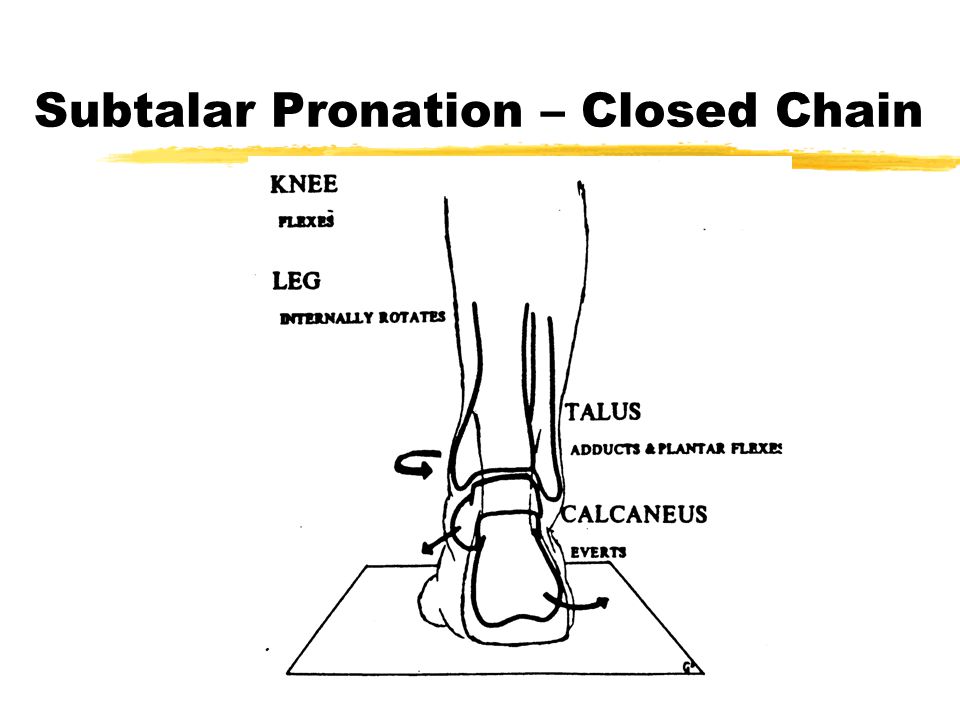
Subtalar Pronation – Closed Chain

8
Subtalar Supination – Closed Chain

9
Subtalar Pronation & Supination Model – Closed Chain

10
Gait Review

11
Subtalar Pronation & Supination during Gait

12
Transverse Tarsal/Midtarsal/Chopart’s Joint Calcaneocuboid Joint Talonavicular Joint

13
Midtarsal Joint Pronation zSTJ unlocks MTJ Supination zSTJ locks up MTJ

14
Midtarsal Joint Motion - Closed Chain PronationNeutralSupination

15
Abnormal Biomechanics zBreakdown of CT zReduced muscle efficiency zChange in muscle function zPoor alignment – Osseous Deformity zDysfunction and Pathology zReduced ability to attenuate GRFs

16
Pronation Closed Chain zCalcaneus eversion (valgus) zTalus adduction (IR - vertical axis) zTalus plantarflexion zTibial IR
 zTalus adduction (IR - vertical axis) zTalus plantarflexion zTibial IR")
17
Normal Pronation in Gait Normal Range: z6 - 10 0 Excessive: z13 0 +

18
Abormal Pronation in Gait zExcessive in magnitude zExcessive in duration zOccurs at wrong time Causes: zIntrinsic deformities zExtrinsic deformities

19
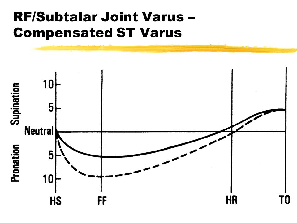
RF/Subtalar Joint Varus zInversion deformity of calcaneus zNo change in relationship of RF on FF Etiology zCongenital/developmental zfailure of talus to derotate

20
RF/Subtalar Joint Varus – Compensated ST Varus

22
Forefoot Varus zMost Common zInsufficiency of 1st ray zDorsiflexed/hypermobile 1st ray zCongenital deformity zInversion of forefoot (metatarsals) relative to rearfoot in STJ neutral
 relative to rearfoot in STJ neutral")
23
Forefoot Varus

24
Forefoot Varus - Compensated

25
Forefoot Varus (Compensated) - Pathomechanics zDuring WA - excessive pronation to get 1st ray on ground zMax. pronation occurs @ HO zPronation remains thru propulsion zFoot never becomes rigid lever zInstability

26
Forefoot Varus - Compensation zProlonged / excessive pronation zCalcaneal valgus zUnlocking of forefoot during propulsion zInsufficient pulley system

27
Forefoot Varus - Pathology zHypermobile 1st ray zExcessive forces on 2 nd MET zProlonged / excessive tibial torsion and/or IR zExcessive anteversion of hip

28
Forefoot Varus - Uncompensated

29
zRigid Foot zLateral ankle sprains zS.I. Joint Dysfunction zITB Dysfunction

30
Subtalar Varus and Compensated Forefoot Varus FF Varus zacquired soft tissue contracture at MTJ z2 0 compensatory pronation for a STJ varus

31
Subtalar Varus and Compensated Forefoot Varus

32
Subtalar Varus and Forefoot Varus, Compensated

33
Ankle Joint Equinus zFixed limitation of DF @ TCJ z< 10 0 of DF when in STJ neutral and knee / Etiology ztight gastrocnemius zspasticity zflattened dome of talus zFx, arthritis, trauma

34
Ankle Joint Equinus

35
Ankle Joint Equinus - Compensated

36
Ankle Joint Equinus - Pathomechanics z pronation 2 0 to DF zloss of ankle rocker ztibia unable to move anterior to talus tibia and talus move anterior to calcaneus zDF of RF at FF z and prolonged pronation during propulsion

37
Compensated Ankle Joint Equinus zExcessive STJ pronation zCalcaneal valgus/eversion zInefficient pulleys zDF of RF on FF

38
Uncompensated Ankle Joint Equinus zGenu Recurvatum zEarly heel rise zExcessive abduction and ER of LE

Similar presentations