Download presentation
Presentation is loading. Please wait.

1
Renal Pathophysiology I
Review of Renal Function Role of the Kidneys in Maintaining Blood Volume and Pressure

2
Main Functions of Kidneys
Regulation of Blood Volume and Pressure (focus of today’s lecture) Regulation of Plasma Composition (focus of Lecture 2) Elimination of Wastes (focus of Lecture 3) Excretion of foreign chemicals Endocrine functions
 Regulation of Plasma Composition. (focus of Lecture 2) Elimination of Wastes. (focus of Lecture 3) Excretion of foreign chemicals. Endocrine functions.")
3
Role of Kidneys in Disease
Innocent bystanders in other problems or pathologies Most often, kidneys help solve these problems Sometimes the kidney can contribute to problems Often damaged by other disease processes Indicator of disease elsewhere in the body

4
The Burden of Kidney Disease
In 2008, nearly 550,000 people were being treated for end stage renal disease. Of these, about 70% were on dialysis. Most are treated at dialysis centers, typically 3 times per week, about 4 hours each time. Cost = $77,000 per patient per year All Americans who need dialysis are covered by Medicare, regardless of age

5
Major Causes of Renal Disease
Diabetes: 205,724 cases per year Hypertension: 133,537 Glomerulonephritis: 83,268 Cystic kidney: 26,094 Urologic disease: 13,065 All other: 86,294

13
From: Physiology of the Kidney and Body Fluids” by R.F. Pitts.

14
Glomerular Filtration Rate (GFR)
Good indicator of renal function Declines with age, but large safety factor Significance of changes: Moderate changes in GFR provide information to kidney about blood volume A significant fall in GFR causes substances that are normally eliminated by the kidneys to remain in the blood = renal failure

15
GFR Declines with Age

16
Determinants of GFR or:
Rate of filtration = hydraulic permeability x surface area x net filtration pressure. or: Rate of filtration = Kf x net filtration pressure. The forces that determine the net filtration pressure are the same Starling Forces that affect all capillaries: Capillary hydrostatic pressure, interstitial hydrostatic pressure, capillary oncotic pressure, interstitial oncotic pressure

18
These determinants can change with injury or disease
Diabetes – deposition of extracellular matrix material decreases Kf. Obstruction of kidney tubule (due to inflammation or scarring) will increase PBS, which will decrease GFR. Autoimmune diseases such as lupus (SLE) involves production of immune complexes that can damage the glomerulus, ultimately decreasing the Kf.
 will increase PBS, which will decrease GFR. Autoimmune diseases such as lupus (SLE) involves production of immune complexes that can damage the glomerulus, ultimately decreasing the Kf.")
19
Regulation of GFR Changes in MAP change renal blood flow
Changes in contraction of renal arterioles can shift the GFR.

21
Regulation of Renal Blood Flow: Tubuloglomerular Feedback
Macula densa cells sense Na+ and Cl - delivery to the distal tubule When BP drops, Na+ and Cl – delivery drop A decrease in Na+ and Cl – delivery dilates the afferent arteriole, helping to raise GFR towards normal. Works in reverse as well: More Na+ and Cl – constricts afferent arterioles

22
Regulation of Sodium and Blood Volume

23
What is the connection between sodium and blood volume?
Sodium is freely filtered at the glomerulus About 65% is reabsorbed in the proximal tubule Another 25% is reabsorbed in the loop of Henle Most of the remaining 10% is reabsorbed in the distal convoluted tubule and collecting duct. Less than 1% winds up in the urine The reabsorption of water follows the reabsorption of sodium, down the osmotic gradient.

25
Sodium Balance Decrease Na+ in urine increased blood volume increase blood pressure

26
Regulation of Sodium Excretion
Most important sensors are ones that detect blood pressure, both inside (the juxtaglomerular apparatus) and outside (the arterial baroreceptors) the kidney. The controlled variable is the amount of sodium excreted in the urine.
 and outside (the arterial baroreceptors) the kidney. The controlled variable is the amount of sodium excreted in the urine.")
27
Regulation of Sodium Excretion
Two Hormone Systems Involved: Renin/Angiotensin/Aldosterone Atrial Natriuretic Peptide (ANP)
")
28
Renin Made by juxtaglomerular cells
An enzyme that splits angiotensinogen to form angiotensin I. Angiotensin I (AI) is then converted to angiotensin II (AII) in the lungs via angiotensin converting enzyme (ACE) Stimuli for release include sympathetic nerve activity, decreased intrarenal BP, decreased delivery of Na+ and Cl- to macula densa
 is then converted to angiotensin II (AII) in the lungs via angiotensin converting enzyme (ACE) Stimuli for release include sympathetic nerve activity, decreased intrarenal BP, decreased delivery of Na+ and Cl- to macula densa.")
30
Secrete renin

31
Actions of Angiotensin II
Release of Aldosterone Vasoconstriction Release of ADH (antidiuretic hormone) Stimulation of Thirst
 Stimulation of Thirst.")
32
Aldosterone Steroid hormone made by adrenal cortex.
Controls activity and/or number of Na+/K+/ATPase pumps in distal tubules and collecting ducts. Increased aldosterone leads to increased reabsorption of Na+ and water (assuming distal tubules and collecting ducts are permeable to water, which is under the control of ADH). Release triggered by angiotensin II, and by high extracellular K+ (more on this later).
. Release triggered by angiotensin II, and by high extracellular K+ (more on this later).")
33
Aldosterone alters the expression of this enzyme

34
Atrial Natriuretic Peptide
Made by atria of the heart Release triggered by increased stretch of atria (indicating increased blood volume) Actions include: Dilates glomerular afferent arterioles, increasing GFR. This increases the amount of sodium filtered, thereby increasing sodium excretion Inhibits Na+ reabsorption in collecting ducts
 Actions include: Dilates glomerular afferent arterioles, increasing GFR. This increases the amount of sodium filtered, thereby increasing sodium excretion. Inhibits Na+ reabsorption in collecting ducts.")
36
Posm = 2 x [Na+(mEq/L)]p + [glucose (mg/dl)]/18 + [urea (mg/dl)]/2.8
Plasma Osmolarity Posm = 2 x [Na+(mEq/L)]p + [glucose (mg/dl)]/18 + [urea (mg/dl)]/2.8 Plasma osmolarity is normally about 295 mOsm/L Sodium, which is normally between mEq/L accounts for most of the osmolarity of the blood. We’ll talk about the rest of what’s in the blood later. Generally, a rise in plasma osmolarity indicates dehydration, at least that’s what your kidney thinks. We’ll discuss exceptions later.
![Posm = 2 x [Na+(mEq/L)]p + [glucose (mg/dl)]/18 + [urea (mg/dl)]/2.8](http://slideplayer.com/slide/5135016/16/images/36/Posm+%3D+2+x+%5BNa%2B%28mEq%2FL%29%5Dp+%2B+%5Bglucose+%28mg%2Fdl%29%5D%2F18+%2B+%5Burea+%28mg%2Fdl%29%5D%2F2.8.jpg "Plasma Osmolarity. Posm = 2 x [Na+(mEq/L)]p + [glucose (mg/dl)]/18 + [urea (mg/dl)]/2.8. Plasma osmolarity is normally about 295 mOsm/L. Sodium, which is normally between mEq/L accounts for most of the osmolarity of the blood. We’ll talk about the rest of what’s in the blood later. Generally, a rise in plasma osmolarity indicates dehydration, at least that’s what your kidney thinks. We’ll discuss exceptions later.")
37
Regulation of Osmolarity
Osmolarity is sensed by osmoreceptors in hypothalamus Controlled variables: Urine volume and osmolarity Thirst and fluid consumption

38
Urine Volume and Osmolarity is Largely Regulated by ADH
ADH = Antidiuretic Hormone = Vasopressin Actions of ADH: Increases reabsorption of water in the distal tubules and collecting ducts. Contraction of arteriolar smooth muscle throughout the body, increasing TPR

39
(ADH release)
")
40
Mechanism of ADH Action
Distal tubules and collecting ducts are normally nearly impermeable to water. In presence of ADH, they become permeable, allowing water to move out of the tubule, down its osmotic gradient. Depending of levels of ADH, the osmolarity of urine can be very high (around 1400 mOsm) or low (100 mOsm). Likewise the volume of the urine can be high or low.
 or low (100 mOsm). Likewise the volume of the urine can be high or low.")
41
Stimuli for ADH Release
An increase in plasma osmolarity (as little as a 2% change) A decrease in blood volume (need about 10% change to trigger release). Sensed by atrial volume receptors and arterial baroreceptors Angiotensin II Certain drugs (nicotine, narcotics) Inhibited by alcohol
 A decrease in blood volume (need about 10% change to trigger release). Sensed by atrial volume receptors and arterial baroreceptors. Angiotensin II. Certain drugs (nicotine, narcotics) Inhibited by alcohol.")
42
Vasopressin = ADH

43
Vasopressin = ADH Also: Alcohol inhibits ADH release

44
Control of Thirst

45
Thirst centers Vasopressin = ADH drinking Return of plasma Volume to normal

46
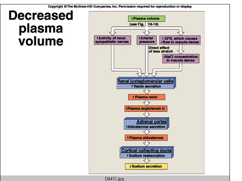
Common Scenario: Hemorrhage Cardiovascular responses help, but cannot correct loss of blood volume

47
Immediate Responses: When plasma volume falls, GFR falls, too Less sodium and water is excreted The reflex increase in sympathetic nerve activity augments this effect.

48
Aldosterone decreases
sodium excretion, which helps retain water

49
ADH is release in response to the fall in BP. It helps retain water and also triggers thirst Thirst centers Vasopressin = ADH drinking Return of plasma Volume to normal

50
Another example of fluid loss:

51
When can this response go wrong?
If there is no true fall in blood pressure, just a decrease in blood flow to the kidney. This could occur if there is atherosclerosis of the renal artery, or an anatomic malformation of this blood vessel. If there is heart failure, blood pressure is low due to failing heart, not blood loss.

52
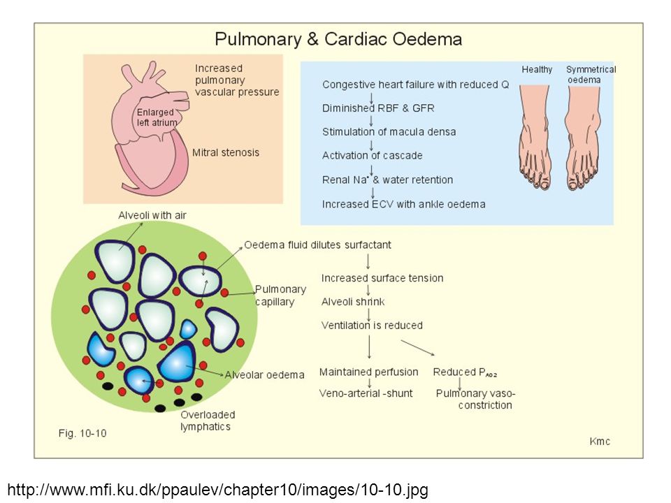
Heart Failure Basic problem is a decrease in blood pressure due to insufficient cardiac output. Kidneys respond as those there has been a hemorrhage – by increasing: Sympathetic nerve activity Renin release ADH release This response can be maladaptive, as excessive amounts of retained fluid cause peripheral and pulmonary edema.

53
Responses to Heart Failure

54
During heart failure, fluid retention helps a little, but can quickly lead to edema

56
Extra slides – Add as needed

57
Osmoreceptors are located in the hypothalamus

58
The Loop of Henle concentrates the tubular fluid.
Urine concentration depends on the plasma ADH levels. ADH determines permeabilitiy of distal tubules and collecting ducts to water.

59
Stimuli for ADH release
Increased plasma osmolarity Decreased plasma volume Angiotensin II Certain drugs (incl. nicotine, narcotics)
")
60
Inhbitors of ADH Release
A decrease in plasma osmolarity An increase in blood volume Atrial Natriuretic Peptide (ANP) (also known as atrial natriuretic factor) Certain drugs (ethanol)
 (also known as atrial natriuretic factor) Certain drugs (ethanol)")
61
Hypovolemia Hypotension Normal blood volume Normal blood pressure ADH release Hypervolemia hypertension Plasma osmolarity (sOsm/L)
")
62
Control of Thirst Activity of osmoreceptors (increased osmolarity stimulates thirst) Activity of blood volume receptors and baroreceptors (a decrease in blood volume or pressure stimulates thirst) Angiotensin II stimulates thirst
 Angiotensin II stimulates thirst.")
63
Thirst centers Vasopressin = ADH drinking Return of plasma Volume to normal

64
Decrease Na+ in urine increased blood volume increase blood pressure

65
When sodium is reabsorbed, water follows down its osmotic gradient

66
Sensing of plasma sodium is mostly through indirect signals that reflect blood volume and pressure
Baroreceptors inside and outside the kidney: Carotid baroreceptors Juxtaglomerular apparatus Macula Densa – senses delivery and reabsorption of chloride (which generally reflects sodium delivery)
")
68
Renin/Angiotensin/Aldosterone System

69
Stimuli for Renin Release
Sympathetic nerve activity Decreased firing of intrarenal baroreceptors Decreased chloride delivery to macula densa

70
Actions of Angiotensin II
Stimulates release of aldosterone Potent vasoconstrictor Stimulates release of ADH Stimulates thirst

71
Aldosterone alters the expression of this enzyme

72
Atrial Natriuretic Peptide (ANP)
Made by the atria of the heart, and released in response to an increase in the stretch of the atria Actions of ANP Dilates glomerular afferent arterioles – increase amount of sodium filtered and thereby excreted Inhibits Na+ reabsorption in collecting tubule

73
Potassium Regulation Most (55%) of the filtered potassium is reabsorbed in the proximal tubule, another 30% is absorbed in the loop of Henle. Depending on diet, potassium may be reabsorbed or secreted in the distal convoluted tubule and the cortical collecting duct.

74
Regulation of potassium secretion
Na+/K+/ATPase A high K+ diet enhances update of K+ into the principal cells (the ones that line the tubule) Aldosterone increases K+ uptake into the principal cells (via Na+/K+/ATPase), and makes the luminal membrane more permeable to K+ K+ secretion is flow dependent – high urine production can lead to K+ deficiency.
 Aldosterone increases K+ uptake into the principal cells (via Na+/K+/ATPase), and makes the luminal membrane more permeable to K+ K+ secretion is flow dependent – high urine production can lead to K+ deficiency.")
75
Aldosterone alters the expression of this enzyme

76
Hypertension and the Kidney
Kidney malfunction is implicated in most cases of hypertension (i.e. the kidney should be able to respond to high BP by increasing salt and water excretion).
.")
77
Drugs to treat hypertension often target the kidney
Diuretics act at various points along the renal tubules to increase water loss in the urine Can lead to potassium depletion Increases sensitivity to heat stress

78
Blood pressure generally increases with age.

79
Hemorrhage

80
Autotransfusion results from a fall in capillary hydrostatic pressure and helps restore blood volume after fluid loss

82
Vasopressin = ADH

83
Heart Failure Basic problem is a decrease in blood pressure due to insufficient cardiac output. Kidneys respond as those there has been a hemorrhage – by increasing: Sympathetic nerve activity Renin release ADH release This response can be maladaptive, as excessive amounts of retained fluid cause peripheral and pulmonary edema.

84
Responses to Heart Failure

85
During heart failure, fluid retention helps a little, but can quickly lead to edema

86
Normal Plasma Values Substance Plasma Concentration Na+
mEq/L K+ 3.5 – 4.5 mEq/L Cl- 95 – 105 mEq/L HCO3 - 24 – 32 mEq/L Glucose 65 – 110 mg/dl (= mmol/L) Blood urea nitrogen (BUN) 8 – 25 mg/dl
 Blood urea nitrogen (BUN) 8 – 25 mg/dl.")
87
Sodium regulation Controlled variable: Sodium excretion in urine
Hormonal systems involved: Renin/Angiotensin/Aldosterone Atrial Natriuretic Peptide

90
Sensing of plasma sodium is mostly through indirect signals that reflect blood volume and pressure
Baroreceptors inside and outside the kidney: Carotid baroreceptors Juxtaglomerular apparatus Macula Densa – senses delivery and reabsorption of chloride (which generally reflects sodium delivery)
")
Similar presentations


 response How the anterior and posterior pituitary.>")
 System>")


