Download presentation
Presentation is loading. Please wait.

1
Injections “101” The Basics on Injection Types, Sites, and Procedures
For Horse, Cattle, Sheep, and Goats

2
What types of injections are there?

3
Subcutaneous “SubQ” What is a subcutaneous injection?
An injection given in the fatty layer of tissue just under the skin. Why are subcutaneous injections given? These injections are given because there is little blood flow to fatty tissue, and the injected medication is generally absorbed more slowly. Diagram from:

4
Intramuscular “IM” What is an intramuscular injection?
An injection given into a muscle. Why are intramuscular injections given? This injection is chosen because of one or more of the following reasons: The amount of medicine to be given. The type of medicine requires it to be given IM. The medicine needs to be faster acting than Sub Q. Diagram from:

5
Intravenous “IV” What is an IV injection: Why are IV injections given?
An injection made directly into the vein Why are IV injections given? Medication reaches parts of the body much faster than other methods of injection. Allow the medicine to reach the heart quickly and circulate through the body extremely fast. Vein Diagram from: injection

6
Intradermal “ID” What is an ID injection? Why are ID injections given?
The introduction of a hypodermic needle into the dermis for the purpose of instilling a substance between the layers of skin, such as a serum or vaccine. Why are ID injections given? These types of injections are often used for conducting skin allergy tests and testing for antibody formation. Diagram from:

7
Locations and Procedures for Giving Injections
Horse

8
Intramuscular Injections
9
Intramuscular There are four main areas for giving intramuscular injections in horses: Neck Region Chest or Pectoral Region Gluteal or Hip Region Hind Leg or Hamstring Region

10
Intramuscular Neck This site is a triangle defined by the nuchal ligaments along the crest of the horse’s neck, the cervical vertebra which form a backward S-shaped curve from the horse’s poll toward the point of the shoulder, and the scapula. Higher toward the crest and you risk hitting the nuchal ligaments, and lower toward the bottom of the neck is were the cervical vertebra and blood vessels are located.

11
Intramuscular Hind Leg or Hamstring Area
Below the point of the horse’s buttocks is another large muscle mass which is a good injection site. It is the preferred injection site for foals because it is one of the larger muscles on a foal’s body. The major drawback to this injection site is that it puts the handler within kicking range of the horse. To find this injection site, drop about 1 inch below the joint of the buttocks and inject anywhere in the large muscle mass along the back of the leg.

12
Intramuscular Chest or Pectoral Region
The pectoral muscles tend to become sore easily and may develop abscesses more easily. Generally are only used when the horse is receiving prolonged treatment and is sore in other injection sites. Thermography of chest abscess

13
Intramuscular Gluteal or Hip Region
The disadvantage to this site is that it has very poor drainage if an abscess develops at the injection site. It can be used for a horse that is sore in all other injection sites. The proper location of this injection site is the intersection of a line between the tail head and point of hip and a line between the top of the croup and the point of the buttocks.

14
Intramuscular The general procedure for an IM injection is to remove the needle from the syringe, set the needle into the muscle, attach the syringe, aspirate to make sure no blood is present, and slowly inject the medication. Some people distract the horse by pinching or tapping the horse’s skin next to the injection site for a few seconds prior to inserting the needle. Photo from: Video: IM Techniques

15
Intravenous Injections
16
Intravenous The site for insertion of the needle should be in the jugular vein, in the middle of the neck, half way between the head and the torso. Photo from:

17
These should only be done by a veterinarian or with the supervision of a veterinarian.
Identify the jugular vein in the middle portion of the neck. Place a finger or hand firmly at the lower portion of the neck over the jugular vein. Watch and feel for swelling in the vein above the constriction. Place needle firmly into the vein, the sharp side of the needle pointed towards the neck. Photos:

18
You should see blood coming out of the hub of the needle
You should see blood coming out of the hub of the needle. Quickly and quietly grab the syringe with preloaded drug in it which has no air bubbles and place it firmly on the hub of the needle and draw up in the syringe. Blood should fill the syringe. Then inject into the vein in a stead by not rapid motion. When material is injected, pull needle and syringe out and place hand over the vein to close’ it. Photo from:

19
Subcutaneous Injections
20
Subcutaneous These are given just beneath the skin.
Simply lift or “tent” the skin on the neck, insert the needle, pull back slightly on the syringe plunger to be sure the needle is not in a blood vessel, and then administer the vaccine.

21
Intradermal Injections

22
Intradermal Given directly into the skin of the neck.
The hair should be clipped to aid in observing reaction. Typically done for allergy testing. Photo from:

23
Safety Procedures Horse
24
Safety Always have a handler when giving a horse and injection. The handler should stand on the same side of the horse as the person giving the shot. Do not tie the horse. The horse may pull back and injure itself. The neck is a relatively safe place to give an injection because you are near the horse’s shoulder. Use extra caution when injecting in the hind quarters because this site puts the handler in kicking range. Information from:

25
Safety If the horse does try to kick, its head should be pulled toward the handler so its hind legs turn away form the handler and the person giving the shot. Injections in the pectoral muscles puts you in a position where you can be easily bitten, struck with a front foot, or run over by the horse. Make sure all drugs are handled properly, given according to directions, and that sterile needles and syringes are used. Information from: Video Link: Photo from: Click here for another video!

26
Locations and Procedures for Giving Injections
Cattle Picture from:

27
Intramuscular Injections

28
Intramuscular Given in the neck area in cattle.
To reduce carcass damage and potential nerve damage, avoid the rear quarters whenever possible. Photo from:

29
Choose muscle tissue of lesser value to consumers for IM injections.
Give IM injections deep into a muscle. Use a needle long enough to penetrate skin, subcutaneous tissue and fat to reach the muscle. The needle should enter the skin perpendicular to the skin surface. Insert the needle into the animal, and attach the syringe to the needle. Check that the needle is not in a blood vessel by pulling back on the plunger and observing for blood flow in the tip of the syringe. If blood appears, remove the needle and put it in a different location at least one inch away form the original injection site. Photo from: Diagram from:

30
Subcutaneous Injections
31
Subcutaneous The best location is half way up the neck in front of the shoulder, or over the ribs well behind the shoulder. Diagram from:

32
Lift a fold of skin to make a skin “tent”
Lift a fold of skin to make a skin “tent”. Insert the needle through one side of the tent at an angel of degrees relative to the surface of the body. Diagram from:

33
Intravenous Injections

34
Intravenous There are two major sites, the jugular vein and the tail vein. The jugular vein is much larger but cattle are often restrained in a neck chute which can make it difficult to safely inject into. The tail vein runs straight down the underside of the tail but is much narrower, so it is harder to inject into but is generally more accessible in dairy cattle.

35
This type of injection should be given by a veterinarian or with the supervision of a veterinarian.
There are many medications that could kill or do serious damage if injected into a vein and run in too quickly. For example, when injecting calcium solutions intravenously, a veterinarian may listen to the heartbeat to gauge the rate of injection by the response of the heart. Without this, sudden deaths may occur.

36
Intradermal Injections

37
Given directly into the skin.
Bovine Tuberculosis tests are administered this way. A cow being tested for tuberculosis. - Subcutaneous and intradermal injection video

38
Safety Procedures

39
Some important considerations are:
Giving the right product at the right time Having adequate restraint of the animal Placing the injection in the right place Using clean techniques and sterile equipment Be careful not to inject yourself. The results could be fatal.

40
Locations and Procedures for Giving Injections
Sheep and Goats

41
Intramuscular Injections
42
Intramuscular The best location is the heavy muscles of the neck.
To reduce carcass damage and potential nerve damage, avoid the rear quarters whenever possible.

43
Insert the needle with a quick thrust
Insert the needle with a quick thrust. Care should be taken to make sure the needle is inserted in the muscle, not just under the skin. Pull back on the plunger to make sure that the needle has not been inserted into a blood vessel. The medication should slowly be injected into the muscle.

44
The leg and loin should be avoided when giving IM injections.
IM injections can cause damage to the muscle tissue (meat).
. v=0.")
45
Intravenous Injections
46
Intravenous The best location is in the jugular vein in the neck.
In lactating dairy goats, the milk vein can sometimes be used to inject small volumes of fluid. Diagram from:

47
Intravenous This type of injection should be given by a veterinarian or with the supervision of a veterinarian. Procedures: Have someone straddle the goat to hold it securely. The holder should elevate the goat’s head up and to the side. Feel for the trachea on the neck. The area between the trachea and the muscles of the neck is the “jugular groove” and is where the jugular vein lies. Put pressure at the bottom of the groove and you will see the groove swell from your finger up to the jaw of the goat. The vein is now filled with blood. Information From: Pictures from:

48
Intravenous Using an 18 to 20 gauge needle, direct it at a 45 degree angle then stab through the skin. Pull back on your syringe and see if there is blood present. If not, adjust the depth (deeper or more shallow) or move up or down the side of the groove until blood is obtained. The presence of blood signifies that the needle is inside the vein. Administer drugs slowly and monitor the animal for evidence of respiratory or cardiac distress. If there is any adverse reaction to the injection, it should be stopped. From: Pictures from:
 or move up or down the side of the groove until blood is obtained. The presence of blood signifies that the needle is inside the vein. Administer drugs slowly and monitor the animal for evidence of respiratory or cardiac distress. If there is any adverse reaction to the injection, it should be stopped. From: Pictures from:")
49
Subcutaneous Injections
Sheep and Goats

50
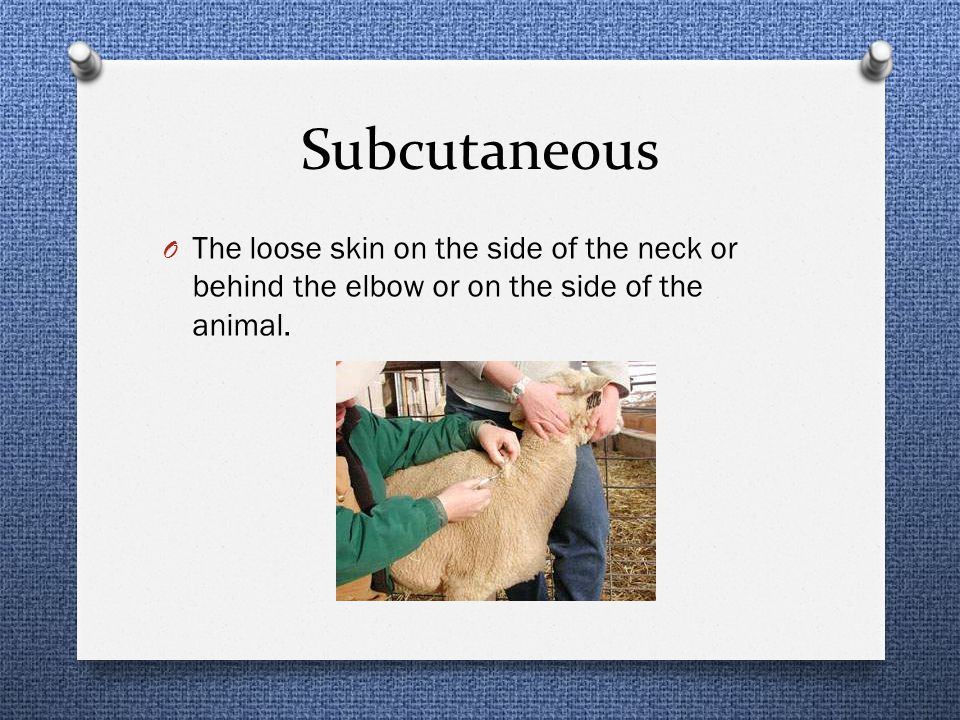
Subcutaneous The loose skin on the side of the neck or behind the elbow or on the side of the animal.
51
Start by making a “tent” with the skin and injecting the solution under the fold of the skin, parallel with the muscle. The medicine should be slowly injected. A ¾ or 1 inch needle should be used.

52
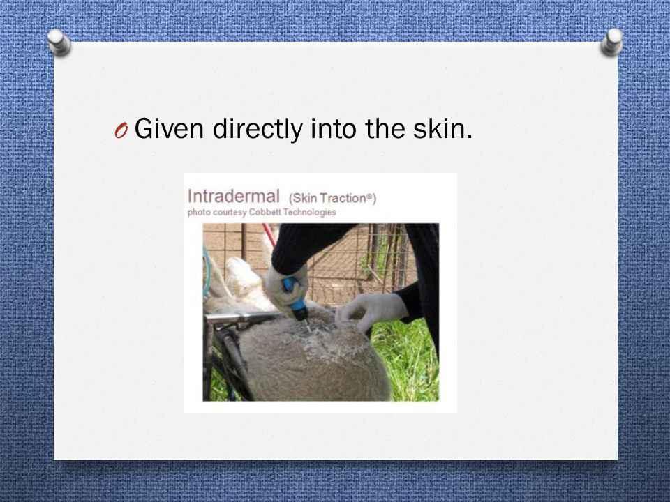
Intradermal Injections
Sheep and Goats

53
Given directly into the skin.
54
Safety Procedures Sheep and Goats
55
Be careful not to inject yourself or others with the vaccine.
Make sure all drugs are handled properly, given according to directions, and that sterile needles and syringes are used. Be sure the animals are properly restrained for the procedures.

56
Consequences of Poor Injection Techniques
What could go wrong? Consequences of Poor Injection Techniques

57
Consequences of Poor Injection Techniques
Treatment failure, if product absorption is delayed or block. Drug residues in meat or milk if the drug cannot be absorbed and metabolized in a timely manner. Animal suffering and incapacitation due to nerve damage or swelling from tissue reactions. Excessive trim at slaughter due to abscess, scarring or broken needles. Shock or death of animal being treated, if medications unintentionally enter the bloodstream. Accidental human injection. From:

58
In Conclusion: Knowledge and proper technique produces high quality livestock and assures the health and safety of all involved! Horse photo from: Cow photo from:

Similar presentations


 中国医科大学护理学院 王健. Medications (three) PARENTERAL MEDICATIONS Nurses given parenteral medications intradermally (ID), subcutaneously (SC or SQ),>")


