Download presentation
Presentation is loading. Please wait.

1
Nose and Pharynx Dr. Sama ul Haque

2
Objectives Discuss the anatomical structure of nose. Define Paranasal sinuses. Describe the anatomical structure of pharynx. Enlist the extrinsic and intrinsic muscles of the pharynx with their nerve supply and actions.

3
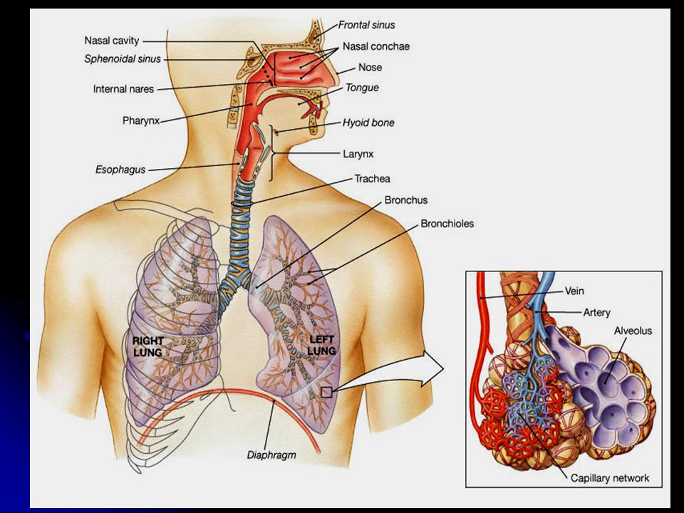
Organization and Functions of the Respiratory System Consists of an upper respiratory tract (nose to larynx) and a lower respiratory tract (trachea onwards). Conducting portion transports air. - includes the nose, nasal cavity, pharynx, larynx, trachea, bronchi and bronchioles. Respiratory portion carries out gas exchange. - composed of small airways called respiratory bronchioles and alveolar ducts as well as air sacs called alveoli.

5
Upper Respiratory Tract Nose Nasal cavity Paranasal sinuses Pharynx (throat) Larynx
 Larynx")
6
Upper Respiratory Tract

7
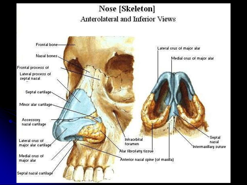
Structure of the Nose Nose, is the only visible part of the respiratory system and serves as the entrance to the respiratory tract The nose has two cavities, separated from one another by a wall called the septum. external (anterior) nares or nostrils, lead to the nasal cavities. The external openings, known as external (anterior) nares or nostrils, lead to the nasal cavities.
 nares or nostrils, lead to the nasal cavities. The external openings, known as external (anterior) nares or nostrils, lead to the nasal cavities..")
9
Nasal Cavity Extends from the external (anterior) nares to the posterior nares (choanae). Extends from the external (anterior) nares to the posterior nares (choanae). Divided into right & left halves by the nasal septum. Divided into right & left halves by the nasal septum. Each half has a: Each half has a: Roof Lateral wall Medial wall (septum) Floor
 nares to the posterior nares (choanae). Divided into right & left halves by the nasal septum. Divided into right & left halves by the nasal septum. Each half has a: Each half has a: Roof Lateral wall Medial wall (septum) Floor.")
10
Roof Narrow & formed (anteroposteriorly) by the: Narrow & formed (anteroposteriorly) by the: 1. Nasal bone & cartilage 2. Frontal bone. 3. Cribriform plate of ethmoid bone 4. Body of sphenoid. Floor Formed by the hard (bony) palate. Formed by the hard (bony) palate. Separates it from the oral cavity. Separates it from the oral cavity. 4 3 2 1 Hard Palate Oral cavity
 palate. Formed by the hard (bony) palate. Separates it from the oral cavity. Separates it from the oral cavity Hard Palate Oral cavity.")
11
Medial Wall (Nasal Septum) Osteo- cartilaginous partition between the two nasal cavities. Formed by: 1.Septal cartilage. 2.Perpendicular plate of ethmoid bone. 3.Vomer.

12
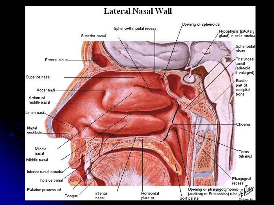
Lateral Wall Shows three horizontal bony projections, the superior, middle & inferior conchae. Shows three horizontal bony projections, the superior, middle & inferior conchae. The cavity below each concha is called a meatus and are named as superior, middle & inferior corresponding to the conchae. The cavity below each concha is called a meatus and are named as superior, middle & inferior corresponding to the conchae. The small space above the superior concha is the sphenoethmoidal (suprameatal) recess. The small space above the superior concha is the sphenoethmoidal (suprameatal) recess. The conchae are covered by respiratory epithelium and thus increase the surface area of the nasal cavity. Inferior concha Superior concha concha middle concha Sphenoethmoidal recess Meati
 recess. The small space above the superior concha is the sphenoethmoidal (suprameatal) recess. The conchae are covered by respiratory epithelium and thus increase the surface area of the nasal cavity. Inferior concha Superior concha concha middle concha Sphenoethmoidal recess Meati.")
13
sphenoidal sinus Superior meatus posterior ethmoidal sinus Middle meatus middle ethmoidal, maxillary, frontal & the anterior ethmoidal sinuses Inferior meatus nasolacrimal duct. The recess & meati receive the openings of the paranasal sinuses & naso-lacrimal duct.

14
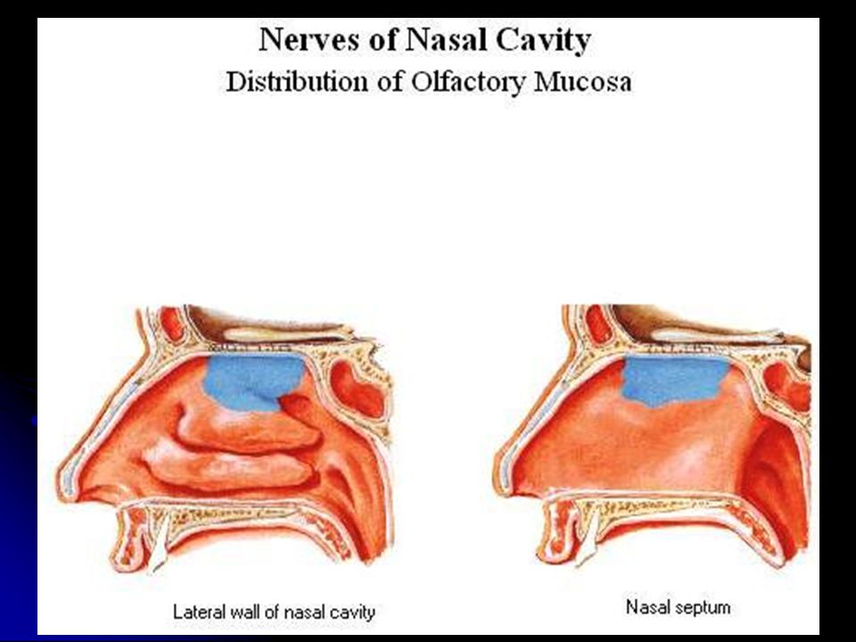
Nose Olfactory mucosa: Mucous membranes that contain smell receptors. Respiratory mucosa: Pseudostratified ciliated columnar epithelium containing goblet cells that secrete mucus which traps inhaled particles.

15
Nerve Supply Olfactory mucosa supplied by olfactory nerves. Nerves of general sensation are derived from ophthalmic & maxillary nerves. Autonomic fibers. Lymphatic Drainage : To the submandibular and the upper deep cervical lymph nodes.

17
Paranasal sinuses

18
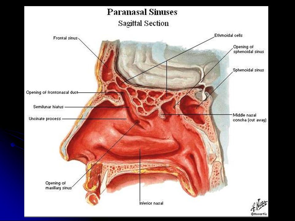
Paranasal Sinuses Air filled cavities located in the bones around the nasal cavity Air filled cavities located in the bones around the nasal cavity Frontal, ethmoidal, sphenoidal, maxillary. Lined by respiratory mucosa which is continuous with the mucosa of the nasal cavity Decrease skull bone weight. Warm, moisten and filter incoming air. Add resonance to voice. Communicate with the nasal cavity by ducts for drainage

21
Drainage of the Paranasal Sinuses Sphenoethmoidal recess: Sphenoidal air sinus Superior meatus: Posterior ethmoidal air sinus Middle meatus : Bulla ethmoidalis: Middle ethmoidal air sinus Hiatus semilunaris: Frontal air sinus Maxillary air sinus Anterior ethmoidal Inferior meatus: Nasolacrimal duct

22
Blood supply of the nasal cavity:-

23
Functions of the Nose Provides an airway for respiration Moistens and warms entering air Filters and cleans inspired air Resonating chamber for speech Detects odors in the air stream

24
Pharynx Common space used by both the respiratory and digestive systems. Commonly called the throat. Walls are lined by a mucosa and contain skeletal muscles that are primarily used for swallowing. Partitioned into three adjoining regions: Nasopharynx Oropharynx Laryngopharynx

25
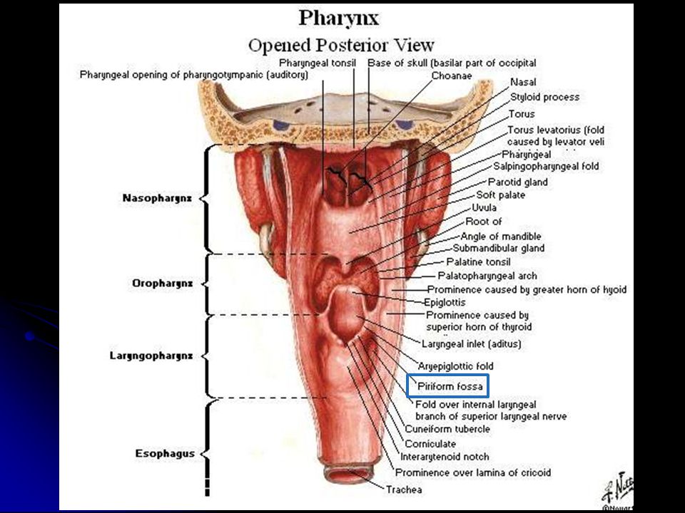
27 Divisions of the Pharynx Divided into three parts: Nasopharynx: Superior part, communicates with the nasal cavity through posterior nasal apertures Oropharynx: Middle part, communicates with the oral cavity through the oro-pharyngeal isthmus Laryngopharynx: Inferior part, communicates with the larynx through the laryngeal inlet

26
Nasopharynx Located directly posterior to the nasal cavity and superior to the soft palate, which separates the oral cavity. Normally, only air passes through. In the lateral walls of the nasopharynx, paired auditory/eustachian tubes connect the nasopharynx to the middle ear. Posterior nasopharynx wall also houses a single pharyngeal tonsil (commonly called the Adenoids).
..")
27
Oropharynx Middle pharyngeal region. Lies immediately posterior to the oral cavity. Common respiratory and digestive pathway through which both air and swallowed food and drink pass. Lymphatic organs here provide the first line of defense against ingested or inhaled foreign materials. Palatine tonsils are on the lateral wall between the arches, and the lingual tonsils are at the base of the tongue.

29
Laryngopharynx Inferior, narrowed region of the pharynx. Terminates at the superior border of the esophagus and the epiglottis of the larynx. Permits passage of both food and air.

30
Piriform fossa A small depression situated on either side of the laryngeal inlet It is a common site for the lodging of foreign bodies. Branches of internal laryngeal & recurrent laryngeal nerves lie deep to the mucous membrane of the fossa and are vulnerable to injury during removal of a foreign body.

32
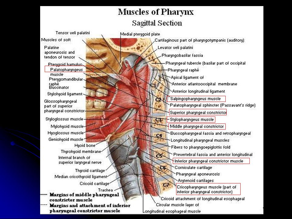
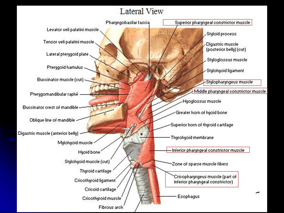
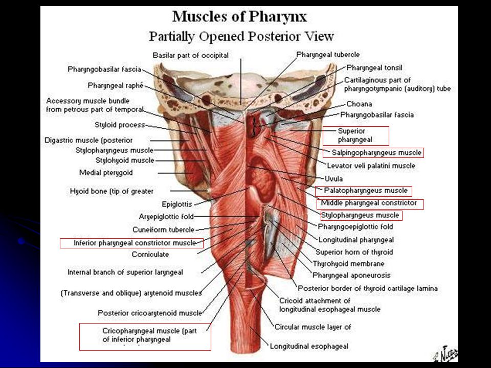
Muscles of Pharynx M S I The muscles of the pharynx are arranged in circular and longitudinal layers Circular (Constrictor) Three muscles, overlap each other: Superior, Middle & Inferior Propel the bolus of food down into the esophagus Longitudinal Muscles Three muscles: Stylopharyngeus Salpingopharyngeus Palatpharyngeous Elevate the larynx & pharynx during swallowing
 Three muscles, overlap each other: Superior, Middle & Inferior Propel the bolus of food down into the esophagus Longitudinal Muscles Three muscles: Stylopharyngeus Salpingopharyngeus Palatpharyngeous Elevate the larynx & pharynx during swallowing")
33
Sensory Nerve Supply Sensory Nerve Supply Nasopharynx: Maxillary nerve Nasopharynx: Maxillary nerve Oropharynx: Glossopharyngeal nerve Oropharynx: Glossopharyngeal nerve Laryngopharynx: Vagus nerve Laryngopharynx: Vagus nerve Motor Nerve Supply : Motor Nerve Supply : All the muscles of pharynx, except the stylopharyngeus, are supplied by the pharyngeal plexus. All the muscles of pharynx, except the stylopharyngeus, are supplied by the pharyngeal plexus. Stylopharyngeus is supplied by the glossopharyngeal nerve Stylopharyngeus is supplied by the glossopharyngeal nerve Pharyngeal plexus A network of nerves (sensory, motor & sympathetic) located on the surface of the middle pharyngeal constrictor muscle, is formed by the: Pharyngeal branches of glossopharyngeal nerve (sensory) Pharyngeal branch of vagus nerve (motor) Sympathetic fibers from superior cervical ganglion (vasomotor)
 located on the surface of the middle pharyngeal constrictor muscle, is formed by the: Pharyngeal branches of glossopharyngeal nerve (sensory) Pharyngeal branch of vagus nerve (motor) Sympathetic fibers from superior cervical ganglion (vasomotor).")
34
Arterial supply : From branches of: Ascending pharyngeal artery Ascending palatine artery Facial artery Maxillary artery Lingual artery The Veins drain into pharyngeal venous plexus, which drains into the internal jugular vein The Lymphatics drain into the: Deep cervical Retropharyngeal & Paratracheal lymph nodes

39
Functions of the Pharynx Provides a passageway for Air & Food Moistens and warms entering air Taste Protection Speech

Similar presentations