Download presentation
Presentation is loading. Please wait.

1
Chapter 16 Respiratory System

2
CopyrightThe McGraw-Hill Companies, Inc
CopyrightThe McGraw-Hill Companies, Inc. Permission required for reproduction or display. Introduction The respiratory system consists of tubes that filter incoming air and transport it into the microscopic alveoli where gases are exchanged. Respiration is defined as the process of inhalation of oxygen and the exhalation of carbon-di-oxide with the help of certain organs of the human body. These organs include the nose, pharynx, larynx, trachea, bronchi and the lungs. These organs together make the human respiratory system.

3
Functions of the Respiratory System
Respiration The most important function of the respiratory system is the process of respiration. The entire process of exchanging gases between the atmosphere and body cells is called and consists of the following: ventilation, gas exchange between blood and lungs, gas transport in the bloodstream, gas exchange between the blood and body cells, and cellular respiration. .

4
Respiration

5
How does the respiratory system function?
1. Inhalation When a person breathes air (which contains oxygen), it passes through the nasal passages containing mucous. This mucous helps in filtering out contaminants like dust, pollen and smoke. The nasal epithelium naturally adds moisture and heat to the air. The larynx prevents food or liquid entering the respiratory tract. The air then passes through the larynx and enters the trachea or the windpipe. Here it gets divided into the two bronchi that connect the trachea to the lungs. The bronchi again gets split into many smaller tubes known as bronchioles. These bronchi end in air sacs, also known as alveoli, which contain blood capillaries. These blood capillaries carry blood which comes through veins from all other parts of the body. Here the carbon dioxide from the blood is exchanged for the oxygen in the alveoli. The blood containing oxygen then goes to the heart where it is later pumped to other parts of the body.
, it passes through the nasal passages containing mucous. This mucous helps in filtering out contaminants like dust, pollen and smoke. The nasal epithelium naturally adds moisture and heat to the air. The larynx prevents food or liquid entering the respiratory tract. The air then passes through the larynx and enters the trachea or the windpipe. Here it gets divided into the two bronchi that connect the trachea to the lungs. The bronchi again gets split into many smaller tubes known as bronchioles. These bronchi end in air sacs, also known as alveoli, which contain blood capillaries. These blood capillaries carry blood which comes through veins from all other parts of the body. Here the carbon dioxide from the blood is exchanged for the oxygen in the alveoli. The blood containing oxygen then goes to the heart where it is later pumped to other parts of the body.")
6
2. Exhalation How does the respiratory system function?
Exhalation in human beings is the process of expelling out of air containing carbon dioxide. The movement of the air while exhalation is through the bronchi, then through the airways and then it passes out through the nose. The exhaled air is completely depleted of oxygen. The lungs are the most important organs of the human respiratory system. There is a muscle located below the lungs known as the diaphragm which also plays an important role in the process of respiration. During inhalation, the diaphragm contracts, creating a vacuum that helps in pulling air into the lungs. On the other hand, during exhalation, the diaphragm relaxes which helps in forcing the air out of the lungs.

7
How does the respiratory system function?
3.Vocalization Vocalization is also one of the major respiratory system functions. Vocalization is the process which enables humans to speak and also to make sound. When the air passes through the pharynx and larynx, it makes the vocal cords in larynx to vibrate which helps in production of sound and speech in humans. 4. Coughing/Sneezing When any foreign particles enter the nasal passages, it can result into irritation. Therefore, expelling out these foreign bodies or irritants is one of the functions of the respiratory system. These irritants are forced out of the respiratory tract through cough or even sneeze

8
To Put It Simply… The principle functions of the respiratory system are: Ventilate the lungs Extract oxygen from the air and transfer it to the bloodstream Excrete carbon dioxide and water vapor Maintain the acid base of the blood Screen and filter air from foreign particles

9
Organs of the Respiratory System
CopyrightThe McGraw-Hill Companies, Inc. Permission required for reproduction or display. The organs of the respiratory tract can be divided into two groups: the upper respiratory tract (nose, nasal cavity, sinuses, and pharynx), and the lower respiratory tract (larynx, trachea, bronchial tree, and lungs).
, and the lower respiratory tract (larynx, trachea, bronchial tree, and lungs).")
10
Nose supported by bone and cartilage, provides an entrance for air in which air is filtered by coarse hairs inside the nostrils Nasal Cavity space posterior to the nose that is divided medially by the nasal septum nasal conchae divide the cavity into passageways that are lined with mucous membrane, and help increase the surface area available to warm and filter incoming air Particles trapped in the mucus are carried to the pharynx by ciliary action, swallowed, and carried to the stomach where gastric juice destroys any microorganisms in the mucus.

11
Paranasal Sinuses Sinuses are air-filled spaces within the maxillary,frontal, ethmoid, and sphenoid bones of the skull. These spaces open to the nasal cavity and are lined with mucus membrane that is continuous with that lining the nasal cavity. The sinuses reduce the weight of the skull and serve as a resonant chamber to affect the quality of the voice.

12
Pharynx aka Throat Larynx common passageway for air and food
aids in producing sounds for speech Larynx an enlargement in the airway superior to the trachea and inferior to the pharynx helps keep particles from entering the trachea and also houses the vocal cords composed of a framework of muscles and cartilage bound by elastic tissue Note: Epiglottis & vestibular folds prevent swallowed material from moving into larynx (can move to cover trachea)
")
13
Larynx continued… Inside the larynx, two pairs of folds of muscle and connective tissue covered with mucous membrane make up the vocal cords The upper pair is the false vocal cords The lower pair is the true vocal cords Changing tension on the vocal cords controls pitch, while increasing the loudness depends upon increasing the force of air vibrating the vocal cords During normal breathing, the vocal cords are relaxed and the glottis is a triangular slit During swallowing, the false vocal cords and epiglottis close off the glottis

14
tracheal wall is supported by 20 incomplete cartilaginous rings
CopyrightThe McGraw-Hill Companies, Inc. Permission required for reproduction or display. Trachea Extends downward anterior to the esophagus and into the thoracic cavity, where it splits into right and left bronchi inner wall of the trachea is lined with ciliated mucous membrane with many goblet cells that serve to trap incoming particles tracheal wall is supported by 20 incomplete cartilaginous rings

15
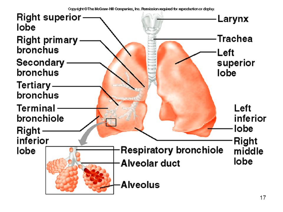
Bronchial Tree consists of branched tubes leading from the trachea to the alveoli begins with the two primary bronchi, each leading to a lung branches of the bronchial tree from the trachea are right and left primarybronchi; these further subdivide until bronchioles give rise to alveolar ducts which terminate in alveoli it is through the thin epithelial cells of the alveoli that gas exchange between the blood and air occurs

16
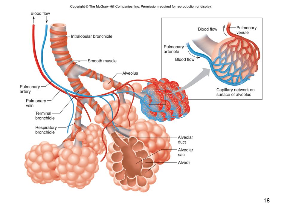
Alveoli Extremely thin-walled sacs covered w/ capillaries
CO2 & O2 move by diffusion across the respiratory membrane About 300 million alveoli in two lungs Size of a tennis court

19
CopyrightThe McGraw-Hill Companies, Inc
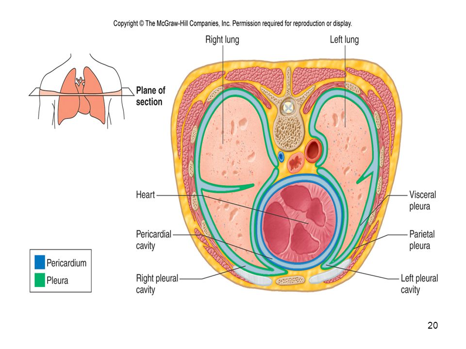
CopyrightThe McGraw-Hill Companies, Inc. Permission required for reproduction or display. Lungs the right and left soft, spongy, cone-shaped lungs are separated medially by the mediastinum and are enclosed by the diaphragm and thoracic cage the bronchus and large blood vessels enter each lung A layer of serous membrane, the visceral pleura, folds back to form the parietal pleura The visceral pleura is attached to the lung, and the parietal pleura lines the thoracic cavity; serous fluid lubricates the “pleura cavity” between these two membranes the right lung has three lobes, the left has two Each lobe is composed of lobules that contain air passages, alveoli, nerves, blood vessels, lymphatic vessels, and connective tissues.

21
Breathing Mechanism Inspiration
CopyrightThe McGraw-Hill Companies, Inc. Permission required for reproduction or display. Breathing Mechanism Ventilation (breathing), the movement of air in and out of the lungs, is composed of inspiration and expiration Inspiration Atmospheric pressure is the force that moves air into the lungs When pressure on the inside of the lungs decreases, higher pressure air flows in from the outside Air pressure inside the lungs is decreased by increasing the size of the thoracic cavity; due to surface tension between the two layers of pleura, the lungs follow with the chest wall and expand Muscles involved in expanding the thoracic cavity include the diaphragm and the external intercostal muscles As the lungs expand in size, surfactant keeps the alveoli from sticking to each other so they do not collapse when internal air pressure is low.
, the movement of air in and out of the lungs, is composed of inspiration and expiration. Inspiration. Atmospheric pressure is the force that moves air into the lungs. When pressure on the inside of the lungs decreases, higher pressure air flows in from the outside. Air pressure inside the lungs is decreased by increasing the size of the thoracic cavity; due to surface tension between the two layers of pleura, the lungs follow with the chest wall and expand. Muscles involved in expanding the thoracic cavity include the diaphragm and the external intercostal muscles. As the lungs expand in size, surfactant keeps the alveoli from sticking to each other so they do not collapse when internal air pressure is low.")
22
CopyrightThe McGraw-Hill Companies, Inc
CopyrightThe McGraw-Hill Companies, Inc. Permission required for reproduction or display. Expiration The forces of expiration are due to the elastic recoil of lung and muscle tissues and from the surface tension within the alveoli Forced expiration is aided by thoracic and abdominal wall muscles that compress the abdomen against the diaphragm

23
Changing Alveolar Volume
Lung recoil Causes alveoli to collapse resulting from Elastic recoil and surface tension Surfactant: Reduces tendency of lungs to collapse Pleural pressure Negative pressure can cause alveoli to expand Pneumothorax is an opening between pleural cavity & air that causes a loss of pleural pressure

24
Compliance Measure of the ease with which lungs & thorax expand
The greater the compliance, the easier it is for a change in pressure to cause expansion A lower-than-normal compliance means the lungs and thorax are harder to expand Conditions that decrease compliance Pulmonary fibrosis Pulmonary edema Respiratory distress syndrome

25
Respiratory Air Volumes and Capacities
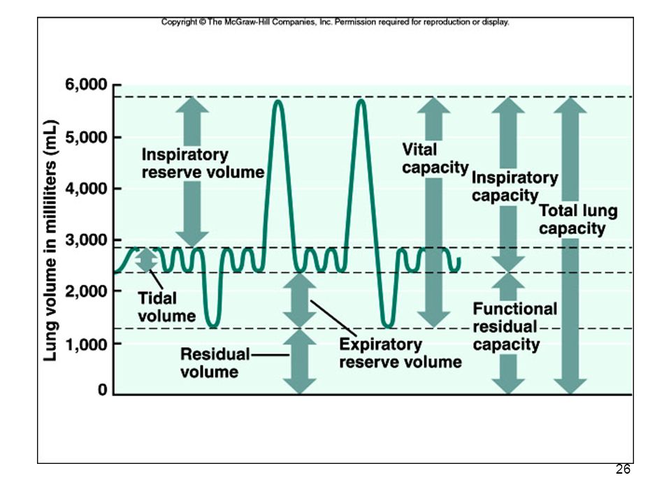
The measurement of different air volumes is called spirometry, and it describes four distinct respiratory volumes One inspiration followed by expiration is called a respiratory cycle; the amount of air that enters or leaves the lungs during one respiratory cycle is the tidal volume During forced inspiration, an additional volume, the inspiratory reserve volume, can be inhaled into the lungs. IRV + TV gives us the inspiratory capacity During a maximal forced expiration, an expiratory reserve volume can be exhaled, but there remains a residual volume in the lungs. Adding the two together gives us the functional reservecapacity Vital capacity is the tidal volume plus inspiratory reserve and expiratory reserve volumes combined Vital capacity plus residual volume is the total lung capacity Anatomic dead space is air remaining in the bronchial tree

27
CopyrightThe McGraw-Hill Companies, Inc
CopyrightThe McGraw-Hill Companies, Inc. Permission required for reproduction or display. Control of Breathing Normal breathing is a rhythmic, involuntary act even though the muscles are under voluntary control Respiratory Center Groups of neurons in the brain stem comprise the respiratory center, which controls breathing by causinginspiration and expiration and by adjusting the rate and depth of breathing

28
Factors Affecting Breathing
Chemicals, lung tissue stretching, an emotional state affect breathing Chemosensitive areas (central chemoreceptors) are associated with the respiratory center and are sensitive to changes in the blood concentration of carbon dioxide and hydrogen ions If either carbon dioxide or hydrogen ion concentrations rise, the central chemoreceptors signal the respiratory center, and breathing rate increases Peripheral chemoreceptors in the carotid sinuses and aortic arch sense changes in blood oxygen concentration, transmit impulses to the respiratory center, and breathing rate and tidal volume increase
 are associated with the respiratory center and are sensitive to changes in the blood concentration of carbon dioxide and hydrogen ions. If either carbon dioxide or hydrogen ion concentrations rise, the central chemoreceptors signal the respiratory center, and breathing rate increases. Peripheral chemoreceptors in the carotid sinuses and aortic arch sense changes in blood oxygen concentration, transmit impulses to the respiratory center, and breathing rate and tidal volume increase.")
29
More Factors Affecting Breathing
An inflation reflex, triggered by stretch receptors in the visceral pleura, bronchioles, and alveoli, helps to prevent over inflation of the lungs during forceful breathing Hyperventilation lowers the amount of carbon dioxide in the blood

30
Alveolar Gas Exchanges
The alveoli are the only sites of gas exchange between the atmosphere and the blood. Alveoli The alveoli are tiny sacs clustered at the distal ends of the alveolar ducts Respiratory Membrane The respiratory membrane consists of the epithelial cells of the alveolus, the endothelial cells of the capillary, and the two fused basement membranes of these layers. Gas exchange occurs across this respiratory membrane

31
Hemoglobin and Oxygen Transport
Oxygen is transported by hemoglobin (98.5%) and is dissolved in plasma (1.5%) Transport of Carbon Dioxide Carbon dioxide is transported as bicarbonate ions (70%) in combination with blood proteins (23%) and in solution with plasma (7%)
 and is dissolved in plasma (1.5%) Transport of Carbon Dioxide. Carbon dioxide is transported as bicarbonate ions (70%) in combination with blood proteins (23%) and in solution with plasma (7%)")
32
Disorders Asthma— spasms of smooth muscle in the bronchioles
Lung cancer Constant irritation produces excess mucous and puts unnecessary stress on the bronchi Alveoli destroyed by WBC’s acting on the irritation Structural cells disappear and cancer cells take over Emphysema— alveolar walls lose their elasticity Some alveoli merge and reduce volume Have to work voluntarily to exhale Bronchitis— inflammation of the bronchi Creates site for infection and increases mucous Pneumonia— infection or inflammation of the alveoli Tuberculosis (TB)— bacterial infection that destroys lung tissue and is replaced by non-elastic connective tissue Respiratory Distress Syndrome (RDS) Lack of surfactant makes breathing difficult Alveoli are sticking together Occurs in infants Pulmonary Embolism blood clot obstructs circulation to lung tissue & tissue dies
— bacterial infection that destroys lung tissue and is replaced by non-elastic connective tissue. Respiratory Distress Syndrome (RDS) Lack of surfactant makes breathing difficult. Alveoli are sticking together. Occurs in infants. Pulmonary Embolism. blood clot obstructs circulation to lung tissue & tissue dies.")
33
Disorders Respiratory Failure Not enough O2 to maintain metabolism
Cannot eliminate enough CO2 Caused by: Drugs Stroke CO poisoning Shock Colds and Flu— viral infections Sudden Infant Death Syndrome (SIDS) Crib death Occurs between 1 week and 12 months Cause is unknown Baby stops breathing Laryngitis— vocal cords Pharyngitis— sore throat Rhinitis— lining of the nose
 Crib death. Occurs between 1 week and 12 months. Cause is unknown. Baby stops breathing. Laryngitis— vocal cords. Pharyngitis— sore throat. Rhinitis— lining of the nose.")
34
Effects of Aging Vital capacity and maximum minute ventilation decrease Residual volume and dead space increase Ability to remove mucus from respiratory passageways decreases Gas exchange across respiratory membrane is reduced

Similar presentations


. 2.Production of sound (vocal cords). 3.Pulmonary ventilation. 4. Inspiration (intercostals muscles lift.>")


