Download presentation
Presentation is loading. Please wait.

1
Lower Airway Larynx Tracheobronchial Tree (TB Tree) Trachea Bronchi
Bronchioles Respiratory Terminal

2
Hyoid Bone Not part of the larynx.
The Hyoid bone is an anchor for the anterior muscles of the neck and is highly mobile. It also attaches to the muscles of the tongue to provide a stable or mobile base as the mobility of the tongue requires.

3
Larynx Voice Box Function Prevents aspiration
Generates sound for speech Conducts air between the pharynx and trachea Creates pressure changes

4
Aspiration Increased Risk of Aspiration Extremes of Age Recent Meal
Table 1 Aspiration Aspiration is the movement of food, liquid, vomit or a foreign substance into the trachea. Aspiration usually involves coughing or choking until the substance is removed if the patient has intact reflexes If large amounts of material or acidic, caustic materials (vomit) are aspirated, lung damage will result Increased Risk of Aspiration Extremes of Age Recent Meal Delayed gastric emptying Trauma Depressed level of consciousness Poor motor control
 are aspirated, lung damage will result. Increased Risk of Aspiration. Extremes of Age. Recent Meal. Delayed gastric emptying. Trauma. Depressed level of consciousness. Poor motor control.")
5
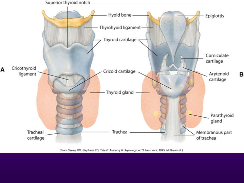
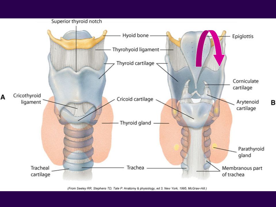
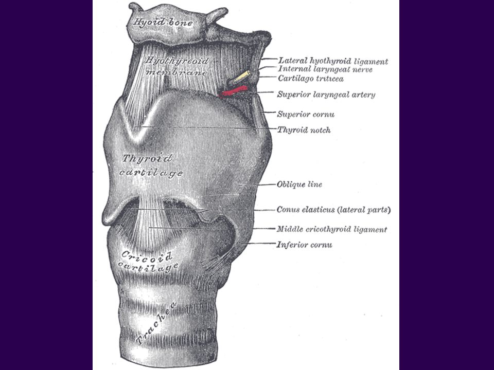
Cartilages of the Larynx
Composed of nine cartilages Three unpaired cartilage Thyroid (Greek for Oblong Shield) Cricoid (Greek for ring) Epiglottitis (Greek for “above glottis”) Three paired cartilages (six total) Arytenoids (Greek for ladle) Corniculates (Latin for horns – cornucopia) Cuneiforms (Latin for wedge)
 Cricoid (Greek for ring) Epiglottitis (Greek for above glottis ) Three paired cartilages (six total) Arytenoids (Greek for ladle) Corniculates (Latin for horns – cornucopia) Cuneiforms (Latin for wedge)")
6
http://coursewareobjects. elsevier
9
2. Arytenoid cartilage 3. Cervical trachea 6. Epiglottic larynx 7. Epiglottis 8. False Vocal Cords 9. Hyoid Bone 12. Subglottic larynx 15. True Vocal Cords

10
Paired Cartilages The Arytenoids, Cuneiforms, and Corniculates are all associated with movement of the vocal cords and are used in phonation.

11
Thyroid Cartilage The largest laryngeal cartilage is the thyroid cartilage “Adam’s Apple” Superior border has a V-shaped notch. Suspended from hyoid bone. Posterior wall is open. The true and false vocal cords are found on the interior of the larynx.

12
Vocal Cords Two pairs of folds that protrude inward:
Upper pair – False cords Lower pair – True cords The space between the vocal cords is called the rima glottidis or glottis Narrowest portion of the adult airway

13
Vocal Cords

14
Vocal Cords Vocal Cord Abduction Vocal Cord Adduction
Cords are opening or moving away from the midline This occurs during inspiration Vocal Cord Adduction Cords are moving toward the midline or coming together This occurs during expiration

16
Epiglottis Spoon-shaped cartilage which prevents aspiration by covering the opening of the larynx during swallowing. The tongue and the epiglottis are connected by folds of mucous membranes which form a small space called the vallecula.

17
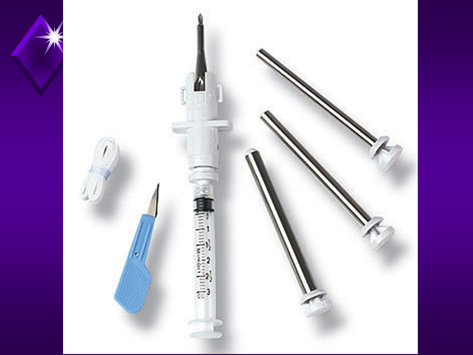
Intubation A device called a laryngoscope is used to visualize the laryngeal structures. It is composed of a handle and one of two types of blades: A curved blade (McIntosh) A straight blade (Miller or Wisconsin)
 A straight blade (Miller or Wisconsin)")
18
A curved laryngoscope blade is inserted into the vallecula during intubation to lift the epiglottis indirectly. A straight laryngoscope blade is used to directly lift the epiglottis during intubation

19
Cricoid Cartilage Resembles signet (class) ring. Inferior to Thyroid.
Only complete ring of laryngeal structures. Inferior border is attached to the first C-shaped tracheal ring. The narrowest portion of the airway in an infant. We use this fact when ventilating infants as infant ET tubes do not have cuffs to seal the trachea.

21
Cricothyroid membrane
Connects the cricoid and thyroid cartilages Is the site for an emergency airway Cricothyrotomy

23
Laryngeal Swelling http://www.rale.ca/Stridor.htm
Swelling (edema) at the glottis, subglottic or supraglottic region can cause stridor Stridor is a high pitched crowing sound usually heard on inspiration from air traveling through a narrowed opening Croup, Epiglottis, Foreign Body
 at the glottis, subglottic or supraglottic region can cause stridor. Stridor is a high pitched crowing sound usually heard on inspiration from air traveling through a narrowed opening. Croup, Epiglottis, Foreign Body.")
24
Laryngospasm A laryngeal reflex which will close the vocal cords inside the larynx Laryngospasm results from Extubations Near drowning Inhalation of noxious substances Smoke inhalation

25
Valsalva Maneuver Forced expiratory effort against a closed glottis to increase intrathoracic pressure (defecation) or to inflate the eustachian tubes and middle ears (“clearing” of the ears on airplanes). The larynx will tightly seal preventing air from escaping during physical work Lifting, pushing, throat-clearing, vomiting, urination, defecation and parturition.
 or to inflate the eustachian tubes and middle ears ( clearing of the ears on airplanes). The larynx will tightly seal preventing air from escaping during physical work. Lifting, pushing, throat-clearing, vomiting, urination, defecation and parturition.")
26
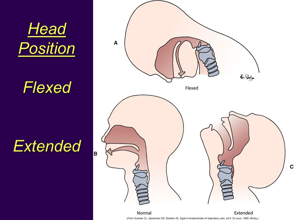
Head Position Flexed Extended
28
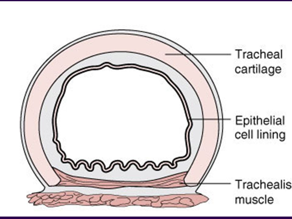
Histology of the Larynx
Above the vocal cords stratified squamous epithelium Below the vocal cords pseudostratified columnar epithelium Trachea to respiratory bronchioles

29
http://coursewareobjects. elsevier
30
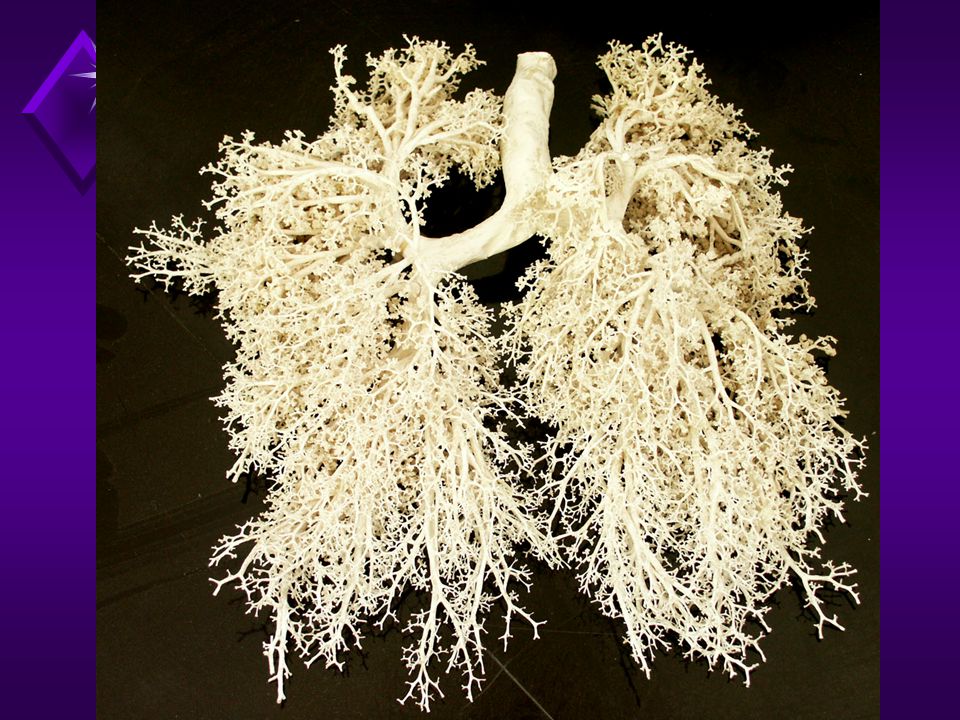
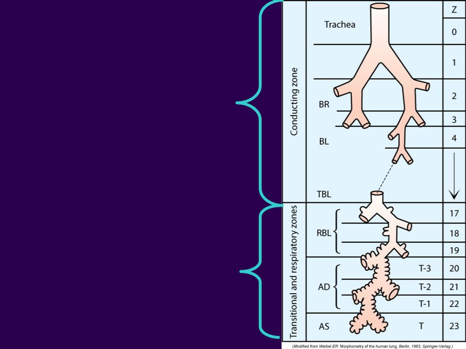
Tracheobronchial Tree
Two Divisions Cartilaginous Airways Primarily conducting airways; no gas exchange. Noncartilaginous Airways Both conducting airways and sites of gas exchange. Dichotomous Branching Each airway divides into two “daughter” branches Each division (bifurcation) gives rise to a new generation of airways As airways divide, they become Shorter Narrower More numerous
 gives rise to a new generation of airways. As airways divide, they become. Shorter. Narrower. More numerous.")
31
http://coursewareobjects. elsevier
33
Cartilaginous Airways
Trachea Main Stem Bronchi Lobar Bronchi Segmental Bronchi Subsegmental Bronchi

34
Lobar Bronchi

35
Trachea Generation 0 11 – 13 cm long and 1.5 – 2.5 cm wide.
Extends from Cricoid cartilage (6th cervical vertebrae) to the 2nd costal cartilage or 5th thoracic vertebrae. C6 – T5 C-shaped cartilages supports the trachea. Posterior wall is contiguous with esophagus.
 to the 2nd costal cartilage or 5th thoracic vertebrae. C6 – T C-shaped cartilages supports the trachea. Posterior wall is contiguous with esophagus.")
36
Trachea The end of the trachea is called the carina.
This is the division of the trachea into the right and left mainstem bronchi. Air is 100% saturated with water vapor and is warmed to 37 °C (body temperature). The carina is located at approximately T5 or the Angle of Louis. The surgical opening into the trachea is called a tracheostomy. 2nd or 3rd tracheal ring.
. The carina is located at approximately T5 or the Angle of Louis. The surgical opening into the trachea is called a tracheostomy. 2nd or 3rd tracheal ring.")
37
http://coursewareobjects. elsevier
38
Main Stem Bronchi Generation 1
Trachea divides into the right and left mainstem bronchi – one for each lung Right Mainstem is wider, shorter and more vertical Branches at a 25 degree angle Left Mainstem Branches at a 40 – 60 angle Infants Both mainstem bronchi form a 55 angle with the trachea

39
Newborn

40
Complications of Intubation
During intubations, if the tube is advanced to far, the tube will usually go into the right mainstem bronchi. Lung inflation will be absent on the left but present on the right. Withdraw tube until bilateral sounds are heard. Failure to hear lung sounds or visualize chest inflation on either side means the tube is probably in the stomach. Extubate the patient and re-attempt the intubation.

41
Aspiration Children who aspirate objects
Foreign body usually lodged in right main stem bronchi secondary to the angle being less acute. Wheezing on right or absent lung sounds (breath sounds).
.")
42
Lobar Bronchi Generation 2
Lobar Bronchi correlate to the number of lobes of the lung. The right mainstem bronchi will divide into the right upper, right middle and right lower lobe bronchi. The left mainstem bronchi will divide into the left upper and left lower lobe bronchi.

43
Segmental Bronchi Generation 3
Correlate with the segments of the lung. There are 10 segmental bronchi on the right. There are 8 segmental bronchi on the left.

44
Subsegmental Bronchi 4th to 9th Generations 1 to 4 mm in diameter
Connective tissue containing: Nerves Lymphatics Bronchial Arteries

46
Non-Cartilaginous Airways
Bronchioles 10th to 15th Generation. 1 mm in diameter. Simple cuboidal epithelium. No cartilage. Terminal Bronchioles Less than 0.5 mm in diameter. No cartilage (lack of support). Cilia and mucous glands disappear. Clara Cells appear Inter-bronchiole connections called Canals of Lambert begin to appear.
. Cilia and mucous glands disappear. Clara Cells appear. Inter-bronchiole connections called Canals of Lambert begin to appear.")
48
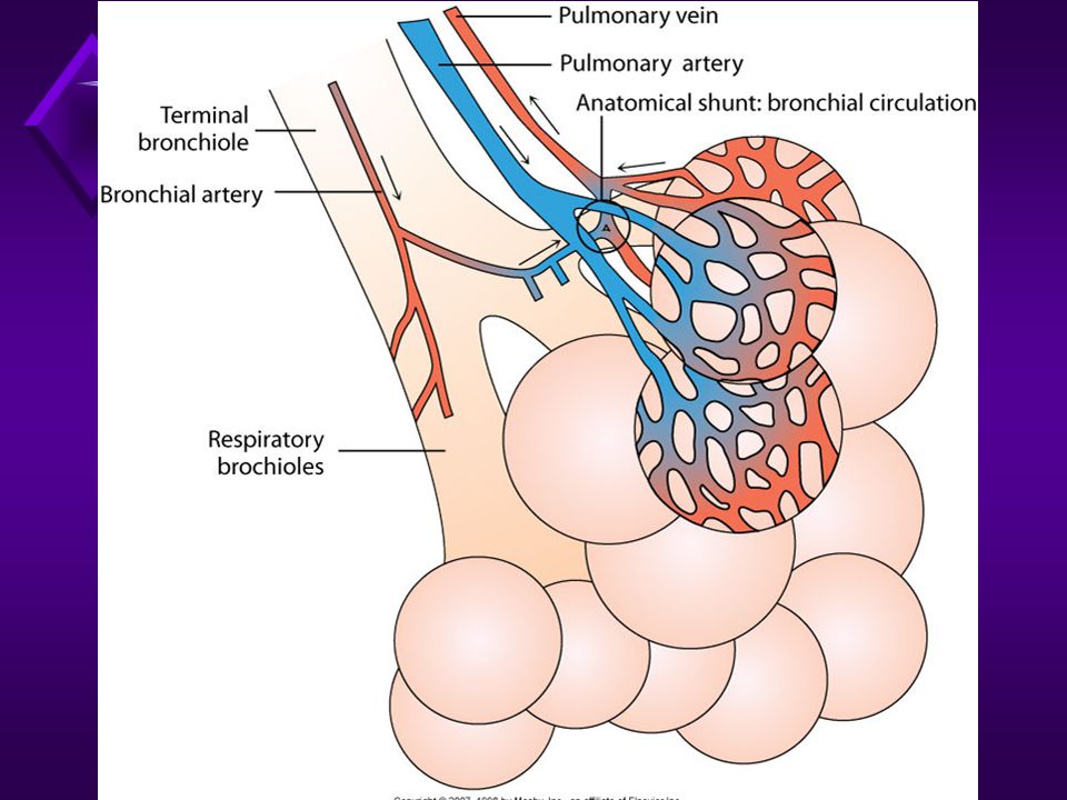
Blood Supply to the Tracheobronchial Tree
Bronchial Blood Supply Bronchial arteries nourish the tracheobronchial tree The arteries arise from the aorta and follow the tracheobronchial tree as far as the terminal bronchioles. Beyond the terminal bronchioles pulmonary arteries & capillaries feed the airways & alveoli. Normal bronchial blood flow is approximately 1% of the cardiac output. Also feed the mediastinal lymph nodes, pulmonary nerves, part of the esophagus and the visceral pleura.

49
Review of TB Tree Trachea Mainstm Bronchi Lobar Bronchi
Segmental Bronchi Subsegmental Bronchi Bronchioles – cartilage disappears Terminal Bronchioles

50
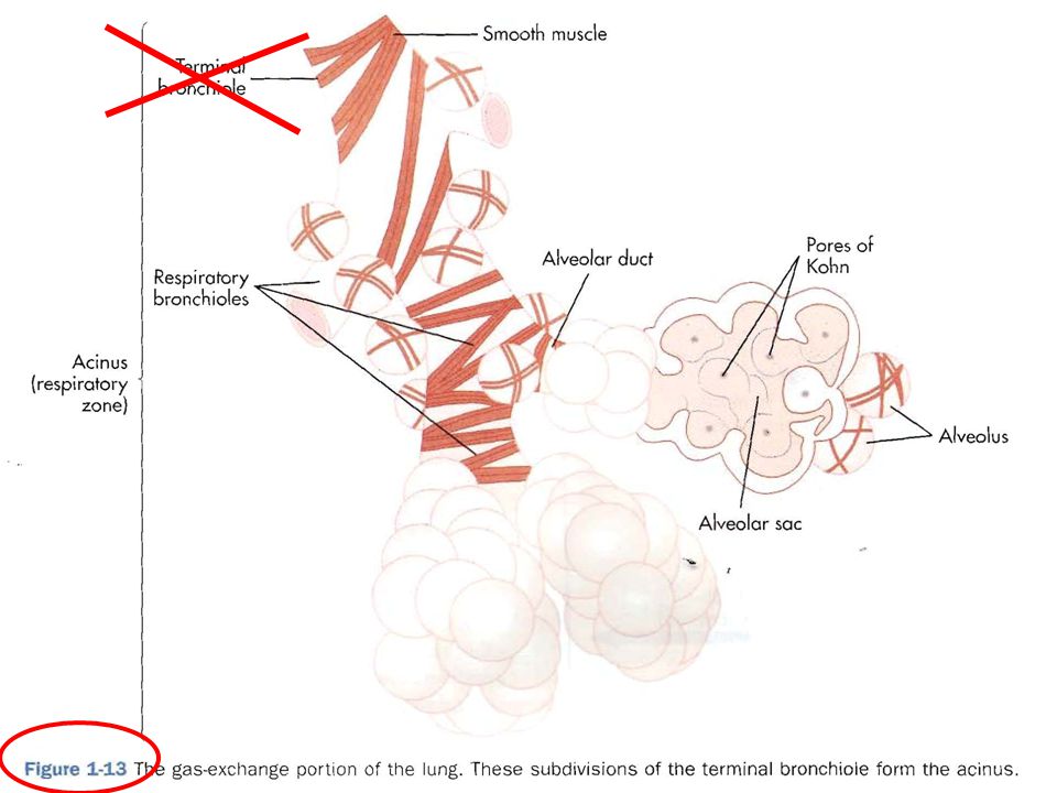
Site of Gas Exchange “The Respiratory Zone”
Consists of the respiratory bronchioles, alveolar ducts, and alveolar sacs, and alveoli. Parenchyma, Acinus or Primary Lobule.

52
Creative Commons Attribution

53
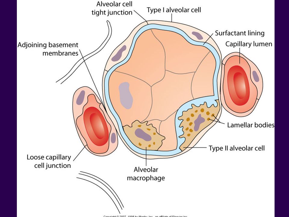
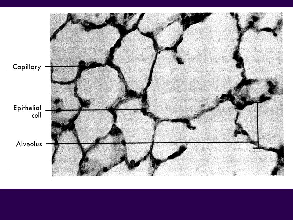
Alveolar Epithelium Two principal cell types: Alveolar Type I Cells
Squamous pneumocyte Broad thin cells. 95% of alveolar surface 0.1 m to 0.5 m thick Alveolar Type II Cells 5% of alveolar surface Cuboidal in shape Responsible for secretion of pulmonary surfactant that reduce surface tension and keep the alveoli stable.

54
Facts about the Lungs There are 300 million alveoli in the lungs.
The surface area of the lungs is square meters (Tennis Court). The lung has 35 times more surface area then the skin.
. The lung has 35 times more surface area then the skin.")
55
Additional Components of Alveolar Epithelium
Pores of Kohn Small holes in the walls of interalveolar septa. 3 m to 13 m in diameter Alveolar Macrophages or Type III alveolar cells. Major role in removing bacteria and other foreign particles. Interstitium Gel-like substance between alveoli-capillary clusters that add support

56
http://coursewareobjects. elsevier
57
http://coursewareobjects. elsevier
59
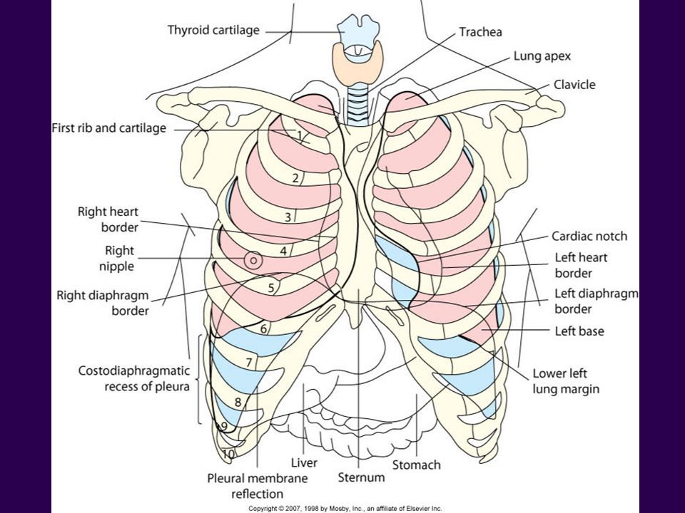
Lung Extends from the diaphragm to 1-2 cm above the clavicles (about the 1st rib). The lung apex is at the top of the lung and is somewhat pointed. The base is broad and concave and lies at about the 6th rib or xiphoid process anteriorly, the 8th rib laterally, and the 11th rib posteriorly. The right lung is larger and heavier than the left.

60
http://coursewareobjects. elsevier
61
Lung Lobes and Segments
Right lung Three Lobes Upper, Middle, Lower Divided by the Horizontal and Oblique fissures. 10 Segments Left lung Two Lobes Upper and Lower Divided by the Oblique fissures. 8 Segments

63
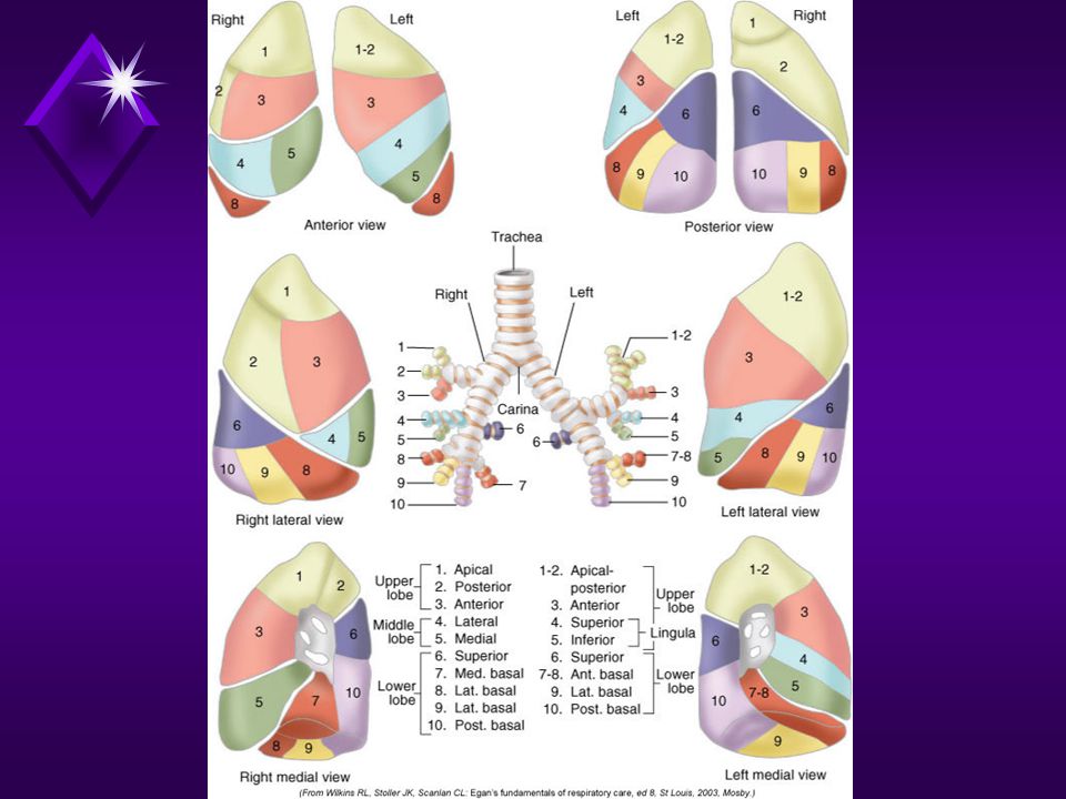
LOBES AND SEGMENTS OF THE LUNGS
RIGHT LUNG LEFT LUNG UPPER LOBE UPPER LOBE (UPPER DIVISION) APICAL SEGMENT ANTERIOR SEGMENT POSTERIOR SEGMENT APICAL/POSTERIOR ANTERIOR MIDDLE LOBE UPPER LOBE (LINGULA) LATERAL MEDIAL SUPERIOR INFERIOR LOWER LOBE ANTERIOR BASAL MEDIAL BASAL LATERAL BASAL POSTERIOR BASAL ANTERIOR/MEDIAL BASAL
 APICAL SEGMENT ANTERIOR SEGMENT. POSTERIOR SEGMENT. APICAL/POSTERIOR. ANTERIOR. MIDDLE LOBE. UPPER LOBE (LINGULA) LATERAL. MEDIAL. SUPERIOR. INFERIOR. LOWER LOBE. ANTERIOR BASAL. MEDIAL BASAL. LATERAL BASAL. POSTERIOR BASAL. ANTERIOR/MEDIAL BASAL.")
64
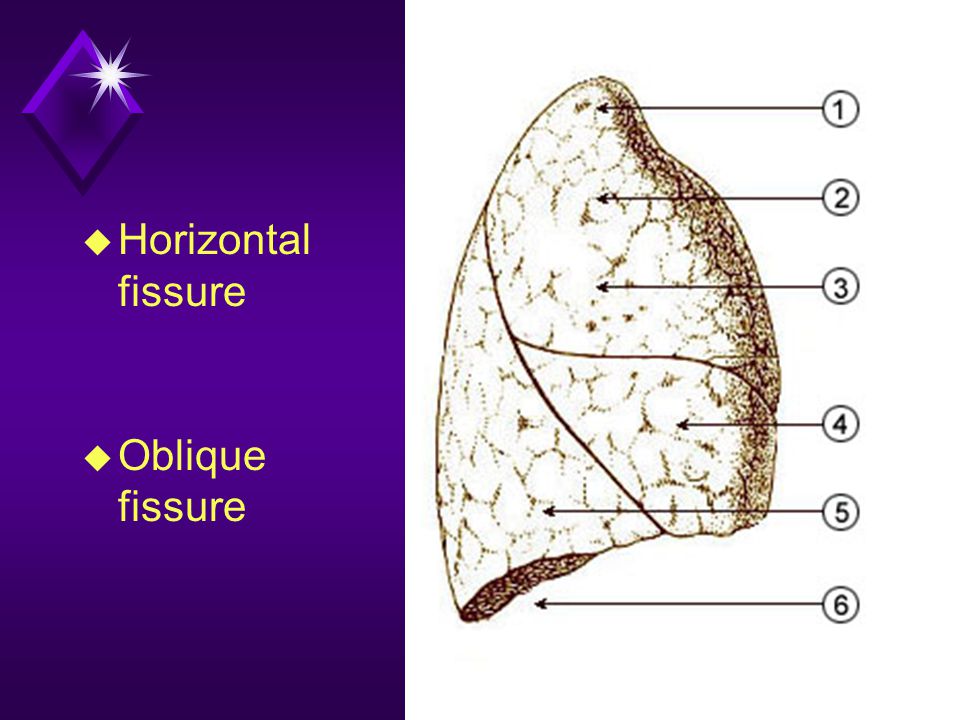
Lung Fissures Oblique Fissure Horizontal or minor Fissure
Found in the left and right lung Separates the upper and lower lobes of both lungs Horizontal or minor Fissure Found only in the right lung Separates the upper and middle lobes

65
Horizontal fissure Oblique fissure
67
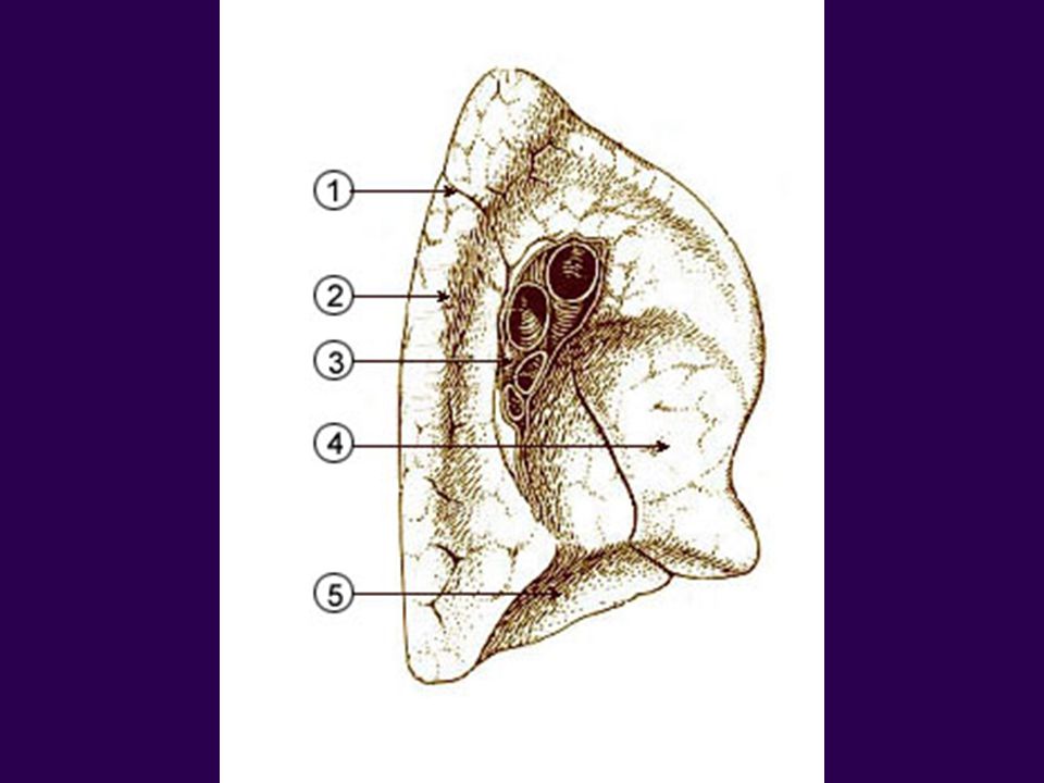
Hilum The hilum is where arteries, veins, bronchi, nerves and lymph vessels enter and leave the lung. It is located on the medial border of the lung.

68
http://upload. wikimedia. org/wikipedia/commons/f/f7/Illu_quiz_lung04
Similar presentations


 nasal cavity.>")

. 2.Production of sound (vocal cords). 3.Pulmonary ventilation. 4. Inspiration (intercostals muscles lift.>")


 bronchus Right main (primary) bronchus Left lung.>")


