Download presentation
Presentation is loading. Please wait.

1
Muscles of the Body

2
http://www. rad. washington
"Copyright University of Washington. All rights reserved including all photographs and images. No re-use, re-distribution or commercial use without prior written permission of the authors and the University of Washington." "Musculoskeletal Images are from the University of Washington "Musculoskeletal Atlas: A Musculoskeletal Atlas of the Human Body" by Carol Teitz, M.D. and Dan Graney, Ph.D." Other copies materials from Marieb, 5th ed., Martini, 6th ed. or online with reference

3
Fascicles Bundles of fibers *

4
Skeletal muscle Epimysium: surrounds whole muscle
Endomysium is around each muscle fiber Perimysium is around fascicle

5
Different arrangements of fascicles

6
Arrangement of fascicles influences movement and power
Skeletal muscles can shorten by about 1/3 of their resting length The more nearly parallel to the axis, the more they can shorten This results in a larger distance of movement The power depends on the total number of fibers Stocky muscles (like bipennate vs parallel) have more fibers, therefore more powerful even though shorten very little
 have more fibers, therefore more powerful even though shorten very little.")
7
Interactions of Skeletal Muscles
Muscles can only pull, they can’t push Actions must be “undone” by a different muscle Muscles that produce opposite movements usually lie on opposite sides of a given joint

8
Interactions, continued
Agonist: prime mover, major responsibility for producing a specific movement Antagonist: oppose or reverse a particular movement Usually contract a little to prevent overshooting the mark or slow the agonist’s action near the end Are being stretched or can remain relaxed when agonist works Antagonists for one movement can be agonists for another

9
Synergists help prime movers
Add a little extra force to the same movement Or reduce undesirable extra movements (e.g. making a fist without flexing at wrist) Fixators: hold a bone firmly so agonist has a stable base on which to move a body part (e.g. fixing scapula when arm moves)
 Fixators: hold a bone firmly so agonist has a stable base on which to move a body part (e.g. fixing scapula when arm moves)")
10
Compartments Contain muscles of similar developmental origin and function Dense fibrous connective tissue separates Upper limb: 2 compartments Lower limb: 3 compartments

11
Naming skeletal muscles (examples)
Location: brachialis is in arm (brachium = arm) Shape: deltoid is triangular (delta = triangle) Size: minimus (smallest), longus (long), brevis (short) Direction of fascicles and fibers: rectus (straight); transversus (right angle) and oblique (oblique) to midline Number of origins: biceps (“two heads”), triceps (“three heads”), quadriceps (“four heads”) Action: “flexor,” “extensor,” “adductor” or “abductor” appear in the name Combinations of the above, e.g. extensor carpi radialis longus
 Shape: deltoid is triangular (delta = triangle) Size: minimus (smallest), longus (long), brevis (short) Direction of fascicles and fibers: rectus (straight); transversus (right angle) and oblique (oblique) to midline. Number of origins: biceps ( two heads ), triceps ( three heads ), quadriceps ( four heads ) Action: flexor, extensor, adductor or abductor appear in the name. Combinations of the above, e.g. extensor carpi radialis longus.")
12
Axial muscles Lie anterior and posterior to body axis (vertebral column) Move trunk; maintain posture Skeletal muscles of thorax, abdomen, and pelvis Many muscles of neck A few muscles in head

13
Limb muscles Arise from ventral region as limb buds
Muscles develop from lateral parts of myotomes In general: dorsal (posterior) muscles become extensors and ventral (anterior) become flexors Lower limb rotates during embryonic development: Extensors on anterior (ventral) side: extend leg at knee, dorsiflex foot at ankle and extend toes Flexors on posterior (dorsal) side: flex leg at knee, plantarflex foot at ankle, flex toes
 muscles become extensors and ventral (anterior) become flexors. Lower limb rotates during embryonic development: Extensors on anterior (ventral) side: extend leg at knee, dorsiflex foot at ankle and extend toes. Flexors on posterior (dorsal) side: flex leg at knee, plantarflex foot at ankle, flex toes.")
14
to use for studying... anterior Text Text

15
posterior

16
16

17
17

18
18

19
19

20
20

21
21

22
Muscles of the Head and Neck
Scalp Muscle: epicranius frontal belly occipital belly gala aponeurotica Muscles of Facial Expression: insert on skin or another muscle Muscles of Mastication (chewing): all have insertions on the mandible Anterior Neck Muscles Posterior Neck Muscles
: all have insertions on the mandible. Anterior Neck Muscles. Posterior Neck Muscles.")
23
23

24
24

26
Extrinsic tongue muscles Pharyngeal constrictors
Mastication: Jaw closure: masseter and temporalis Side to side grinding: pterygoids Buccinator: compresses cheek “glossus” = tongue Deep chewing muscles Extrinsic tongue muscles Pharyngeal constrictors Tongue itself (instrinsic muscles): digestive tract section
: digestive tract section.")
27
Muscles of the Anterior Neck
Above hyoid (suprahyoid): form floor of oral cavity, anchor tongue, elevate hyoid, move larynx superiorly during swallowing Below hyoid (infrahyoid): depress hyoid and larynx during swallowing and speaking Right side (of slide) is deeper than left Sternocleodomastoid
: form floor of oral cavity, anchor tongue, elevate hyoid, move larynx superiorly during swallowing. Below hyoid (infrahyoid): depress hyoid and larynx during swallowing and speaking. Right side (of slide) is deeper than left. Sternocleodomastoid.")
28
Neck Posterior neck Anterolateral neck
Splenius’ (capitis and cervicis) extend head Anterolateral neck Scalenes elevate first 2 ribs
 extend head. Anterolateral neck. Scalenes elevate first 2 ribs.")
29
Deep muscles of back Quadratus lumborum Erector spinae
Right side: deeper Quadratus lumborum (lateral flexion) Erector spinae (extend back): Iliocostalis Longissimus Spinalis Labeled cervicis, thoracics, lumborum depending on where they are
 Erector spinae. (extend back): Iliocostalis. Longissimus. Spinalis. Labeled cervicis, thoracics, lumborum depending on where they are.")
30
Per Marieb… (worthwhile to know)
“During full flexion (i.e. when touching fingertips to floor), erector spinae are relaxed and strain is borne entirely by ligaments of back; on reversal of the movement, these muscles are initially inactive, and extension is initiated by hamstring muscles of thighs and gluteus maximus muscles of buttocks. As a result of this peculiarity, lifting a load or moving suddenly from a bent over position is potentially injurious to muscles and ligaments of back and intervertebral discs; erector spinae muscles readily go into painful spasms following injury to back structures.”
, erector spinae are relaxed and strain is borne entirely by ligaments of back; on reversal of the movement, these muscles are initially inactive, and extension is initiated by hamstring muscles of thighs and gluteus maximus muscles of buttocks. As a result of this peculiarity, lifting a load or moving suddenly from a bent over position is potentially injurious to muscles and ligaments of back and intervertebral discs; erector spinae muscles readily go into painful spasms following injury to back structures.")
31
Deep muscles of the thorax: breathing
Lift rib cage: inspiratory Intercostals Short: rib to rib Diaphragm prime mover of inspiration Depress rib cage: in forced expiration Floor of thoracic cavity: when flattens, air moves in

32
Anterior Chest Muscles
Superficial: sternocleidomastoid pectoralis major Deeper: pectoralis minor serratus anterior subclavius

33
Muscles of the abdominal wall
From more superficial to deep: External oblique Internal oblique Transversus abdominis Nearer midline: Rectus abdominis Note inguinal ligament- from anterior superior iliac spine to pubic symphysis: lower border of external oblique rolls up on itself to form it * * The rectus abdominis is the medial pair of muscles; it is ensheathed by the aponeurosis of the lateral muscles, which don’t come to the midline

34
Muscles of the abdominal wall from the side

35
Muscles moving the scapula
trapezius levator scapulae rhomboids posterior

36
9 Muscles crossing shoulder joint: movement of arm (humerus)
Three most powerful of the nine and prime movers: pectoralis major latissimus dorsi deltoid

37
Rotator cuff supraspinatus, infraspinatus, subscapularis, teres minor
remaining 2muscles : teres major and coracobrachialis

38
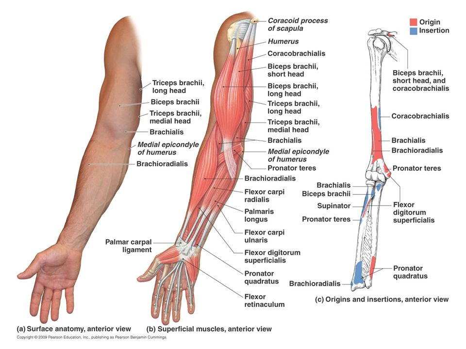
Forearm extensors (posterior)
Triceps brachii Anconeus helps

39
Forearm flexors (anterior)
Brachioradialis Brachialis Biceps brachii 3 muscle on right from this site:

40
Retinaculae (retinaculum, singular)
“retainers,” “wrist bands”, “ankle bracelets” Bands of fascia holding tendons of wrist and ankle in place (prevent “bow-stringing”) Tendons covered by slippery tendon sheets
 Tendons covered by slippery tendon sheets.")
41
Forearm muscles: movement of wrist, hand and fingers
Many arise from distal humerus Cross elbow, wrist and finger joints Minimal action at elbow At wrist joint: flexion, extension, abduction and adduction of the hand At finger joints: mostly just flex and extend (other movements- by small muscles in the hand itself)
")
42
Forearm muscles, continued
Two compartments (each with superficial and deep muscle layers) Anterior = flexor compartment ( except includes 2 pronators) Most originate from a common tendon on the medial epicondyle of humerus Posterior = extensor compartment (except includes supinator and brachioradialis) Many arise from a common tendon from lateral epicondyle of humerus Most muscles that move the palm and fingers are located in the forearm, not the hand itself They operate by tendons like strings with puppets There are some small muscles in the hand itself
 Anterior = flexor compartment ( except includes 2 pronators) Most originate from a common tendon on the medial epicondyle of humerus. Posterior = extensor compartment (except includes supinator and brachioradialis) Many arise from a common tendon from lateral epicondyle of humerus. Most muscles that move the palm and fingers are located in the forearm, not the hand itself. They operate by tendons like strings with puppets. There are some small muscles in the hand itself.")
43
Anterior wrist pronator and flexors
Origin on medial epicondle of humerus: pronator teres, flexor carpi radialis, palmaris longus, flexor carpi ulnaris, flexor digitorum superficialis Pronator teres Palmaris longus

44
Deep anterior hand muscles (some)
Flexor pollicis longus Flexor digitorum profundus (only muscle that flexes DIPs)
")
45
Individually Flexor digitorum superficialis
(this is right arm, anterior) Flexor digitorum profundus Flexor pollicis longus Pronator quadratus
 Flexor digitorum profundus. Flexor pollicis longus. Pronator quadratus.")
46
Extensor carpi radialis (longus & brevis)
origin on lateral epicondyl of humerus Extensor carpi radialis (longus & brevis) Extensor digitorum Extensor carpi ulnaris See individually in next slide
 Extensor digitorum. Extensor carpi ulnaris. See individually in next slide.")
47
Superficial extensors
Extensor carpi radialis longus Extensor digitorum Extensor carpi ulnaris Extensor carpi radialis brevis

48
Deep posterior muscles
Extensor pollicis longus & brevis Abductor pollicis longus Supinator Extensor indicis

49
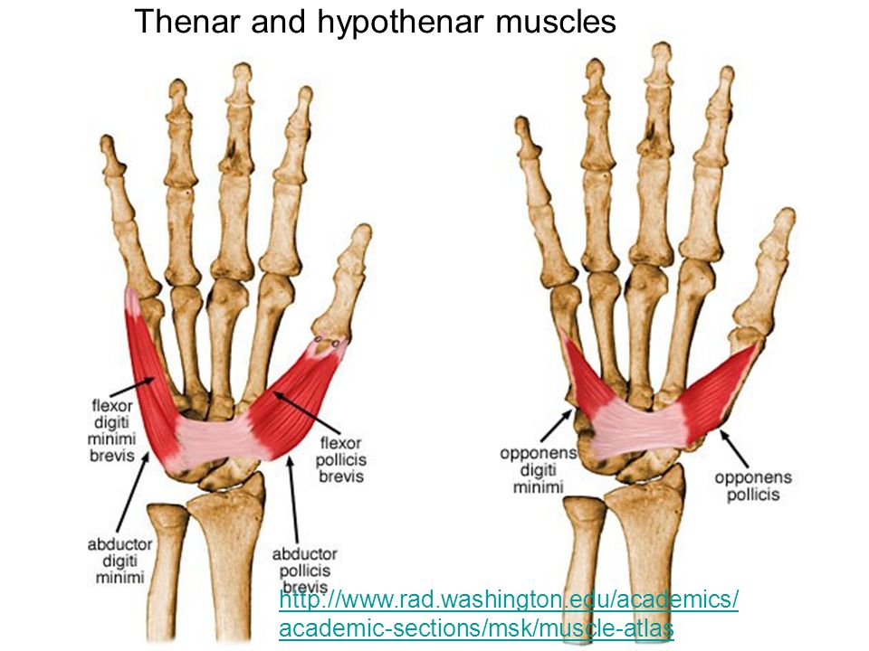
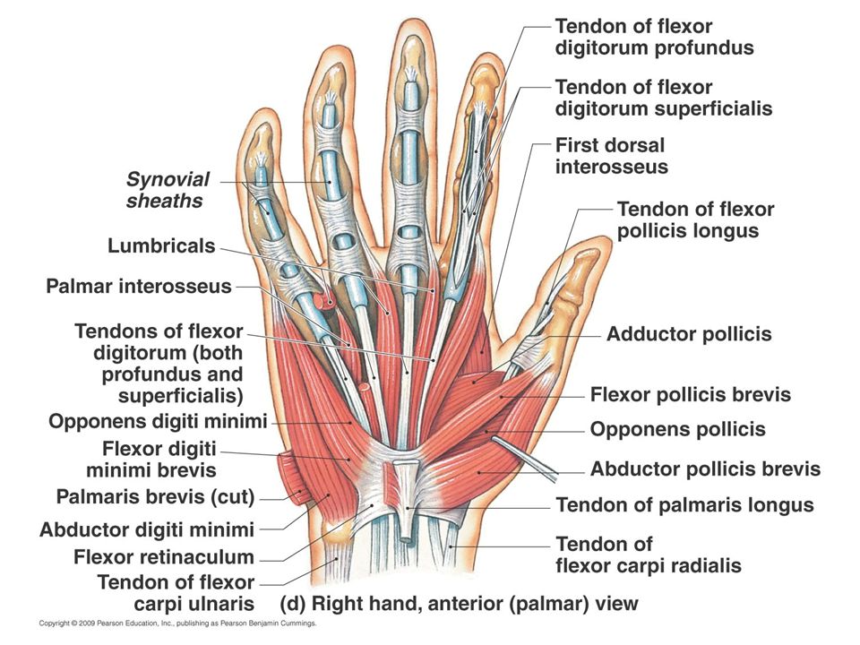
Hand Te Thenar Hypothenar Midpalmar Lumbricals Interossei

50
Thenar and hypothenar muscles
52
Right forearm, anterior view, from superficial to deep

54
Right forearm, posterior, from superficial to deep

58
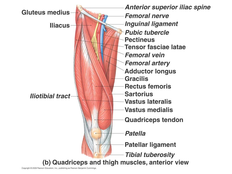
Muscles crossing the hip and knee joints
Three groups separated by fascia; all three enclosed by deep fascia of thigh (fascia lata) 1. Anterior Flex femur at hip; extend leg at knee (e.g. foreswing phase of walking) 2. Posterior Mostly extend thigh and flex leg (backswing phase of walking) 3. Adductor (medial) Move thigh only, not leg
 1. Anterior. Flex femur at hip; extend leg at knee (e.g. foreswing phase of walking) 2. Posterior. Mostly extend thigh and flex leg (backswing phase of walking) 3. Adductor (medial) Move thigh only, not leg.")
59
Muscles that flex thigh at hip
Originate from vertebral column and pelvis and pass anterior to hip joint Sartorius Iliopsoas Tensor fasciae lata Rectus femoris (only quad with origin on pelvis) Pectineus (medial compartment)
 Pectineus (medial compartment)")
60
Muscles that flex thigh at hip: individually (go between last slide and this one)
Iliopsoas Pectineus Tensor fascia lata Sartorius Rectus femoris Inserts on tibial tuberosity via patellar tendon

61
Thigh extensors (posterior)
Arise posterior to hip joint Gluteus maximus Hamstrings (cross hip and knee joints: extend thigh & flex knee) Biceps femoris Semitendinosus Semimembranosus (antagonists of quads) _______
 Biceps femoris. Semitendinosus. Semimembranosus. (antagonists of quads) _______.")
62
Hamstrings cross hip and knee joints: extend thigh and flex knee
Biceps femoris long head Biceps femoris short head Semitendinosus Semimembranosus

63
Abductors of thigh Buttocks muscles that lie lateral to hip joint
Gluteus medius Gluteus minimus (under medius) Tensor fascia lata
 Tensor fascia lata.")
64
Thigh abductors Buttocks muscles that lie lateral to the hip joint
Gluteus medius Gluteus minimus

65
Lateral rotators Piriformis
Piriformis laterally rotates hip; also helps abduct hip if it is flexed Piriformis Also shown are other rotators and the gluteus muscles

66
Adduction of thigh Muscles originate medial to hip joint Gracilis
Adductor magnus Adductor longus Adductor brevis Pectineus

67
Thigh adductors (originate medial to hip joint)
Adductor magnus Pectineus Adductor brevis Gracilis Adductor longus

68
Knee extensors Quadraceps femoris – the only extensors of the leg (lower leg) at the knee Rectus femoris (only quad with origin on pelvis) Vastus lateralis Vastus intermedius Vastus medialis Antagonized by hamstrings Rectus femoris

69
Quadriceps Vastus lateralis, intermedius, and medialis
Rectus femoris (only quad with origin on the pelvis) Quadriceps Vastus lateralis, intermedius, and medialis Note “o” and “i” _________ Insert: tibial tuberosity via patellar ligament
 Quadriceps. Vastus lateralis, intermedius, and medialis. Note o and i _________. Insert: tibial tuberosity via patellar ligament.")
70
Leg (lower leg) 3 compartments Movements at joints: Anterior Posterior
Lateral Movements at joints: Ankle Dorsiflex Plantarflex Intertarsal Inversion of foot Eversion of foot Toes Flex (point) Extend
 Extend.")
71
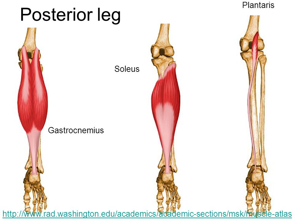
Posterior compartment of leg
Superficial: these plantarflex foot Gastrocnemius Soleus Plantaris

72
Posterior leg Plantaris Soleus Gastrocnemius
73
Posterior leg continued
Deep Popliteus Flexor digitorum longus Flexor hallucis longus Tibilialis posterior

74
Deep posterior leg Popliteus Flexor digitorum longus
Flexor hallucis longus Tibialis posterior

75
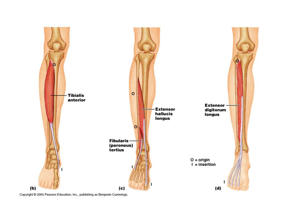
Anterior leg extensors
Mainly extend toes and dorsiflex foot Tibialis anterior Extensor digitorum longus Extensor hallucis longus

77
More pics Extensor hallucis longus Tibialis anterior
Extensor digitorum longus

78
Lateral compartment of leg
Fibularis (peroneus) longus: to first metatarsal and cuneiform Fibularis (peroneus) brevis: to fifth metatarsal
 longus: to first metatarsal and cuneiform. Fibularis (peroneus) brevis: to fifth metatarsal.")
84
Sole – third (deepest) layer
 layer")
85
Addendum: some rotator cuff tests (FYI)
FIGURE 3. Supraspinatus examination ("empty can" test). The patient attempts to elevate the arms against resistance while the elbows are extended, the arms are abducted and the thumbs are pointing downward. FIGURE 2. Apley scratch test. The patient attempts to touch the opposite scapula to test range of motion of the shoulder. (Left) Testing abduction and external rotation. (Right) Testing adduction and internal rotation.
. The patient attempts to elevate the arms against resistance while the elbows are extended, the arms are abducted and the thumbs are pointing downward. FIGURE 2. Apley scratch test. The patient attempts to touch the opposite scapula to test range of motion of the shoulder. (Left) Testing abduction and external rotation. (Right) Testing adduction and internal rotation.")
86
FIGURE 4. Infraspinatus/teres minor examination
FIGURE 4. Infraspinatus/teres minor examination. The patient attempts to externally rotate the arms against resistance while the arms are at the sides and the elbows are flexed to 90 degrees. FIGURE 5. Neer's test for impingement of the rotator cuff tendons under the coracoacromial arch. The arm is fully pronated and placed in forced flexion. FIGURE 6. Hawkins' test for subacromial impingement or rotator cuff tendonitis. The arm is forward elevated to 90 degrees, then forcibly internally rotated.

Similar presentations


>")

