Download presentation
Presentation is loading. Please wait.

1
Chest X-ray Interpretation
Dr C. Lokubalasooriya

2
Introduction Single most common basic examination Low cost examination
More information

3
X-rays were first discovered accidentally by Wilhelm Conrad Röntgen in 1895.
X-rays are waves of electromagnetic energy that have a shorter wavelength than normal light He discovered that these new invisible rays could pass through most objects that casted shadows including human tissue but not human bones and metals. Within a year of the discovery many scientists replicated the experiment Röntgen performed and began using it in clinical settings In 1901 Röntgen won the first Nobel Prize in Physics

4
Essentials Before Getting Started
Identification Exposure Overexposure Underexposure Sex of Patient Male Female

5
Essentials Before Getting Started
Path of x-ray beam PA AP Patient Position Upright Supine

6
Essentials Before Getting Started
Breath Inspiration Expiration

7
Systematic Approach Bony Framework Soft Tissues Lung Fields and Hila
Diaphragm and Pleural Spaces Mediastinum and Heart Abdomen and Neck

8
Systematic Approach Bony Fragments Ribs Sternum Spine Shoulder girdle
Clavicles

9
Systematic Approach Soft Tissues Breast shadows Supraclavicular areas
Axillae Tissues along side of breasts

10
Systematic Approach Lung Fields and Hila Hilum Lungs Blood vessels
Pulmonary arteries Pulmonary veins Lungs Linear and fine nodular shadows of pulmonary vessels Blood vessels 40% obscured by other tissue

11
Systematic Approach Diaphragm and Pleural Surfaces Diaphragm
Dome-shaped Costophrenic angles Normal pleural is not visible Interlobar fissures

12
Systematic Approach Mediastinum and Heart Heart size on PA Right side
Inferior vena cava Right atrium Ascending aorta Superior vena cava

13
Systematic Approach Mediastinum and Heart Left side Left ventricle
Left atrium Pulmonary artery Aortic arch Subclavian artery and vein

14
Systematic Approach Abdomen and Neck Abdomen Neck Gastric bubble
Air under diaphragm Neck Soft tissue mass calcifications

15
Pitfalls to Chest X-ray Interpretation
Poor inspiration Over or under penetration Rotation Forgetting the path of the x-ray beam

16
Lung Anatomy Trachea Carina Right and Left Pulmonary Bronchi
Secondary Bronchi Tertiary Bronchi Bronchioles Alveolar Duct Alveoli

17
Lung Anatomy Right Lung Left Lung Superior lobe Middle lobe
Inferior lobe Left Lung

18
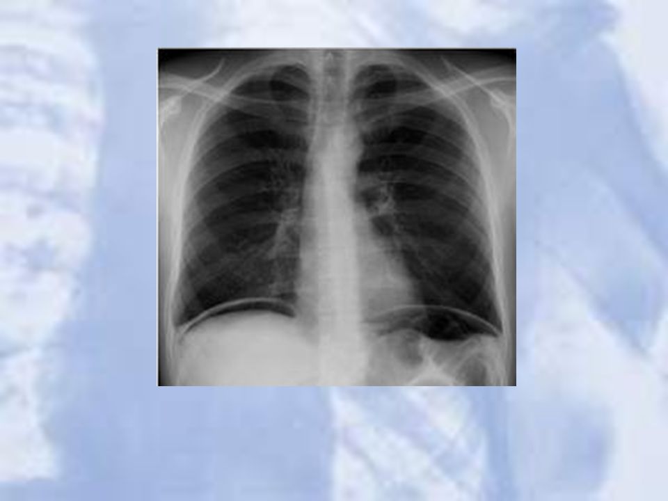
Lung Anatomy on Chest X-ray
PA View: Extensive overlap Lower lobes extend high Lateral View: Extent of lower lobes

19
Lung Anatomy on Chest X-ray
The right upper lobe (RUL) occupies the upper 1/3 of the right lung. Posteriorly, the RUL is adjacent to the first three to five ribs. Anteriorly, the RUL extends inferiorly as far as the 4th right anterior rib
 occupies the upper 1/3 of the right lung. Posteriorly, the RUL is adjacent to the first three to five ribs. Anteriorly, the RUL extends inferiorly as far as the 4th right anterior rib.")
20
Lung Anatomy on Chest X-ray
The right middle lobe is typically the smallest of the three, and appears triangular in shape, being narrowest near the hilum

21
Lung Anatomy on Chest X-ray
The right lower lobe is the largest of all three lobes, separated from the others by the major fissure. Posteriorly, the RLL extend as far superiorly as the 6th thoracic vertebral body, and extends inferiorly to the diaphragm. Review of the lateral plain film surprisingly shows the superior extent of the RLL.

22
Lung Anatomy on Chest X-ray
The lobar architecture of the left lung is slightly different than the right. Because there is no defined left minor fissure, there are only two lobes on the left; the left upper

23
Lung Anatomy on Chest X-ray
Left lower lobes

24
The Normal Chest X-ray PA View: Aortic arch Pulmonary trunk
Left atrial appendage Left ventricle Right ventricle Superior vena cava Right hemidiaphragm Left hemidiaphragm Horizontal fissure

25
The Normal Chest X-ray Lateral View: Oblique fissure
Horizontal fissure Thoracic spine and retrocardiac space Retrosternal space

26
The Silhouette Sign An intra-thoracic radio-opacity, if in anatomic contact with a border of heart or aorta, will obscure that border. An intra-thoracic lesion not anatomically contiguous with a border or a normal structure will not obliterate that border.

27
Putting It All Together

29
Understanding Pathological Changes
Most disease states replace air with a pathological process Each tissue reacts to injury in a predictable fashion Lung injury or pathological states can be either a generalized or localized process

30
Liquid Density Liquid density Increased air density Generalized
Localized Diffuse alveolar Diffuse interstitial Mixed Vascular Infiltrate Consolidation Cavitation Mass Congestion Atelectasis Localized airway obstruction Diffuse airway obstruction Emphysema Bulla

31
Stages of Evaluating an Abnormality
1. Identification of abnormal shadows 2. Localization of lesion 3. Identification of pathological process 4. Identification of etiology 5. Confirmation of clinical suspension Complex problems Introduction of contrast medium CT chest MRI scan

32
Putting It Into Practice

33
Case 1

35
A single, 3cm relatively thin-walled cavity is noted in the left midlung. This finding is most typical of squamous cell carcinoma (SCC). One-third of SCC masses show cavitation
. One-third of SCC masses show cavitation.")
36
Case 2

38
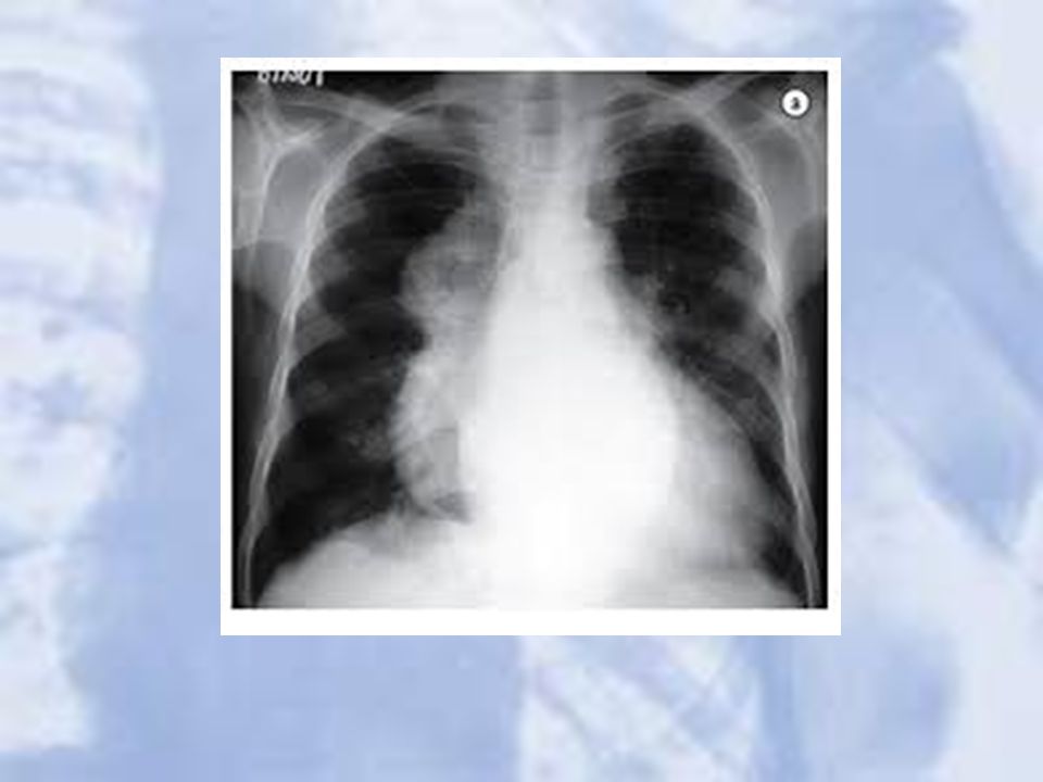
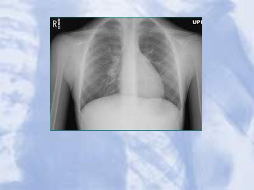
LUL Atelectasis: Loss of heart borders/silhouetting
LUL Atelectasis: Loss of heart borders/silhouetting. Notice over inflation on unaffected lung

39
Case 3

41
Right Middle and Left Upper Lobe Pneumonia

42
Case 4

44
Cavitation:cystic changes in the area of consolidation due to the bacterial destruction of lung tissue. Notice air fluid level.

45
Cavitation

46
Case 5

48
Tuberculosis: bilateral upper lobe consolidations

49
Case 6

51
Bronchial Harmatoma: popcorn calcifications with defined margins

52
Case 7

54
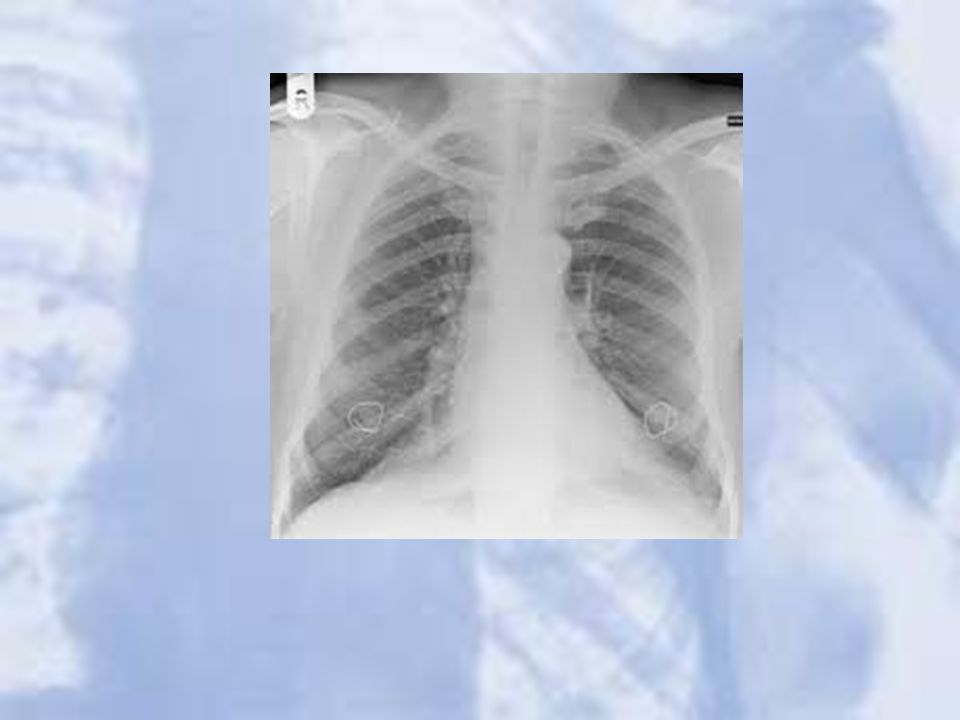
Nipple shadow: bilateral symmetrical lower zone opacity.

55
Case 8

57
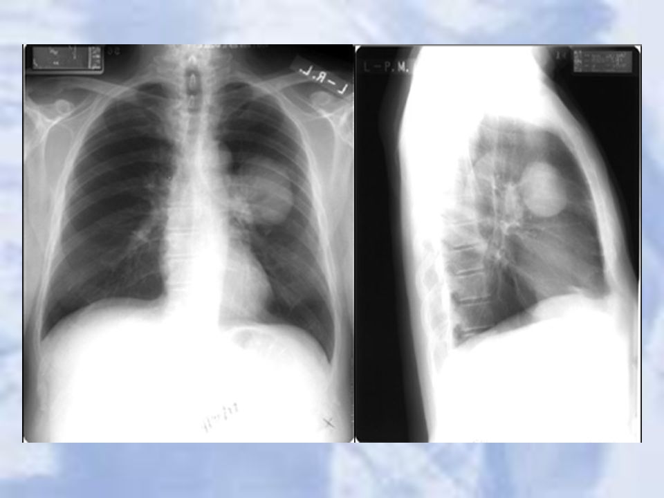
Extra medullary haemopoasis: enlarged paravertebral soft tissues
D/D neurofibroma

58
Case 9

60
Dextracardia : gastric air bubble seen under right hemi diaphram.

61
Case 10

63
Chest wall lesion: arising off the chest wall and not the lung

64
Case 11

66
Pleural effusion: Note loss of left hemidiaphragm
Pleural effusion: Note loss of left hemidiaphragm. Fluid drained via thoracentesis

67
Case 12

69
Lung Mass

70
Case 13

72
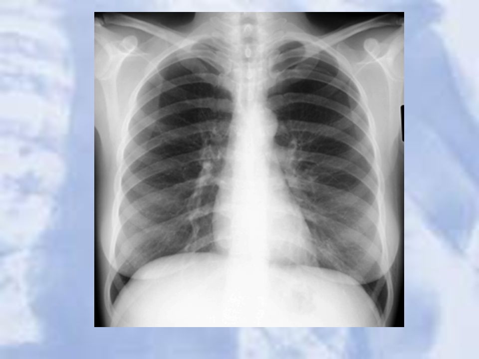
Small Pneumothorax: LUL

73
Case 14

75
Right Middle Lobe Pneumothorax: complete lobar collapse

76
Case 15

78
Metastatic Lung Cancer: multiple nodules seen

79
Case 16

81
Mycetoma: left upper zone intracavitatory lesion

82
Case 17

84
Perihilar mass: Hodgkin’s disease

85
Case 18

87
Widened Mediastinum with illdefined aortic outline: Aortic Dissection

88
Case 19

90
Achalasia cardia: dilated oesophagus.

91
Case 20

93
Pneumo mediatinum : the lucent line adjascent to the pericardium continues in the diaphramatic surface:

94
Case 21

96
Sarcoidosis : bilateral symmetrical hilar LNs

97
Case 22

99
Cystic bronchiectasis : bilateral multiple cystic air spaces with fluid level

100
Case 23

102
Gas under diaphram : lucent gas shadow under the diaphram

103
Questions?

Similar presentations


 –Partial.>")


