Download presentation
Presentation is loading. Please wait.

1
Introduction to Thoracic Radiology
Dr. Meghan Woodland September 30, 2010.

2
Indications Coughing Dyspnea / Tachypnea Heart Murmur, Collapse
Primary or Secondary Neoplasia Check for metastasis Thoracic Trauma Chest Wall Mass Exercise Intolerance, Weight Loss

3
Technical Factors Potential for Movement High inherent contrast
Respiration Decrease mAs High inherent contrast High kVp Collimation Should include thoracic inlet to diaphragm Center over the heart Pull thoracic limbs forward High kVp and low mAs will reduce the naturally high contrast in the thorax and increase the lung detail visible. Radiographic techniques: the dog By Joe P. Morgan, John Doval, Valerie Samii

4
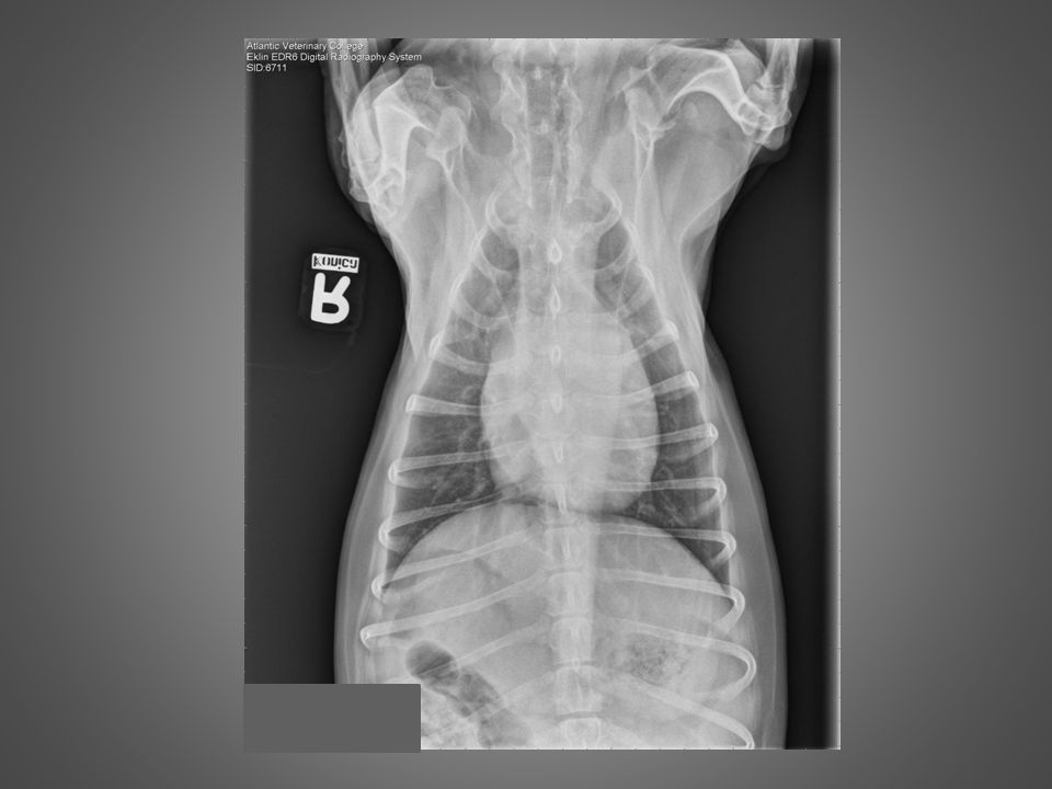
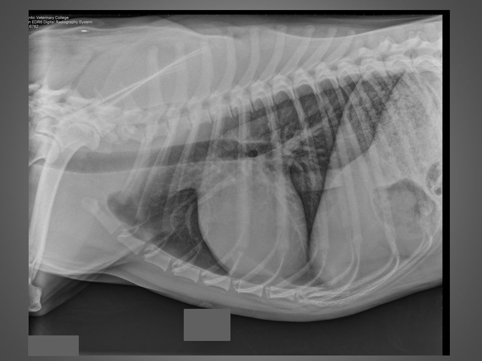
Normal lateral thorax

5
Patient is rotated Abdomen should be collimated out

6
Determining the Phase of Respiration
Always expose at peak inspiration Maximizes lung contrast Better visualization of pulmonary parenchyma Less compression of lungs by diaphragm Inspiratory lateral view: Caudodorsal aspect of lung is caudal to T12 Increased aeration of accessory lung lobe Separation of cardiac silhouette and diaphragm Inspiratory VD/DV view: Diaphragmatic cupola caudal to mid-T8 Tips of lung caudal to T10 In anesthetised patients, it may be necessary to manually inflate the lungs. As well, in sedated/anesthetised patients, you should obtain the DV/VD view before the laterals, because anaesthesia-induced atelectasis may arise quickly after induction. This is especially true in large or overweight dogs.

7
Inspiratory vs. Expiratory Lateral
Notice size of triangle

8
Inspiratory vs. Expiratory VD
Easy to see the difference in well visualized lung

9
DV vs. VD DV Best view to evaluate cardiac silhouette and caudal pulmonary vessels Less stressful for the patient Diaphragm rounded See small amounts of pleural air VD Best view to evaluate lungs Heart appears elongated Flat diaphragm – Mickey Mouse ears See small amounts of pleural fluid

10
DV VD

11
DV vs. VD

12
Normal VD Thorax

13
Normal DV Thorax

14
Cranial thorax has been collimated out of image.

15
Radiograph should be centered at the thorax.

16
Patient is rotated!

17
Right vs. Left Lateral Caudal Vena Cava enters the right diaphragmatic crus Right Lateral Better cardiac detail R crus forward See CVC go into it Left Lateral Heart appears round L crus forward See Cava go past Caudal vena cava

18
Left or Right Lateral? Right lateral

19
Left or Right Lateral? Left lateral

20
The Effects of Lateral Recumbency
Lung lesions (mass, nodule, infiltrate) may only be seen on a single view Only the non-dependent (up) lung can be critically evaluated Dependent lung loses aeration (atelectasis) Increased opacity Silhouettes with lesions Dependent lung loses aeration because of the increased pressure from mediastinal structures (especially the heart) and the dependent crus of the diaphragm. This is even more increased in anesthetised patients.
 may only be seen on a single view. Only the non-dependent (up) lung can be critically evaluated. Dependent lung loses aeration (atelectasis) Increased opacity. Silhouettes with lesions. Dependent lung loses aeration because of the increased pressure from mediastinal structures (especially the heart) and the dependent crus of the diaphragm. This is even more increased in anesthetised patients.")
21
Sedation Induced Atelectasis

22
Interpretation of Thoracic Radiographs
Systematic approach is crucial Heart (Cardiac Silhouette) Lungs Mediastinum Pleural space Chest wall Bones, Abdomen, Neck Structures seen on thoracic radiographs. Also evaluate for radiographic quality. Don’t forget the extrathoracic structures.
 Lungs. Mediastinum. Pleural space. Chest wall. Bones, Abdomen, Neck. Structures seen on thoracic radiographs. Also evaluate for radiographic quality. Don’t forget the extrathoracic structures.")
23
Normal Cardiac Silhouette
Size is subjective Lateral views: Dog = 2 ½ - 3 ½ intercostal spaces Cat = 2 – 2 ½ intercostal spaces VD/DV views: 65% the width of the thorax Objective: Buchanan method Vertebral heart scale Cardiac silhouette includes: pericardium, pericardial fluid, myocardium (including epicardium and endocardium), the origins of major vessels and blood. Size of the cardiac silhouette on radiographs is breed dependent. Barrel-chested dogs such as the Bulldog, Yorkshire Terrier and Dachshund have a relatively large cardiac silhouette on lateral radiographs. Deep-chested dogs, such as the Doberman, have taller, more slender cardiac silhouettes. Vertebral heart scale: On the lateral radiograph, the distance between the ventral aspect of the carina and the cardiac apex is taken as length. The width is the maximum width of the heart perpendicular to the length line. The number of vertebral lengths is taken for length and width, beginning at T4. Dogs: normal = 9.2 – 10.2 (>10.5 is cardiomegaly) Cats: normal = 7.2 – 7.8 (>8.1 is cardiomegaly)
, the origins of major vessels and blood. Size of the cardiac silhouette on radiographs is breed dependent. Barrel-chested dogs such as the Bulldog, Yorkshire Terrier and Dachshund have a relatively large cardiac silhouette on lateral radiographs. Deep-chested dogs, such as the Doberman, have taller, more slender cardiac silhouettes. Vertebral heart scale: On the lateral radiograph, the distance between the ventral aspect of the carina and the cardiac apex is taken as length. The width is the maximum width of the heart perpendicular to the length line. The number of vertebral lengths is taken for length and width, beginning at T4. Dogs: normal = 9.2 – 10.2 (>10.5 is cardiomegaly) Cats: normal = 7.2 – 7.8 (>8.1 is cardiomegaly)")
24
Clock Face 11-1 Aortic Arch 1-2 Main Pulmonary Trunk 2-3 Left Auricle
2-5 Left Ventricle 5-9 Right Ventricle 9-11 Right Atrium Centrally – Left Atrium

26
Lateral View Make a Plus sign Bermuda triangle Left atrium
Right atrium Main pulmonary artery Aortic Arch Left atrium Left Ventricle Right Ventricle

27
Thoracic and Pulmonary Vessels
Aorta Caudal Vena Cava Cranial pulmonary vessels Proximal third rib Caudal pulmonary vessels Where crosses 9th rib Veins are ventral and central Artery, bronchus, vein ABV’s

28
Trachea, Bronchial Tree
Trachea ends at the carina Then splits to the main stem bronchi followed by the lobar bronchi Tracheal rings can mineralize (age) Decreased tracheal diameter Tracheal narrowing (stenosis, extramural compression) Tracheal hypoplasia Tracheal collapse Expiratory lateral views are utilized to examine a patient for tracheal collapse. Tracheal hypoplasia is common in certain brachiocephalic breeds, such as the bulldog.
 Decreased tracheal diameter. Tracheal narrowing (stenosis, extramural compression) Tracheal hypoplasia. Tracheal collapse. Expiratory lateral views are utilized to examine a patient for tracheal collapse. Tracheal hypoplasia is common in certain brachiocephalic breeds, such as the bulldog.")
29
Lungs Normal anatomy 1 4 2 5 3 6 7 Left Right
Cranial (cranial subsegment) 1 Cranial (caudal subsegment) 2 Caudal 3 Right Cranial 4 Middle 5 Caudal 6 Accessory 7 1 4 2 5 3 6 7
 1. Cranial (caudal subsegment) 2. Caudal 3. Right. Cranial 4. Middle 5. Caudal 6. Accessory")
31
The Mediastinum Cranial, middle, caudal compartments
Routinely visible structures: Cardiac silhouette, trachea, caudal vena cava, aorta, +/- thymus, +/- esophagus Cranioventral mediastinal reflection Caudoventral mediastinal reflection Aka phrenopericardiac ligament Left side on VD radiograph Thymus is visible in young dogs. Cranial = pre-cardiac Middle = cardiac Caudal = post-cardiac

32
Mediastinal Reflection
Caudoventral mediastinal reflection

33
Extrathoracic Structures
Sternum Vertebrae Ribs Adjacent soft tissues Diaphragm

34
Pectus excavatum: congenital sternal anomaly.

35
The Diaphragm Cupola Right and left crura
Cranioventral convex portion Right and left crura Attach to cranioventral border of L3 and body of L4 May cause irregularity on these surfaces Appearance depends on centering of X-ray beam

36
The Diaphragm Right versus left lateral

38
The End

Similar presentations