Download presentation
Presentation is loading. Please wait.

2
The Human Respiratory Tract

3
1.nostrils: opening to the nasal passages 2. nasal passages: lined with a mucus membrane bearing cilia (warms, moistens, and filters incoming air) 3. pharynx (throat)--cavity in back of mouth
 3. pharynx (throat)--cavity in back of mouth.")
4
4. glottis: windpipe or trachea opening epiglottis--muscular flap covering the glottis- -prevents food from entering the windpipe 5. larynx (voice box) upper part of the windpipe containing sound producing vocal cords 6. trachea: (windpipe)--about 4 inches long & 1 inch in diameter --supported by rings of cartilage --lined with a ciliated mucus membrane which filters incoming air
 upper part of the windpipe containing sound producing vocal cords 6. trachea: (windpipe)--about 4 inches long & 1 inch in diameter --supported by rings of cartilage --lined with a ciliated mucus membrane which filters incoming air.")
5
7. bronchi: two main branches of trachea which enter lungs --lined with cartilage and ciliated 8. bronchioles: smallest air tubes which end at the alveoli or air sacs --cartilage ring support "fades out” from this point on 9. alveoli (air sacs): site of respiratory gas exchange by diffusion --resemble bunches of grapes --moist surface aids exchange-- each alveolus is surrounded by capillaries & gas exchange occurs with them (oxygen in--carbon dioxide out)
: site of respiratory gas exchange by diffusion --resemble bunches of grapes --moist surface aids exchange-- each alveolus is surrounded by capillaries & gas exchange occurs with them (oxygen in--carbon dioxide out).")
8
Purpose and Function of the System The purpose of getting in oxygen is to get it through our bloodstream to the cells of our body. At our cells it combines with glucose food to form usable ATP energy (cell respiration).
..")
9
** The capillaries which surround the alveoli are involved in gas exchange between the blood and the alveoli. In the blood, oxygen is carried by the red blood cell as oxyhemoglobin. Oxygen, which is loosely bound to the hemoglobin, diffuses into the cells where it is used during aerobic cellular respiration.

10
** The end products of aerobic cellular respiration, water and carbon dioxide diffuse into the blood. Then these waste gases are released from the lungs.

12
diaphragm: sheet of muscle separating the thoracic (chest) cavity from the abdominal cavity Breathing - the mechanical part of respiration... involves both the diaphragm and rib cage muscles

13
Some Respiratory System Malfunctions

14
pneumonia: alveoli fill with fluids and mucus resulting in coughing and breathing difficulties

15
asthma: allergic reaction in which bronchiole contractions create breathing difficulties

16
emphysema: air sacs lose their elasticity and breakdown resulting in a loss of respiratory surface in the lungs --shortness of breath is the chief symptom (smoking frequently leads to this disorder)
")
17
What happens when… We breathe in? -diaphragm moves downward, creates a “low pressure” in our thoracic cavity. -air is forced into (we don’t suck it into) our lungs, filling any available empty space. We breathe out? -diaphragm pushes upwards, creating a high pressure in our thoracic cavity. -air is forced out through our nose and mouth, leaving a “low” that can be refilled (hopefully immediately)
 our lungs, filling any available empty space. We breathe out. -diaphragm pushes upwards, creating a high pressure in our thoracic cavity. -air is forced out through our nose and mouth, leaving a low that can be refilled (hopefully immediately).")
19
The Circulatory System- Transport The absorption and circulation of materials throughout an organism

20
The Human (Mammalian) Heart Structure 1. septum: muscular central wall dividing the mammalian heart into two halves 2. atria: thin walled upper heart chambers which receive blood pump blood to the ventricles 3. Ventricles: muscular thick walled chambers which pump blood from the heart -- the lower chambers 4. Valves - prevent the backward flow of blood in the heart

22
The heart functions as a double pump beating in unison. 1. Deoxygenated blood--relatively low in oxygen 2.Oxygenated blood--relatively high in oxygen ** Heartbeat is stimulated by an electrochemical impulse. Pacemaker - in the right atrium - initiates the heartbeat

24
Pathway of a Red Blood Cell through the Heart

25
1. Inferior & superior vena cava 2. Right atrium 3. valve 4. Right ventricle 5. valve 6. Pulmonary arteries (BLOOD TO THE LUNGS – GAS EXCHANGE) … 7. Pulmonary veins 8. Left Atrium 9. valve 10. Left ventricle 11. valve 12. Aorta
 … 7. Pulmonary veins 8. Left Atrium 9. valve 10. Left ventricle 11. valve 12. Aorta.")
26
A = aorta F = left ventricle B = pulmonary arteries G = right ventricle C = pulmonary veins H = valve D = left atrium I = vena cavae E = valve J = right atrium

27
Some Blood Functions Why do we need blood?????????

28
** In general, the blood is a fluid tissue helping to maintain homeostasis for all cells in the body. Transport of needed substances to body cells. (oxygen, amino acids, glucose, fatty acids, glycerol, salts, etc.)
.")
29
2. Transport of wastes from cells. (urea, water, carbon dioxide in the form of the bicarbonate ion) 3. Helps to maintain a constant body temperature. 4. Aids the body in fighting disease.
 3. Helps to maintain a constant body temperature. 4. Aids the body in fighting disease..")
30
Blood Composition Plasma 55% (liquid part of the blood); Blood Cells 45%
; Blood Cells 45%")
31
BLOOD CELL TYPES Red Blood Cells: –most numerous –biconcave disc shaped –smaller than white blood cells, larger than platelets –no nucleus when mature –produced in the red marrow of long bones –destroyed in the liver and spleen –contain the iron protein compound HEMOGLOBIN whose chief function is to combine with oxygen and carry it to the cells

32
** Carbon monoxide has 40X the affinity of oxygen for hemoglobin--breathing it may lead to death

33
White Blood cells --largest blood cells--several different types --about 8,000 per drop of blood --most are formed in the bone marrow or in the lymph tissue --most protect the body against diseases by forming antibodies or engulfing bacteria

34
Main White Blood Cell Types 1.Phagocytes-- engulf bacteria and viruses by phagocytosis --able to leave the bloodstream and move between the cells of the body by squeezing through the capillary walls

35
2. Lymphocytes--produce antibodies which clump bacterial poisons or bacteria (antigens) (antigens--foreign substances in the body)
 (antigens--foreign substances in the body).")
36
3. Platelets --smallest blood cells (fragments) --150,000 to 300,000 per drop of blood --needed for clotting
 --150,000 to 300,000 per drop of blood --needed for clotting.")
37
Clotting Process Involves a series of enzyme controlled reactions resulting in the formation of protein fibers that trap blood cells and form a clot.

39
Blood Vessels 1. Arteries --carry blood away from the heart --usually spurt blood when cut --all except the pulmonary artery carry oxygenated blood --thick walled and elastic pulse: expansion and contraction of the artery walls in response to the heartbeat

41
Veins --carry blood toward the heart --contain valves --closer to the body surface than the arteries --all except the pulmonary vein carry deoxygenated blood --thinner, less muscular and elastic than arteries --depend upon muscle and diaphragm movements for blood flow

43
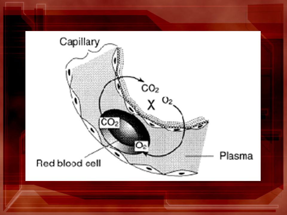
Capillaries --most numerous vessels --connect arteries to veins --microscopic, one cell thick walls --site of much exchange between the blood and the intracellular fluid (lymph) by diffusion
 by diffusion")
44
Lymph vessels -have walls one cell thick -present around all body cells -Lymph composition is similar to that of blood except for the absence of RBC and some plasma proteins. -chief site of material exchange with the tissues

45
Major lymph vessels have lymph nodes which contain phagocytic white blood cells which filter bacteria and dead cells from the lymph. X = lymph nodes Valves are present in some lymph vessels-- aiding in the movement of the lymph. Respiratory movements also aid lymph flow.

46
Malfunctions and disorders of the heart and blood vessels Hypertension (High Blood Pressure) --caused by a narrowing of the arterioles resulting in an increased resistance to the flow of blood--increases the strain on the heart
 --caused by a narrowing of the arterioles resulting in an increased resistance to the flow of blood--increases the strain on the heart")
47
Causes implicated: 1. excess sodium intake 2. stress 3. cigarettes (nicotine) 4. saturated fats 5. alcohol & caffeine 6. obesity 7. heredity & aging No cure--may be treated by medication & diet. "Silent killer"--millions don't know they have it
 4. saturated fats 5. alcohol & caffeine 6. obesity 7. heredity & aging No cure--may be treated by medication & diet. Silent killer --millions don t know they have it.")
48
Angina pectoris --pain in the chest which radiates into the left shoulder and arm --occurs especially when physical exertion results in a lack of oxygen supply to the heart muscle --caused by a reduction of blood supply due to partial blockage(s) of coronary arteries
 of coronary arteries")
49
Coronary thrombosis - heart attack --caused by a blood clot in a coronary artery that stops circulation to part of the heart muscle --attack is fatal if much heart muscle is involved

Similar presentations