Download presentation
Presentation is loading. Please wait.

1
The Face

2
The Face Shape of face depend on : - facial skeleton - disposition of soft tissue - type of face **Facial growth during child hood as the paranasal sinus develop and permanent teeth erupt.

3
Type of face There are two types of face:- 1- Leptoprosopic face
- long , narrow - protruding maxilla - retruding mandible 2- Euryprosopic face - upper part of face is less prominent - nose is short - eyes are wide set - cheek bones are usually more prominent.

4
Sexual Variation of face
- face show sexual dimorphism although facial types are similar until the age of year. - face of female attains its mature form earlier than the male. - male face tend to be more protuberant, bulky and coarse. - nose of female have a concave to straight profile. - supra orbital ridge of male overhang the face, those of female are at same level as inferior orbital margin and cheek bones.

5
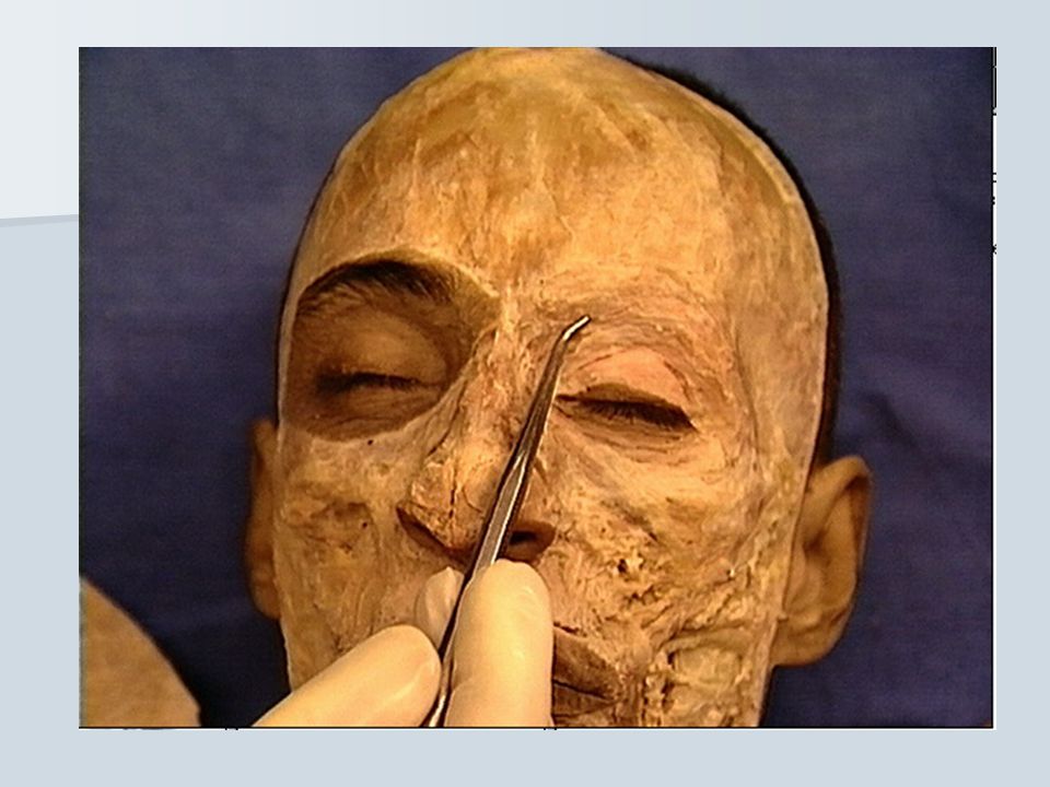
**On reflection the skin of the face, the following main structures are revealed:
1- muscles of facial expression 2- facial nerve 3- cutane. branches of trigeminal nerve and great auricular n. 4- parotid gland and duct 5- buccal pad of fat 6- facial L.N.

14
Skin of face: - Thin - Vascular - Movable
- Abundly supplied with sebaceous and sweat gland - No deep fascia - So it especially adaptable for surgical plastic operation. c

15
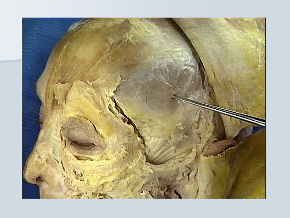
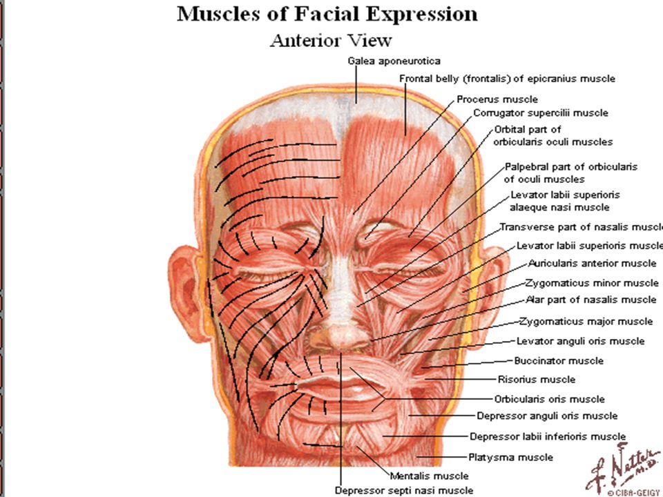
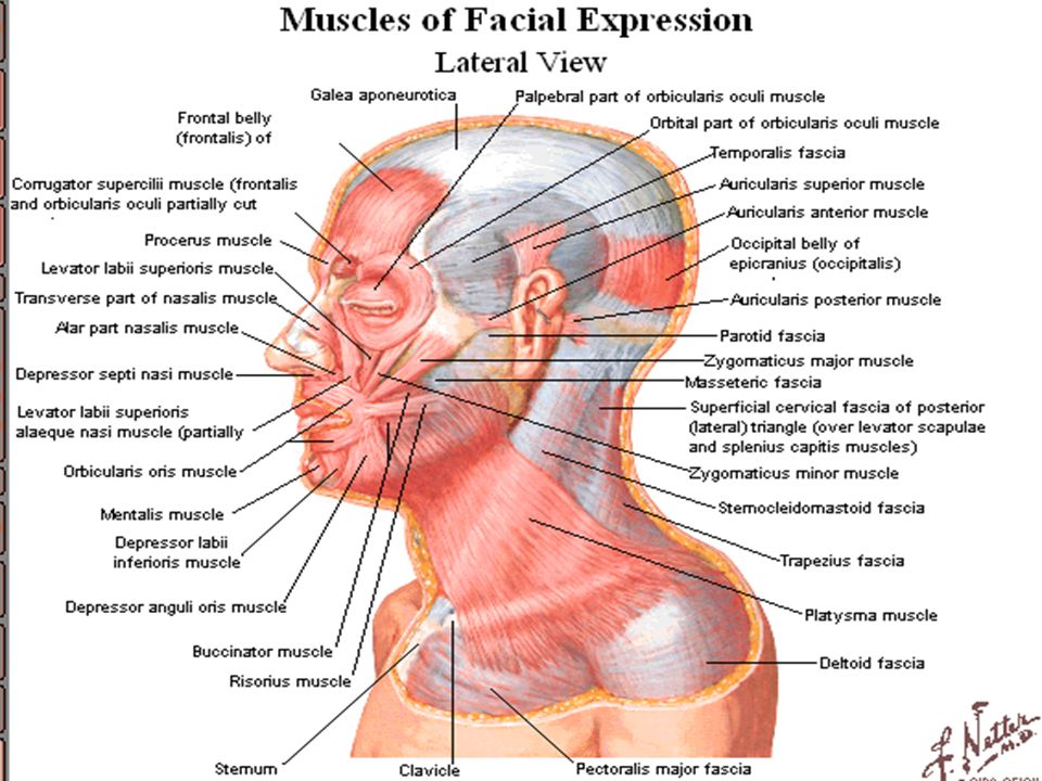
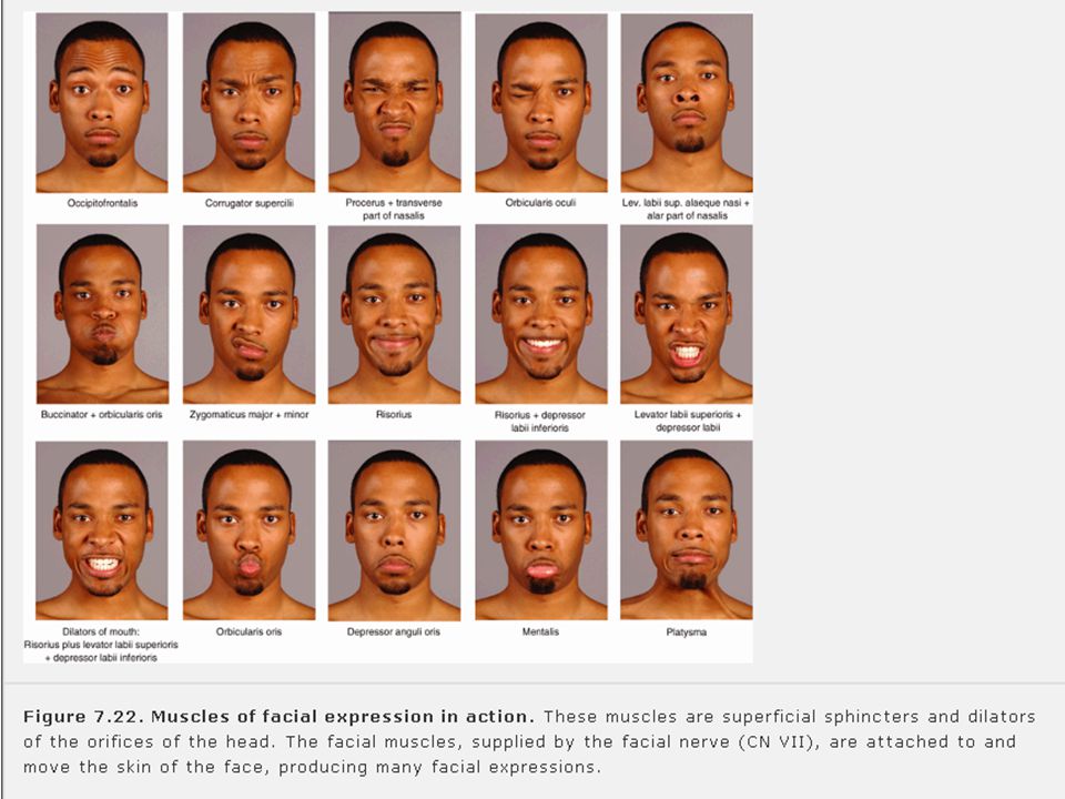
Muscles of the face *Characterized by subcutaneous location move the skin and change facial expression. *Placed around orifice of eye (palpebral fissure), ear nose and mouth ( oral fissure). * All muscle of face developed from 2nd pharyngeal arch which supply by facial nerve.
, ear nose and mouth ( oral fissure). * All muscle of face developed from 2nd pharyngeal arch which supply by facial nerve.")
20
2 orbicalaris : orbiculais ocul
orbiculais oris 2 associated with nose : procerus compressor nares(in infant) 2 associated with nose : zygomaticus minor Zygomatoe area : zygomaticus major(m.of smiling) 2 elevator of upper lip : levator labii superioris alaeque nasi Levator labii superioris 2 muscle with angle of mouth :levator anguli oris Depressor angel oris 2 muscles with lower lip: risorius Depressor labii inferioris 2 muscle associated with chin and cheek: Mentalis buccinator muscle
 2 associated with nose : zygomaticus minor. Zygomatoe area : zygomaticus major(m.of smiling) 2 elevator of upper lip : levator labii superioris alaeque nasi. Levator labii superioris. 2 muscle with angle of mouth :levator anguli oris. Depressor angel oris. 2 muscles with lower lip: risorius Depressor labii inferioris. 2 muscle associated with. chin and cheek: Mentalis. buccinator muscle.")
22
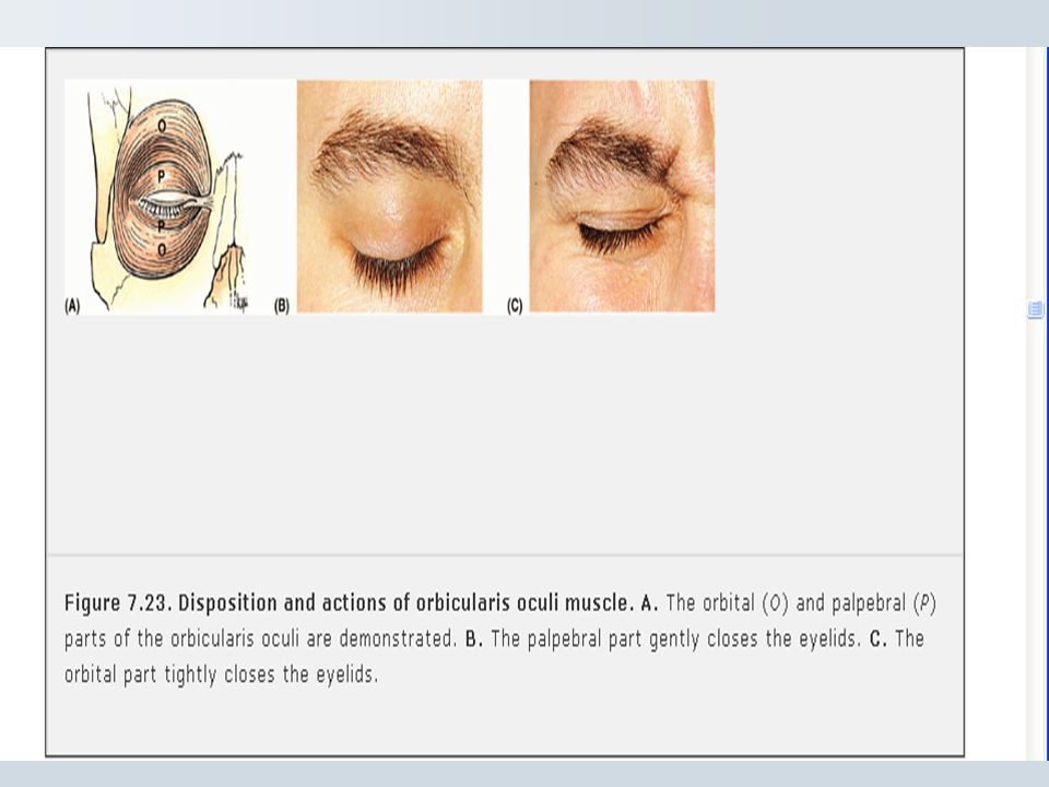
Orbicularis Oculi: 3 parts:
1- Orbital part: strongly close of eye protecting it from dust, bright light , mingling with fiber of frontalis muscle. 2- Palpebral part: gently close the eye lid as in blinking or in sleep to keep the cornea from drying 3- Lacrimal part: posterior border of lacrimal fossa to lid , its function to dilate lacrimal sac so that fluid discharge from conjunctiva to lacrimal sac.

23
Buccinator:- * Attached to alveolar process of maxilla, mandible opposite molar tooth to ptergomandibular raphe. * Active in smile. * Keep check taut, preventing it from folding and being injured during chewing * Mingle medially with orbicularis oris * Aid in mastication * Used during whistling , sucking and blowing

25
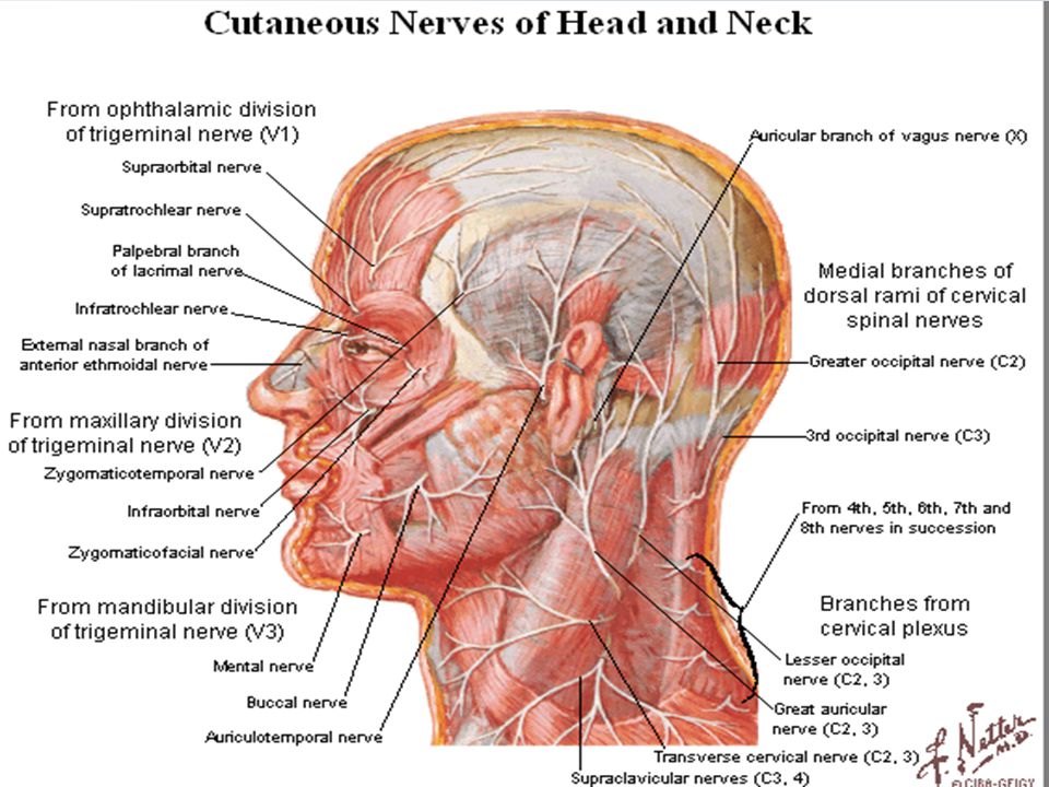
Nerve Supply of the face
Face have both motor and sensory * Motor: nerve derived from facial nerve (nerve of second pharyngeal arch) * Sensory: innervations is primarly from three division of trigeminal nerve (CN V) with exception of small area over angle of mandible and parotid gland which supply by great auricular nerve(C2,C3). * Motor nerve of the face:- - facial nerve to muscle of facial expression - motor root of mandibular nerve to muscle of mastication
 * Sensory: innervations is primarly from three division of trigeminal nerve (CN V) with exception of small area over angle of mandible and parotid gland which supply by great auricular nerve(C2,C3). * Motor nerve of the face:- - facial nerve to muscle of facial expression. - motor root of mandibular nerve to muscle of mastication.")
31
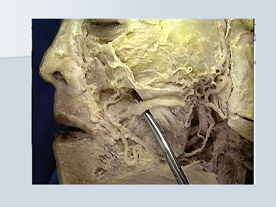
Facial nerve:- - leave the skull at stylomastoid foramen give off posterior auricular nerve - send fiber to :- - Stapedius muscle - Stylohyoid muscle - Posterior belly of digastric muscle - Scalp muscles - Auricular muscle - Platysma muscle - Buccinator

32
* Provides secretary fiber to salivary and sensory (taste) fibers to anterior 2/3 of tongue.
* Enter parotid isthmus and passes between superior and deep loops of gland. * It is superficial to external carotid artery and retromandibular vein so may be injured in operation in parotid region . * Terminal branches appear at margin of parotid (antromedial surface of gland).
.")
33
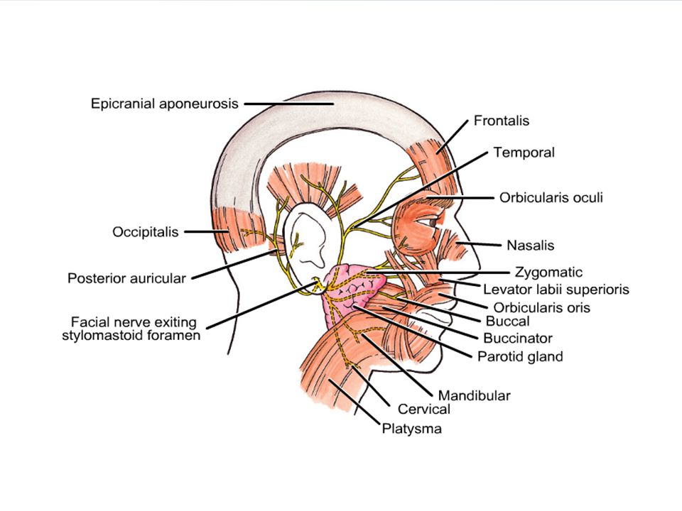
Five terminal branches of facial nerve
1- Temporal branch- frontalis- orbicularis oculi(upper). 2- Zygomatic branch – orbicularis oculi:(lower) 3- Buccal branch- Buccinator- orbicularis oris. 4- Mandibular- (marginal) passes along lower border of mandible crossing facial artery and vein and submundibular L.N. 5- Cervical branch – platysma
. 2- Zygomatic branch – orbicularis oculi:(lower) 3- Buccal branch- Buccinator- orbicularis oris. 4- Mandibular- (marginal) passes along lower border of mandible crossing facial artery and vein and submundibular L.N. 5- Cervical branch – platysma.")
34
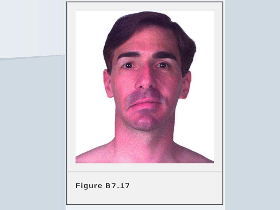
Facial Nerve Injury Non-traumatic cause of facial parlay is inflammation of facial nerve near stylomastoid foramen so patient has; 1- Can not close his eyes and palpebral fissure appear wider, lacrimal fluids drips on cheeks laterally, drying of cornea. 2- Patient van not whistle, blow or chew effictualey so food will accumulate between cheeks and gum so patient use his finger to remove food due to parlysis of buccintor muscle. 3- Displacement of corner of mouth , so food and saliva dripling outside of mouth.

35
Facial Nerve Pulsy has many causes:
1- Idiopathic (Belly pulsy) Exposure to cold (30 to 50 years). 2- Complication of surgery in Parotid gland. 3- Dental manipulation-vaccination. 4- Infection of middle ear .
 Exposure to cold (30 to 50 years). 2- Complication of surgery in Parotid gland. 3- Dental manipulation-vaccination. 4- Infection of middle ear .")
36
Injury to Branch of facial nerves
1- By stab wound- gunshots. 2- Injury at birth. 3- Injury of temporal bones. 4- Surgical approach to sub mandibular gland- resulting in dropping of corner of mouth.

38
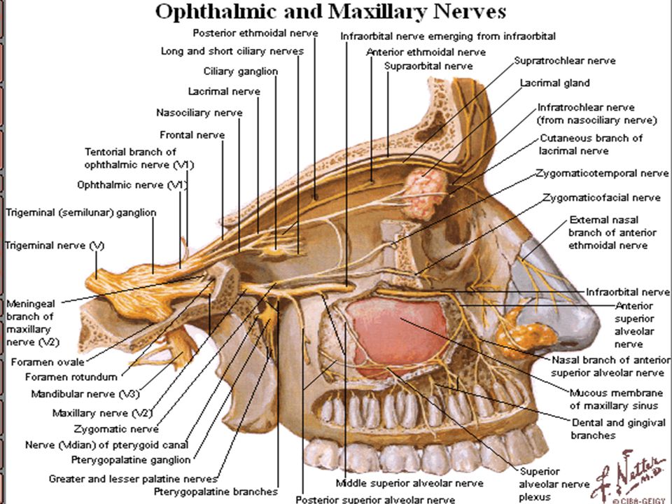
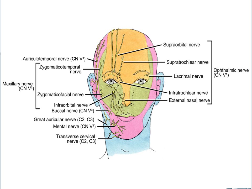
Sensory Nerves of face Trigeminal nerve (5) V1- Ophthalmic nerve :
Forehead – upper eyelid- conjunctiva of eye- side of nose 5 Branches 1-Lacrimal nerve 2- Supraorbital nerve 3- Supratrochler nerve 4- Infratochlear nerve 5- External nasal nerve

41
V2- Maxillary nerve: Side of nose- lower eyelid-check-upper lip-lateral side of orbital opening 3 Branches 1- Infraorbital nerve 2- Zygomatico facial nerve 3- Zygomatico temporal nerve

44
V3- Mandibular Nerve: Lower lip- lower part of face- temporal region –part of auricle 3Branches 1- Mental nerve-inferior alveolar nerve 2- Buccal nerve- Mucous membrane of check. 3- auriculo temporal nerve-accompany superficial temporal vessels

45
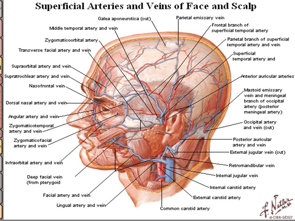
1- Facial Artery: - from external carotid artery
- winds its way to inferior border of mandible - interior to massetor so artery lies superficial deep to platysma - cross mandible, buccinator, maxilla to medial canthus of eye - lies deep to Zygomaticus major, levator labii superioris.

48
- lies fingerbirth lateral to angle of mouth.
- give superior,inferior labial arteries - ascend alongside of nose joined dorsal nasal branches of ophthalmic artery. - terminal branch of facial artery is called angular artery.

49
2- Superficial temporal artery: transverse facial artery.
3- Maxillary artery: mental artery-buccal artery- infra orbital artery 4- Opthlmic artery: supra orbital artery- supra trochlear artery- lacrimal artery – dorsal nasal artery- external nasal artery

51
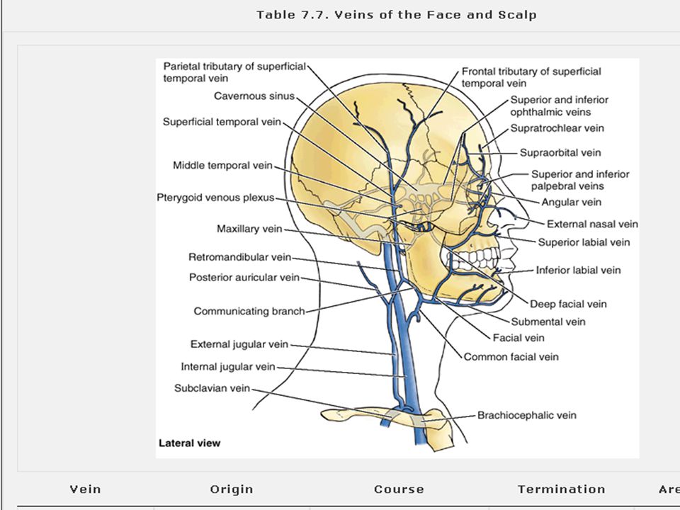
Venous drainage of face
* Is formed by union of supra orbital and supra trochlear. * Connected to superior ophthalmic vein which connected facial vein to cavernous sinus. * Join anterior division of retro mandibular vein. * Drain into internal jugular vein directly or indirectly. * Taking a less tortuous but more superficial course.

52
Lymphatic drainage of face:
1- submundibular L.N.- forehead- anterior part of face. 2- Buccal L.N. 3- Parotid L.N. lateral part of face- lateral part of eyelid. 4- Submental L.N.-central part of lower lip, skin of chin.

54
Veins of the face Supra temporp v. supratrochele v. + +
Maxillary v supraorbital v. Angular v. Retromandibular v. Ant Facial v. Post. + Post. Amicular v common facial v. Ext. jugular v int. jugular v.

55
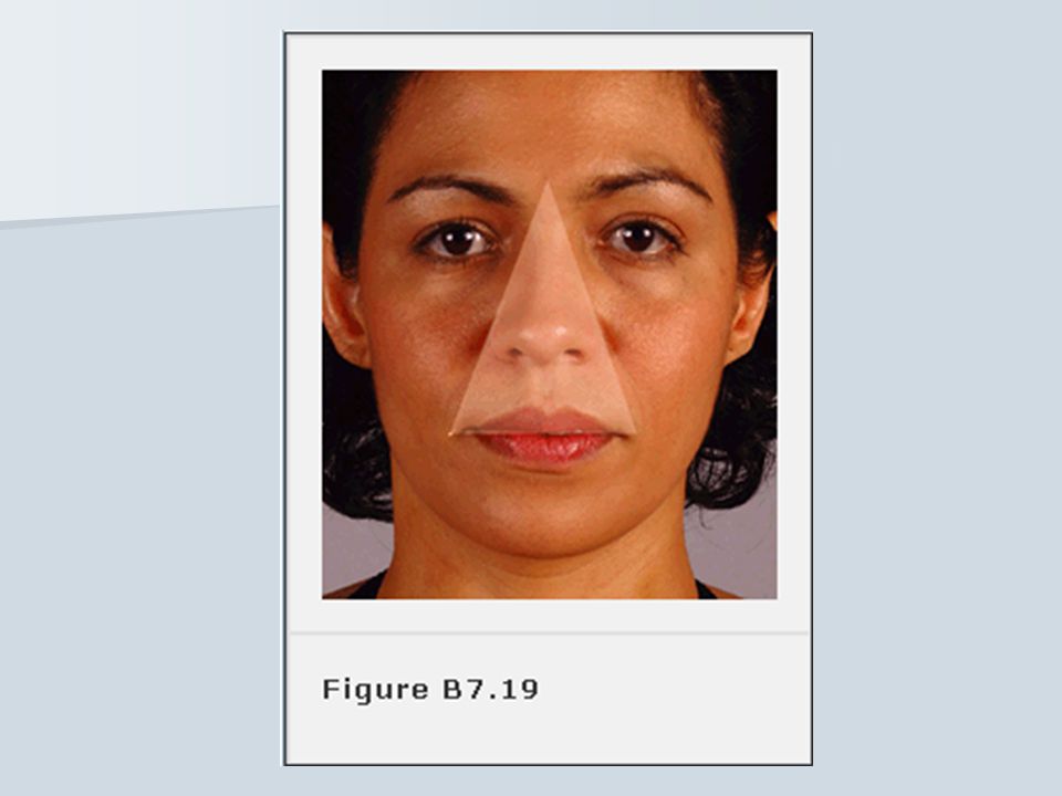
Dangerous Area of face * Is triangle bounded by lines join root of nose with angle of mouth. * Venus drainage from this area enter angular veins which communicate with cavernous sinus. * Therefore boil, carbuncle in this region produce cavernous sinus thrombosis.

58
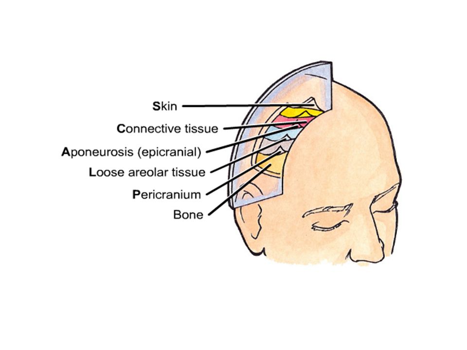
The Scalp *Consist of five layers of soft tissue covering calvaria:-
S=skin C= C.T. A= Aponeurotic L= loose C.T. (dangerous area) P= Pericranium * Frontalis muscle has no bony attachments.
 P= Pericranium. * Frontalis muscle has no bony attachments.")
63
Lymphatic Drainage of the scalp
Nerve of the scalp - Trigeminal nerve - Cervical plexus C2-C3 ( great auricular, lesser occipital, greater occipital) Artery of the scalp 1- external carotid artery – occipital – posterior – auricular – superficial temporal 2- internal carotid artery – supra trochlear- supra orbital. Lymphatic Drainage of the scalp - there is no L.N. in the scalp - superficial ring of L.N. (submental , submandibular, parotid, retro auricular and occipital L.N.
 Artery of the scalp. 1- external carotid artery – occipital – posterior – auricular – superficial temporal. 2- internal carotid artery – supra trochlear- supra orbital. Lymphatic Drainage of the scalp. - there is no L.N. in the scalp. - superficial ring of L.N. (submental , submandibular, parotid, retro auricular and occipital L.N.")
64
Quiz Q1/ Could you explain the following:
1- In Bells palsy there is decrease of lacrimation 2- Loss of tast in the anterior 2/3 of tongue 3- Painful sensitivity to sound 4- Deviation of the lower jaw and tongue Q2/ What are the efferent and afferent limbs of cornel blink reflex (closing of the eyes) Q3/ Death may result from bilateral severance of which of the following nerve ? A- Trigeminal nerve B- Facial nerve C- Vague nerve D- Spinal accessory nerve E- Hypoglossal nerve
 Q3/ Death may result from bilateral severance of which of the following nerve A- Trigeminal nerve. B- Facial nerve. C- Vague nerve. D- Spinal accessory nerve. E- Hypoglossal nerve.")
Similar presentations


 .>")
