Download presentation
Presentation is loading. Please wait.

1
An introduction to the ILO Radiological Classification of the PneumoconiosesILO Radiological Classification Professor Neil White

2
Objective of the session To provide an introduction to the radiology of pneumoconioses; Background reading material –Guidelines for the use of the ILO International Classification of Radiographs of Pneumoconioses Home Study material –Guidelines for the use of the ILO International Classification of Radiographs of Pneumoconioses –ROLDS CD-ROM “The Symposium” “The Simulation”

3
The “ILO Classification” ILO International Classification of Radiographs of Pneumoconioses (1980) Primarily an epidemiological tool. ILO Classification not intended to define pathological entities, take into account working capacity, nor imply a definition of pneumoconiosis for compensation purposes.

4
ILO International Classification of Radiographs of Pneumoconioses. 22 standard films Comparison with these films enables the reader to categorise the appearances in a standardised fashion –Parenchymal –Pleural –Other abnormalities

5
ILO Classification: Quality 1 = acceptable 2 = minor defect 3 = major defect but still interpretable 4 = unreadable

6
ILO Classification: Abnormal? If any appearances of pleura or parenchyma are consistent with pneumoconiosis proceed. Use symbols and comments for any appearances that are definitely not pneumoconiosis. If it is probable that all appearances are the result of other aetiology do not classify. If the appearances might be due to pneumoconiosis record, but note what other aetiology was considered.

7
Small Opacities - type Rounded p < 1.5 mm q 1.5 to < 3 mm r 3 to < 10 mm Irregular s < 1.5 mm t 1.5 to < 3 mm u 3 to < 10 mm An opacity is rounded if its greatest diameter is less than 1.5 times its smallest diameter.

8
Small Opacities - type Source: Guidelines for use of ILO Classification, Revised Edition, 1980.

9
Small Opacities - type Decide on the predominant type of opacity Mark that type e.g. “q” Decide whether most opacities are of that type or not. If virtually all opacities are that size and shape then record the symbol twice e.g. q/q If another size or shape also seen then record this as a second letter e.g. q/r

10
Small Opacities - type Source: Guidelines for use of ILO Classification, Revised Edition, 1980.

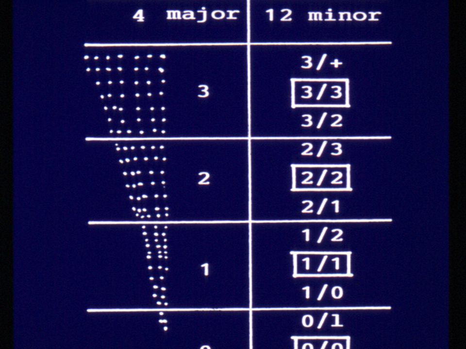
11
Small Opacities - profusion Four point scale 0 = normal 1, 2, 3 = abnormal (increasing numbers of small opacities) Four point scale extended to a 12 point scale. 0/- 0/0 0/1 = normal 1/0 1/1 1/2 2/1 2/2 2/3 3/2 3/3 3/+

12
Small opacities - profusion Source: Guidelines for use of ILO Classification, Revised Edition, 1980.

13
Small Opacities – profusion Visually integrate the small opacities seen. The distinction between 0/1 and 1/0 crucial – 0/1 is normal, 1/0 is not. Select out the closest standard film. If the film corresponds closely to a standard film, then use that full category e.g. 2/2. If less profuse use 2/1, if more profuse use 2/3.

16
Small Opacities – profusion Pneumoconiosis is usually, but not always symmetrical. If there is significant regional difference in profusion apply the following: –If any zone is 3 or more sub-categories less profuse than the most profuse zone then do not include that zone in the decision on recording profusion.

17
Small Opacities - zones Lung fields divided into six zones Affected zones marked Source: Guidelines for use of ILO Classification, Revised Edition, 1980.

18
Large opacities (P.M.F.) A = greatest diameter >10 mm but 10 mm but sum of greatest diameters < 50 mm B = one or more opacities with sum of greatest diameter > 50 mm but < area of right upper zone C = combined area > right upper zone (Note: the lower border of the RUZ is at the lower margin of the anterior end of the second rib on a non- lordotic or kyphotic film.)
 A = greatest diameter >10 mm but 10 mm but sum of greatest diameters < 50 mm B = one or more opacities with sum of greatest diameter > 50 mm but < area of right upper zone C = combined area > right upper zone (Note: the lower border of the RUZ is at the lower margin of the anterior end of the second rib on a non- lordotic or kyphotic film.)")
19
Large Opacities (P.M.F.) Source: Guidelines for use of ILO Classification, Revised Edition, 1980.
 Source: Guidelines for use of ILO Classification, Revised Edition, 1980.")
20
Large Opacities (P.M.F.) Source: Parkes
 Source: Parkes")
21
Pleural abnormalities Circumscribed (plaques) or diffuse Site (R/L) – both sides are recorded separately Width a < 5 mm b maximum width > 5 and < 10 mm c maximum width > 10 mm Extent 1 < ¼ lateral projection of chest wall 2 > ¼ but < ½ lateral projection of chest wall 3 > ½ lateral projection of chest wall Pleural Calcification Diaphragms and chest walls recorded separately
 or diffuse Site (R/L) – both sides are recorded separately Width a < 5 mm b maximum width > 5 and < 10 mm c maximum width > 10 mm Extent 1 < ¼ lateral projection of chest wall 2 > ¼ but < ½ lateral projection of chest wall 3 > ½ lateral projection of chest wall Pleural Calcification Diaphragms and chest walls recorded separately")
22
Symbols Use of symbols is obligatory. When symbols are used it is understood that they are preceded by an appropriate word or phrase e.g. “suspect” “changes suggestive of” “opacities suggestive of” etc.

23
Symbols ax = coalesence of small pneumoconiotic opacities (can be used with A, B, C, of classification) bu = bulla(e) ca = cancer of lung or pleura cn = calcification in small pneumoconiotic opacities co = abnormality of cardiac size or shape cp = cor pulmonale cv = cavity
 bu = bulla(e) ca = cancer of lung or pleura cn = calcification in small pneumoconiotic opacities co = abnormality of cardiac size or shape cp = cor pulmonale cv = cavity")
24
Symbols di = marked distortion of intrathoracic organs ef = effusion em = definite emphysema (usually COPD) es = eggshell calcification of hilar and/or mediastinal lymph nodes fr = fractured rib(s) hi = enlarged lymph nodes, hilar and/or mediastinal ho = honeycomb lung
 es = eggshell calcification of hilar and/or mediastinal lymph nodes fr = fractured rib(s) hi = enlarged lymph nodes, hilar and/or mediastinal ho = honeycomb lung")
25
Symbols id = ill defined diaphragm (> 1/3 of 1 hemi- diaphragm ih = ill defined heart outline (> 1/3 of left heart outline) kl = septal lines (Kerley B lines) od = other disease pi = interlobular fissure pleural thickening px = pneumothorax rp = rheumatoid pneumoconiosis (Caplan’s syndrome) tb = tuberculosis
 kl = septal lines (Kerley B lines) od = other disease pi = interlobular fissure pleural thickening px = pneumothorax rp = rheumatoid pneumoconiosis (Caplan’s syndrome) tb = tuberculosis")
26
ILO: Short form Datum van X-straal opname: Date of X-ray plate: 0/0 0/1 p q r s t u A B C tba tbu cv hi es hv 1/0 1 2 3 ef pla plw plc em oth Comments :______________________________________ Reader:_________________ Date read:_______________ Name:____________ I.D. No._______

27
ILO: NIOSH/ ACR Form

28
MBOD/ NCOH Form

Similar presentations


 –Partial.>")


