Download presentation
Presentation is loading. Please wait.

1
Embryology - development of Conotruncal region
Dr Julian Johny Thottian

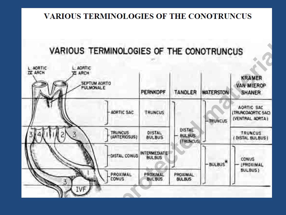
2
Introduction The Conus- also known as Infundibulum (Keith 1909 ) The Conotruncus comprises collectively two myocardial sub segments, the Bulbus cordis and the Truncus arteriosus BULBUS CORDIS –refers to the ventricular outflow tract TRUNCUS ARTERIOSUS- embryologic precursor of great arteries. D A Goor etal
 The Conotruncus comprises collectively two myocardial sub segments, the Bulbus cordis and the Truncus arteriosus. BULBUS CORDIS –refers to the ventricular outflow tract. TRUNCUS ARTERIOSUS- embryologic precursor of great arteries. D A Goor etal.")
3
EARLY HEART

4
Definitions Conotruncus -The conotruncus is the outflow region of the developing heart It consists of: Conus cordis and Truncus Arteriosis Conus- Inferior to the aortic and pulmonary valves Truncus- Superior to the valves that is continuous with the ventral aorta (aortic sac).
.")
6
Bulbus cordis- also known as the conotruncus lies ventral to primitive ventricle.
Together with primitive ventricle it forms the ventricle of the formed heart. Keith (1909) The developing two main truncal cushions and the underlying two conal cushions are perfectly aligned and no line of demarcation between the two is identifiable in mammals (Van Mierop and Patterson, 1980)
 The developing two main truncal cushions and the underlying two conal cushions are perfectly aligned and no line of demarcation between the two is identifiable in mammals. (Van Mierop and Patterson, 1980)")
7
The net result is…. Proper alignment of the outlet septum with ventricular trabecular septum with membraneous septum in between. Proper posterior alignment of left outflow tract with left ventricle and aorto mitral continuity. Mechanism – differential growth and apoptosis.

8
Secondary heart field Area of the ventral pharyngeal mesoderm (Kelly et al, 2001; Mjaadvedt et al, 2001; Waldo et al, 2001) - Pre cardiac Splanchnic Mesodermic -region providing myocardial precursor cells, which migrate to the Out flow Tract area of the developing cardiac tube, where they build up the Conotruncal myocardium as well as smooth muscle cells joining the caudal portion of the aortic sac (Waldo et al, 2005).
 - Pre cardiac Splanchnic Mesodermic -region providing myocardial precursor cells, which migrate to the Out flow Tract area of the developing cardiac tube, where they build up the Conotruncal myocardium as well as smooth muscle cells joining the caudal portion of the aortic sac (Waldo et al, 2005).")
9
The topography of the SHF.
Figure 1. The topography of the SHF. A and B, Lateral whole-mount view and transverse section of an embryo carrying an Fgf10 lacZ transgene at the linear heart tube stage (mouse embryonic day 8.5). SHF cells (blue) are located in pharyngeal mesoderm in the dorsal pericardial wall, adjacent to pharyngeal endoderm.5 C and D, Lateral whole-mount and transverse section after cardiac looping (mouse embryonic day 9.5). Fgf10 transgene expression is now evident in the OFT and dorsal pericardial wall (arrowheads). E and F, Neural crest cells, visualized with a Wnt1-Cre R26R transgene108 (E, blue) or anti-Tbx3 antibodies79 (F, green), invade the pharyngeal region and become positioned between SHF cells in the dorsal pericardial wall and pharyngeal endoderm, where they are closely apposed with Fgf10 transgene expressing mesodermal cells (F, red). E is reproduced from Jiang et al108 with permission. Scale bars: 100 μm (B, D, and E); 50 μm (F). Rochais F et al. Circulation Research 2009;104: Copyright © American Heart Association
. SHF cells (blue) are located in pharyngeal mesoderm in the dorsal pericardial wall, adjacent to pharyngeal endoderm.5 C and D, Lateral whole-mount and transverse section after cardiac looping (mouse embryonic day 9.5). Fgf10 transgene expression is now evident in the OFT and dorsal pericardial wall (arrowheads). E and F, Neural crest cells, visualized with a Wnt1-Cre R26R transgene108 (E, blue) or anti-Tbx3 antibodies79 (F, green), invade the pharyngeal region and become positioned between SHF cells in the dorsal pericardial wall and pharyngeal endoderm, where they are closely apposed with Fgf10 transgene expressing mesodermal cells (F, red). E is reproduced from Jiang et al108 with permission. Scale bars: 100 μm (B, D, and E); 50 μm (F). Rochais F et al. Circulation Research 2009;104: Copyright © American Heart Association.")
11
Molecular aspect SHF expressed NKx2.5 and Gata4 transcription factors. NKx2.5- and Gata4 SHF-committed cells join and incorporate themselves into the outflow tract of the primary heart tube, these cells undergo terminal myocardial differentiation under the induction of the local primary myocardial Bmp2 factor (Waldo et al 2001)
")
12
Wnt, fibroblast growth factor, bone morphogenetic protein, Hedgehog, and retinoic acid are all involved in signalling. SHF contributes to the outflow tract (OFT), right ventricle, and inflow region
, right ventricle, and inflow region.")
13
Illustration showing the core features of the Wnt, Fgf, Bmp, Hh, and Notch signaling pathways.
Figure 2. Illustration showing the core features of the Wnt, Fgf, Bmp, Hh, and Notch signaling pathways. This simplified schema summarizes the key steps from ligand binding to target gene transcription for each pathway. Wnt/β-catenin signaling: binding of Wnt ligands to the Frizzled-LRP5/6 receptor complex allows the activation of the intracellular effector Disheveled (DSH), which results in the uncoupling of β-catenin from a multiprotein degradation complex composed of adenomatous polyposis coli (APC), axin, and glycogen synthase kinase (GSK)3β. β-Catenin then translocates to the nucleus and, in association with LEF/TCF, activates target gene transcription. β-Catenin also exists in a cadherin bound form and regulates cell–cell adhesion. Wnt ligand binding can be blocked by binding to secreted frizzled-related molecules (FRZB). Noncanonical Wnt signaling: at least 2 divergent signaling pathways are involved in noncanonical Wnt signaling downstream of DSH. Activated DSH promotes small G protein (Rac and Rho) activation, leading to the activation of c-Jun N-terminal kinase (JNK) and Rho-associated kinase (ROCK). Ultimately, the ATF/CREB complex is activated and results in target gene transcription. The second pathway, independent of DSH, results in intracellular Ca2+ release and activation of the Ca2+/calmodulin-dependent kinase 2 (CamK2) and the protein kinase (PK)C pathways. Fgf signaling: binding of Fgf ligands leads to the autophosphorylation of the Fgf receptor tyrosine kinase, allowing interaction of the FRS2α docking protein and subsequent activation of the GRB2/SOS (growth factor receptor-bound protein 2/Son of sevenless) complex. Activated SOS, a guanine nucleotide exchange factor, in turn, activates the small G protein RAS, triggering a cascade of phosphorylation leading to the successive activation of RAF, MEK, and mitogen-activated protein kinase (ERK) kinases. p-ERK phosphorylates target transcription factors (including Ets family members) to activate gene expression. Sprouty blocks the phosphorylation cascade by interacting with the FRS2a/GRB2/SOS complex. Bmp signaling: Bmp ligands belong to the transforming growth factor (TGF)β family of signaling molecules and bind to a receptor complex composed of type 1 and type 2 Bmp receptors. On receptor activation, Smad proteins (Smad1/5/8) are phosphorylated and associate with a coactivator Smad (Smad 4). The resulting Smad complex enters the nucleus and activates target gene expression. Bmp signaling can be inhibited by the secreted proteins Noggin (NOG) and Chordin (CHORD) and intracellular Smad proteins (Smad 6/7). Hh signaling: in the absence of Hh ligand, the Patched receptor inhibits activity of the transmembrane protein Smoothened (Smo). The COS2/Fu/SuFu (Costal-2/Fused/Suppressor of fused) complex is thus able to convert the transcription factor Gli into a repressor form (GLIR). In the presence of Hh ligand, Hh binds to Patched and thereby releases the inhibition of SMO. SMO blocks the COS2/FU/SuFu complex, leading to the generation of the activator form of GLI (GLIA) that translocates to the nucleus to drive target gene transcription. Notch signaling: interaction of the Notch receptor with transmembrane ligand proteins (Ser, Serrate; Del, Delta; Jag, Jagged) in a neighboring cell triggers the proteolytic cleavage of Notch by tumor necrosis factor (TNF)α-converting enzyme (TACE) and γ-secretase. This cleavage releases the Notch intracellular domain (NICD) and allows its translocation to the nucleus. NICD forms a transcriptional complex with recombination signal binding protein (RBP)-Jκ to activate target genes. Rochais F et al. Circulation Research 2009;104: Copyright © American Heart Association
, which results in the uncoupling of β-catenin from a multiprotein degradation complex composed of adenomatous polyposis coli (APC), axin, and glycogen synthase kinase (GSK)3β. β-Catenin then translocates to the nucleus and, in association with LEF/TCF, activates target gene transcription. β-Catenin also exists in a cadherin bound form and regulates cell–cell adhesion. Wnt ligand binding can be blocked by binding to secreted frizzled-related molecules (FRZB). Noncanonical Wnt signaling: at least 2 divergent signaling pathways are involved in noncanonical Wnt signaling downstream of DSH. Activated DSH promotes small G protein (Rac and Rho) activation, leading to the activation of c-Jun N-terminal kinase (JNK) and Rho-associated kinase (ROCK). Ultimately, the ATF/CREB complex is activated and results in target gene transcription. The second pathway, independent of DSH, results in intracellular Ca2+ release and activation of the Ca2+/calmodulin-dependent kinase 2 (CamK2) and the protein kinase (PK)C pathways. Fgf signaling: binding of Fgf ligands leads to the autophosphorylation of the Fgf receptor tyrosine kinase, allowing interaction of the FRS2α docking protein and subsequent activation of the GRB2/SOS (growth factor receptor-bound protein 2/Son of sevenless) complex. Activated SOS, a guanine nucleotide exchange factor, in turn, activates the small G protein RAS, triggering a cascade of phosphorylation leading to the successive activation of RAF, MEK, and mitogen-activated protein kinase (ERK) kinases. p-ERK phosphorylates target transcription factors (including Ets family members) to activate gene expression. Sprouty blocks the phosphorylation cascade by interacting with the FRS2a/GRB2/SOS complex. Bmp signaling: Bmp ligands belong to the transforming growth factor (TGF)β family of signaling molecules and bind to a receptor complex composed of type 1 and type 2 Bmp receptors. On receptor activation, Smad proteins (Smad1/5/8) are phosphorylated and associate with a coactivator Smad (Smad 4). The resulting Smad complex enters the nucleus and activates target gene expression. Bmp signaling can be inhibited by the secreted proteins Noggin (NOG) and Chordin (CHORD) and intracellular Smad proteins (Smad 6/7). Hh signaling: in the absence of Hh ligand, the Patched receptor inhibits activity of the transmembrane protein Smoothened (Smo). The COS2/Fu/SuFu (Costal-2/Fused/Suppressor of fused) complex is thus able to convert the transcription factor Gli into a repressor form (GLIR). In the presence of Hh ligand, Hh binds to Patched and thereby releases the inhibition of SMO. SMO blocks the COS2/FU/SuFu complex, leading to the generation of the activator form of GLI (GLIA) that translocates to the nucleus to drive target gene transcription. Notch signaling: interaction of the Notch receptor with transmembrane ligand proteins (Ser, Serrate; Del, Delta; Jag, Jagged) in a neighboring cell triggers the proteolytic cleavage of Notch by tumor necrosis factor (TNF)α-converting enzyme (TACE) and γ-secretase. This cleavage releases the Notch intracellular domain (NICD) and allows its translocation to the nucleus. NICD forms a transcriptional complex with recombination signal binding protein (RBP)-Jκ to activate target genes. Rochais F et al. Circulation Research 2009;104: Copyright © American Heart Association.")
14
NEURAL CREST CELLS Crest develops from - Dorsal neural tube It overlaps the vagal neural crest and migrates to populate the pharyngeal arches 3, 4 and 6 (producing structures in the head) and to the heart, forming connective tissue that separates the great vessels of the heart. Other Migration Locations: Pharyngeal arches and Truncus arteriosus , aorticopulmonary septum and the smooth muscle of great arteries. Anterior of the aorta to become the four pre-aortic ganglia (celiac ganglion, superior mesenteric ganglion, inferior mesenteric ganglion and aortical renal ganglia)
 and to the heart, forming connective tissue that separates the great vessels of the heart. Other Migration Locations: Pharyngeal arches and Truncus arteriosus , aorticopulmonary septum and the smooth muscle of great arteries. Anterior of the aorta to become the four pre-aortic ganglia (celiac ganglion, superior mesenteric ganglion, inferior mesenteric ganglion and aortical renal ganglia)")
15
Role of neural crest cells
Neural crest cells modulate the SHF cells. It plays a role in elongation of the OFT. Ablation of these cells cause failure of migration of SHF cells to conotruncus. (Kelly et al 2002) They provide the cells for entire conotruncal septum
 They provide the cells for entire conotruncal septum.")
16
Cardiac neural crest cells

17
Sequential events related to conotruncal septum

19
Embryo – Aged days

20
Explanation The heart tube is convoluted to forms five straight segments (limbs), and in-between them, four curves. The proximal segment of the heart tube (starting at the venous end) is the A-V canal. It is oriented posteroanteriorly. First curve or proximal bend- the heart tube makes a 90 degree turn toward the right to become the proximal transverse limb, or the interventricular foramen.
, and in-between them, four curves. The proximal segment of the heart tube (starting at the venous end) is the A-V canal. It is oriented posteroanteriorly. First curve or proximal bend- the heart tube makes a 90 degree turn toward the right to become the proximal transverse limb, or the interventricular foramen.")
21
Curve 2 the heart tube makes a 90° turn cephalically to become the ascending limb .
Curve 3 the heart tube turns in 90degree medially to form the distal transverse limb. Curve 4 the heart tube turns in 90degree toward the back of the embryo to form the terminal limb, which is cephalad and parallel to the A-V canal. Each curve has a Greater and a Lesser curvature The Lesser curvature of curve 2 is the Conoventricular Flange

23
Conotruncus and ostium bulbi
Border between meta ampulla and conus is the Ostium bulbi, or the conoventricular junction. On the right the Ostium bulbi is the transition from the trabeculated ventricular endocardium to the smooth conal endocardium On the left the Ostium bulbi is lower edge of conoventricular flange.

25
Shift or rotation of ostium bulbi
Ostium bulbi shifts toward the left to cephalically and override the IVF. This critical process provides the conus with an access to the left ventricle.

26
Truncal and conal cushions

27
Conotruncal ridges The conotruncal ridges are arranged in a spiral course, like riflings of a gun barrel.

28
Two main opposing dextrosuperior and sinistroinferior truncal endocardial cushions appear.
Occupying respectively a dorsal and a ventral oblique position, these cushions extend from the junction between the aortic sac and the truncus arteriosus down to the beginning of the conus, where they align with the dextrodorsal and sinistroventral conal cushions, respectively (Van Mierop and Patterson, 1980).
.")
29
Aortic truncus Pulmonary truncus

30
Truncal rotation The truncus rotates about ° in a counterclockwise direction This counterclockwise rotation (torsion) of the truncus, which follows the earlier counterclockwise rotation of the ostium bulbi, unwinds the coiled course of the conotruncal ridges As a result, the aortic truncus is transferred to the same side as the aortic conus (left side) and the aortic and pulmonary trunks become coiled, this situation is seen in the definitive heart.
 of the truncus, which follows the earlier counterclockwise rotation of the ostium bulbi, unwinds the coiled course of the conotruncal ridges . As a result, the aortic truncus is transferred to the same side as the aortic conus (left side) and the aortic and pulmonary trunks become coiled, this situation is seen in the definitive heart.")
31
Absorption of conus Marked shortening of the conus and the equivalent lengthening of the aorta and pulmonary arteries. The aortic conotruncus is reduced in length from 700 to 400 microns The length of the pulmonary conotruncus is reduced from 880 microns to 600 microns D A Goor et al

32
Absorption of the bilateral proximal conuses brought the distal conus septum toward the ventricular septum, and absorption of the distal aortic conus accounts for the fibrous continuity between the aortic and mitral valves.

33
The truncus is continuous distally with the aortic sac (ventral aorta) which is devoid of endocardial cushions. At the same time, the septum aortopulmonale grows from the dorsal wall of the aortic sac toward the truncal septum to fuse with it As a result of the fusion of these two septa the aortic sac is divided into the ascending aorta and the pulmonary artery .

34
Muscular elements arising from the right
ventricle invade the conus septum. Once the conus septum is muscularized it receives the anatomic appearance of the crista supraventricularis.

35
Crista supraventricularis

36
Summarize Effect of conus absorption-
1. “Migration“ of the distal conus septum toward the heart where it assumes its definitive position in the interventricular septum 2. Additional absorption of the distal aortic conus accounts for the fibrous continuity seen in the mature heart between the aortic and mitral valves

37
Inversion of conotruncus- 2 stages
Stage1 – Inversion of ostium bulbi at same time of looping Stage 2- Rotation of truncus which occurs after the formation of septum aortopulmonale. Ostium bulbar rotation causes the anatomic concordance between the left ventricle and the proximal aortic conus Truncal torsion in similar manner and bring the semilunar valves to the same sides as their proximal conuses and unwinding the spiral course of the conotruncal ridges.

38
Basis for conotruncal defects
Ward et al. (2005) and Ward and Kirby (2006) emphasize that a short outflow tract, through SHF ablation and through experimental NC ablation and with consequent low SHF cellular output to the conotruncal region, does not allow a normal conotruncal rotation. PTA, tetralogy of Fallot (TF), pulmonary atresia with ventricular septal defect (VSD), and double-outlet right ventricle (DORV) as a consequence of the primary short conotruncal morphology.
 and Ward and Kirby (2006) emphasize that a short outflow tract, through SHF ablation and through experimental NC ablation and with consequent low SHF cellular output to the conotruncal region, does not allow a normal conotruncal rotation. PTA, tetralogy of Fallot (TF), pulmonary atresia with ventricular septal defect (VSD), and double-outlet right ventricle (DORV) as a consequence of the primary short conotruncal morphology.")
39
Contd… Myocardialisation of the ridges gives a zippering effect resulting in fusion. Fusion occurs in a distal to proximal direction during the sixth week, allowing for cleavage of the aorta and pulmonary trunk The spiralling nature of the ridges causes the pulmonary trunk to twist around the aorta.

40
Formation of the Aorta and Pulmonary Artery

41
Truncal septation and development of arterial valves
The external wall of the truncal myocardium creates some sort of rim known as myocardial cuff, which appears to cover the root of the aortic sac (Thompson and Fitzharris, 1979) Dextrosuperior and sinistroinferior truncal endocardial cushions extend from the junction between the aortic sac and the truncus arteriosus down to the beginning of the conus, where they align with the dextrodorsal and sinistroventral conal cushions, respectively (Van Mierop and Patterson, 1980).
 Dextrosuperior and sinistroinferior truncal endocardial cushions extend from the junction between the aortic sac and the truncus arteriosus down to the beginning of the conus, where they align with the dextrodorsal and sinistroventral conal cushions, respectively (Van Mierop and Patterson, 1980).")
42
FORMATION OF SEMILUNAR VALVES
The mesenchymal truncal septum undergoes a complex differentiation process leading to the formation of the right and left pulmonary valve cusps and of the right coronary and left coronary aortic valve cusps. Two additional intercalated truncal endocardial swellings appear to occupy a parietal position on the right and on the left side of the truncus arteriosus. After the normal counterclockwise conotruncal rotation, the right intercalated cushion becomes the posterior noncoronary aortic valve cusp and the left intercalated cushion becomes the anterior pulmonary valve cusp.

43
Semilunar Valve Formation

44
Development of sinus of valsalva
Caudal elongation of the aortic sac, concomitantly with the downward retraction of the truncus arteriosus, is the one that allows the development of the intrapericardial portions of the great arteries and of the arterial walls of the sinuses of Valsalva

45
MOLECULAR ASPECT Sox4 and NF-Atc transcription factors -mainly involved in this developmental phase Sox4 regulates the normal development and fusion of the truncal endocardial cushions. [Schilham et al. (1996) and Ya et al. (1998)] Targeted disruption of the NF-Atc gene produced absence of both arterial valves. Ranger et al. (1998)
 and Ya et al. (1998)] Targeted disruption of the NF-Atc gene produced absence of both arterial valves. Ranger et al. (1998)")
46
Conotruncal anomalies
TOF DORV TGA PTA

47
TOF Conal septum deviates anteriorly- faulty partition of conotruncal septum. Abnormal conal rotation takes place Mal rotation of trunco-bulbar ridges causes misalignment of septum and straddling of aorta over VSD Another mechanism-hypoplasia and under development of the pulmonary infundibulum causes infundibular hypoplasia. Van Praagh et al {Amj Card 1970;26:25-33}

48
Truncus arteriosus Due to incomplete or failed septation of the embryonic truncus arteriosus Aortopulmonary and interventricular defects are believed to represent an abnormality of conotruncal septation. Van Praagh {Amj Card 1965 ;16; }

49
TGA Arrest of both proximal & distal conal rotation lead to the transposition group of diseases, in which the aorta is dextroposed on the right side of the pulmonary artery & has no continuity with left ventricle D A Goor et al

50
Contd.. Faulty absorption of the conus
Absent leftward shift of the conoventricular junction account for the variability of transposition

51
DORV Impaired morphogenesis of either the outflow portion (conotruncus) or the conoventricular flange Abnormal connection between the muscular ventricular septum and the conus septum And hence sub aortic flow path from right ventricle Originally both vessels arise from RV and if no conoventricular shifting occurs then DORV (Manner et al , Thorac cardiovasc surg 1995 )
")
52
THANK YOU

53
Book references Moss & Adams` Heart Disease in infants, children and adolescents. Vol 2 Development of conotruncus , 906,911,1040,1102 Avery Diseases of newborn. William Tauesch. Development of conotruncus. Pg 972 Langman`s essential embryology. Thomas W Sadler Conotruncal inversions Pg 49 Clinical recognition of congenital heart disease by Joseph Perloff. Diagnosis of conotruncal anomalies

54
Article references 1. DA Goor and Walton Lillihei Circulation 1972;46: 2. Francesca Rochais et al Circulation 2009;104 : 3. Angelo Restivo et al THE ANATOMICAL RECORD PART A 288A:936–943 (2006) 4. Keith A The Hunterian lectures on malformations of the heart. Lancet 12:359–364. 5. Mjaadvedt CH et al The outflow tract of the heart is recruited from a novel heart forming field. Dev Biol 238:97–109.
 4. Keith A The Hunterian lectures on malformations of the heart. Lancet 12:359– Mjaadvedt CH et al The outflow tract of the heart is recruited from a novel heart forming field. Dev Biol 238:97–109.")
55
6. Waldo K et al Conotruncal myocardium arises from a secondary heart field. Development 128:3179–3188. 7. Ward C, Kirby ML The secondary heart field: understanding conotruncal defects from a developmental perspective. Curr Cardiol Rev 2:5–20. 8. Van Praagh {Amj Card 1965 ;16; } 9. Van Mierop LHS et al Pathogenesis of persistent truncus arteriosus in light of observations made in a dog embryo with the anomaly. Am J Cardiol 41:755–762.

56
QUIZ

57
QUESTION 1 SHF CELLS ARE DERIVED FROM Pharyngeal ectoderm
Pharyngeal mesoderm Pharyngeal endoderm Dorsal pericardial wall

58
QUESTION 2 NO: OF TRUNCAL AND CONAL CUSHIONS 2 & 4 4 & 2 3 & 4 4 & 3

59
QUESTION 3 MAIN SIGNALING PATHWAYS IN OUTFLOW TRACT DEVELOPMENT INCLUDE ALL EXCEPT. WNT FGF SHH NOTCH TBX5

60
QUESTION 4 NUMBER OF ROTATIONS THAT TAKE PLACE IN THE CONOTRUNCAL REGION A . 3 B . 4 C . 2 D . 1

61
QUESTION 5 SHF CONTRIBUTES TO ALL EXCEPT RV LV OUTFLOW TRACT
INFLOW REGION

62
QUESTION 6 WHICH GENE IS INVOLVED IN TRUNCAL VALVE FORMATION Sox 4
Tbx5 Fgf 10 Shh

63
QUESTION 7 SEPTUM AORTOPULMONALE IS DERIVED FROM Shf & Ncc Fhf
Septum transversus Dorsal pericardial wall

64
QUESTION 8 CATCH 22 INCLUDES ALL EXCEPT 1. Cardiac anomalies
2. Hyperparathyroidism 3. Thymic hypolasia 4. Cleft palate 5. Abnormal facies

65
QUESTION 9 DEVELOPMENT OF THE CONOTRUNCUS OCCURS FROM 2-4 weeks

66
QUESTION 10 WHICH IS NOT A NORMAL DEVELOPMENTAL PROCESS
AV shift to right Ostio bulbar shift to left Clockwise rotation of truncus Counter clockwise rotation of ostium bulbi

Similar presentations


 Week 5, right oblique view.>")
, UNSEPARATED VENTRICLE (18), LIVER (12), UMBILICAL VEIN (17), TRANSVERSE.>")


